. 2018 Oct 1;59(12):4812-4820.
doi: 10.1167/iovs.18-25061.

Clinical Features of a Retinopathy Associated With a Dominant Allele of the RGR Gene

Affiliations

Clinical Features of a Retinopathy Associated With a Dominant Allele of the RGR Gene

Rola Ba-Abbad et al. Invest Ophthalmol Vis Sci. .

Abstract

Purpose: We describe the clinical features in two pedigrees with dominantly inherited retinopathy segregating the previously reported frameshifting mutation, c.836dupG (p.Ile280Asn*78) in the terminal exon of the RGR gene, and compare their haplotypes to that of the previously reported pedigree.

Methods: The probands were ascertained at West Virginia University Eye Institute (WVU) and Moorfields Eye Hospital (MEH) through next generation sequencing (NGS) and whole genome sequencing (WGS) respectively. Clinical data included visual acuity (VA), visual fields, fundus autofluorescence (FAF), optical coherence tomography (OCT), and electroretinography (ERG). Haplotype analysis was performed using Sanger sequencing of the DNA from the molecularly ascertained individuals from the three pedigrees.

Results: Nine heterozygous mutation carriers were identified in two families. Four carriers were asymptomatic; five carriers had variable VA reduction, visual field constriction, and experienced difficulty under dim illumination. Fundus examination of the asymptomatic carriers showed diffuse or reticular pigmentation of the retina; the symptomatic carriers had chorioretinal atrophy. FAF imaging showed widespread signal loss in advanced retinopathy, and reticular hyperautofluorescence in mild cases. OCT showed loss of outer retinal lamina in advanced disease. ERG showed moderate-to-severe rod-cone dysfunction in two symptomatic carriers; and was normal in three asymptomatic carriers. A shared haplotype flanking the mutation of up to 6.67 Mb was identified in both families. Within this region, 1.27 Mb were shared with the first family reported with this retinopathy.

Conclusions: The clinical data suggest a variable and slow degeneration of the RPE. A shared chromosomal segment surrounding the RGR gene suggests a single ancestral mutational event underlying all three families.


Figures

Figure 1
Figure 1
Pedigrees of the families from WVU and MEH-GC4177. Subjects with history of blindness in WVU: I, II, and MEH II, III, and IV are shown as solid symbols. In both pedigrees, all the genotyped members (denoted as M/WT for heterozygotes and WT/WT for those with wild type alleles) were examined and retinal images were obtained. Subjects WVU: IV-2, IV-5; and MEH-GC4177: V-2, VI-1 had distinct retinal changes on imaging but no visual symptoms. Arrows indicate the proband in each family.
Figure 2
Figure 2
Retinal images for the carriers of the c.836dupG mutation in RGR from the WVU pedigree. Color fundus photographs (A, D, G, J), FAF (B, E, H, K), and OCT scans (C, F, I, L) for individuals from the WVU pedigree harboring the c.824dupG mutation in RGR, each column shows images for one individual. (A–C) Images for patient III-8 showing: (A) diffuse chorioretinal atrophy, with partial preservation of the fovea (note the lack of intraretinal pigment migration); (B) FAF of the posterior pole showing an island of preserved, yet mottled signal; (C) OCT shows loss of the ellipsoid zone (EZ) in the entire scan except for a small area under the fovea, with marked attenuation of the outer nuclear layer (ONL); the choroidal blood vessels are nearly absent. (D–F) images for patient III-6 showing: (D) extensive peripapillary atrophy, and an isolated patch of intraretinal pigmentation (green arrow); (E) FAF shows loss of AF extending in the nasal retina, and a reticular pattern in the posterior pole with foveal sparing; (F) OCT shows an intralaminar bridge (yellow arrow) demarcating the edge of the preserved outer retina; focal excrescence of the RPE band is shown in the region of the reticular changes (cyan arrow). (G–I, J–L) Images for patients IV-5 and IV-6, showing peripapillary atrophy and reticular pigmentation (G, J), peripapillary hypoautofluorescence and reticular changes nasal to the optic disc (H, K), sharp demarcation on OCT between the atrophic and preserved retina (I, L, see cyan arrow in I).
Figure 3
Figure 3
Retinal images for the carriers of the c.836dupG mutation in RGR from the MEH pedigree. Color fundus photographs (A, D, G), FAF (B, E, H), and OCT images (C, F, I) for individuals from the GC4177 family carrying the c.824dupG mutation in RGR. (A–C) Images for the proband IV-1 showing (A) diffuse chorioretinal atrophy, patches of intraretinal pigmentation (green arrows) are noted in the temporal retina; (B) FAF shows diffuse loss of AF except for an island with scalloped edges temporal to the fovea; (C) OCT shows outer retinal tubulation (ORT; red arrow). (D–F) Images for patient V-1. (D) Deep reticular pigmentation in the nasal retina. (E) FAF shows peripapillary atrophy and reticular changes in the nasal retina, note the edge nasal to the fovea corresponding to the subtle irregularity of the ellipsoid zone and RPE bands on OCT ([F], cyan arrow). (G–I) Fundus images for the asymptomatic carrier V-2 showing (G) peripapillary atrophy and reticular pigmentation nasal to the optic disc and temporal to the macula. (H) Outer retinal atrophy extending toward the fovea (cyan arrow), and reticular AF pattern nasal to the disc. (I) OCT showing attenuation of the ONL and EZ nasal to the fovea. Note the ORT temporal to optic nerve head. (J–L) fundus images of the asymptomatic carrier VI-1. (J) peripapillary atrophy and diffuse deep retinal pigmentation anterior to the vascular arcades, better visualized on FAF (K). (L) OCT showing absence of the ONL and EZ temporal to the optic nerve head; note also the ORT (cyan arrow).
Figure 4
Figure 4
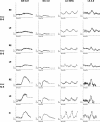
ERG for three carriers of the c.824dupG mutation in RGR: data for WVU: III-6, WVU: III-8, and WVU: IV-5, are presented with responses recorded from a healthy 58-year control in the bottom for comparison. Note that the recording was performed according to the previous ISCEV standard protocols, which have been updated after the recordings were obtained. The WVU age-matched normative data are given in Supplementary Table S2. DA 0.01: dark adapted response to a dim flash stimulus (0.01 cd.s.m−2); DA 3.0: dark adapted response to a bright flash stimulus (3.0 cd.s.m−2); LA 30Hz: light adapted response to a 30 Hz flicker stimulus with intensity 3.0 cd.s.m−2; LA 3.0: light adapted response to a flash stimulus (3.0 cd.s.m−2). Note the electrical noise resulting from the mains interference.
Figure 5
Figure 5
ISCEV-standard ERG for two carriers of the c.824dupG mutation in RGR: GC4177: V-2 and GC4177: VI-1, together with responses recorded from a healthy control in the bottom for comparison. RE, right eye; LE, left eye. DA 0.01: dark adapted response to a dim flash stimulus (0.01 cd.s.m−2); DA 10.0: dark adapted response to a bright flash stimulus (10.0 cd.s.m−2); LA 30Hz: light adapted response to a 30 Hz flicker stimulus with intensity 3.0 cd.s.m−2; LA 3.0: light adapted response to a flash stimulus (3.0 cd.s.m−2); PERG: pattern ERG response to a standard 15° field. The full field and PERG for VI-1 are normal. The right eye of V-2 underwent extended dark adaptation before ERG testing to investigate if the RGR mutation affects the visual cycle. The amplitude of the dark-adapted response to DA 10.0 from the right eye is higher than that from the left eye, but this difference is not clinically significant. The scotopic and photopic full field ERGs from both eyes are normal. PERG shows mild reduction of the P50 amplitude in the left eye, with normal peak time.

References

    1. Bech-Hansen NT, Naylor MJ, Maybaum TA, et al. Mutations in NYX, encoding the leucine-rich proteoglycan nyctalopin, cause X-linked complete congenital stationary night blindness. Nat Genet. 2000;26:319–323. - PubMed
    1. Dryja TP, McGee TL, Reichel E, et al. A point mutation of the rhodopsin gene in one form of retinitis pigmentosa. Nature. 1990;343:364–366. - PubMed
    1. Kohl S, Zobor D, Chiang WC, et al. Mutations in the unfolded protein response regulator ATF6 cause the cone dysfunction disorder achromatopsia. Nat Genet. 2015;47:757–765. - PMC - PubMed
    1. Chakarova CF, Papaioannou MG, Khanna H, et al. Mutations in TOPORS cause autosomal dominant retinitis pigmentosa with perivascular retinal pigment epithelium atrophy. Am J Hum Genet. 2007;81:1098–1103. - PMC - PubMed
    1. Jiang M, Pandey S, Fong HK. An opsin homologue in the retina and pigment epithelium. Invest Ophthalmol Vis Sci. 1993;34:3669–3678. - PubMed

Publication types

MeSH terms

Substances