Review
. 2018 Oct 1;59(12):4856-4870.
doi: 10.1167/iovs.18-24957.

Ocular Clocks: Adapting Mechanisms for Eye Functions and Health

Affiliations
Review

Ocular Clocks: Adapting Mechanisms for Eye Functions and Health

Marie-Paule Felder-Schmittbuhl et al. Invest Ophthalmol Vis Sci. .

Abstract

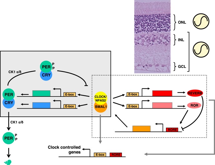
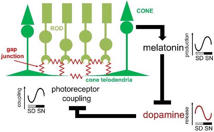
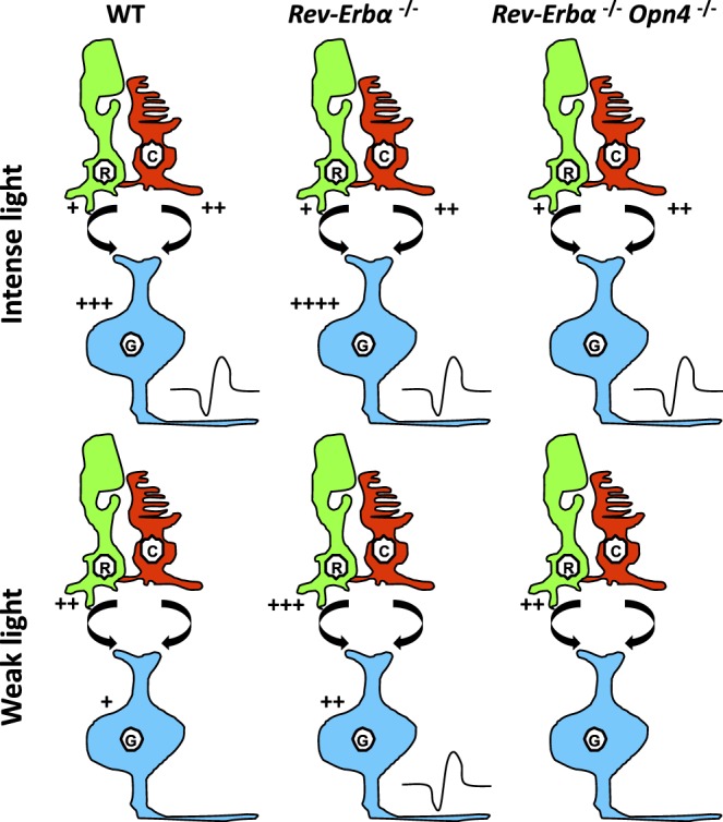
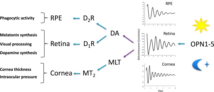
Vision is a highly rhythmic function adapted to the extensive changes in light intensity occurring over the 24-hour day. This adaptation relies on rhythms in cellular and molecular processes, which are orchestrated by a network of circadian clocks located within the retina and in the eye, synchronized to the day/night cycle and which, together, fine-tune detection and processing of light information over the 24-hour period and ensure retinal homeostasis. Systematic or high throughput studies revealed a series of genes rhythmically expressed in the retina, pointing at specific functions or pathways under circadian control. Conversely, knockout studies demonstrated that the circadian clock regulates retinal processing of light information. In addition, recent data revealed that it also plays a role in development as well as in aging of the retina. Regarding synchronization by the light/dark cycle, the retina displays the unique property of bringing together light sensitivity, clock machinery, and a wide range of rhythmic outputs. Melatonin and dopamine play a particular role in this system, being both outputs and inputs for clocks. The retinal cellular complexity suggests that mechanisms of regulation by light are diverse and intricate. In the context of the whole eye, the retina looks like a major determinant of phase resetting for other tissues such as the retinal pigmented epithelium or cornea. Understanding the pathways linking the cell-specific molecular machineries to their cognate outputs will be one of the major challenges for the future.


Figures

Figure 1
Figure 1
The molecular clock pathways and retinal clocks. Schematic representation of the transcriptional/translational feedback loops model for the molecular clock. The BMAL1/CLOCK (or BMAL1/NPAS2) dimer activates transcription of the Per and Cry genes upon binding to the Ebox sequences in their promoters. In turn, PER and CRY proteins form heterodimers able to inhibit transcriptional activity of BMAL1/CLOCK, thus turning down their own transcription. Meanwhile, these factors undergo posttranslational modifications, in particular, phosphorylation of PER proteins by the Casein Kinases 1δ or 1ε, signaling for ubiquitination and proteasomal degradation, and then allowing the cycle to restart. BMAL1/CLOCK likewise activates the expression of Rev-Erb and Ror genes, which products respectively repress and activate transcription of the Bmal1 gene at retinoic acid-related orphan receptor binding elements (RORE) sites. This generates an additional loop interlocked with the previous one, all together contributing to the robustness of the clockwork. The presence of Ebox and/or RORE sequences throughout the genome supports the rhythmic regulation of a set of target genes (CCG) for BMAL1/CLOCK, BMAL1/NPAS2, REV-ERB, and ROR transcription factors. Clock gene expression dynamics over the 24-hour cycle conforming to this model have been described in the ONL and in the inner retina (INL + GCL) in several ex vivo studies, as symbolized next to the eosin/hematoxylin stained transversal section of a rat retina shown in the upper-right corner of the figure.
Figure 2
Figure 2
Overview of photoreceptor gap junction coupling and the melatonin/dopamine pathway. Gap junctions are located at photoreceptor terminals. Melatonin production is under the control of a clock within the photoreceptor layer and high at night. Melatonin suppresses dopamine release, and it is the nocturnal decrease in dopamine release and the subsequent decrease in activity of dopamine D2/4 receptors on photoreceptors that increases photoreceptor coupling. SD, subjective day; SN, subjective night.
Figure 3
Figure 3
Hypothetical mechanism explaining the effect of Rev-Erbα knockout on nonvisual responses. In wild-type mice, output of ipRGCs (G, blue) in response to light information is an integrated product of all three photoreceptor types, rods (R, green), cones (C, red), and ipRGCs. At sufficient light intensity, input from any/all of these sources can trigger firing (symbolized by the black curve close to ipRGCs), whereas at low light intensity, stimulation is insufficient, although the cells are partly depolarized and exhibit resting potentials close to threshold. However, removal of Rev-Erbα leads to increased rod input (shortened a-wave latencies) and/or increased melanopsin input (higher expression levels, higher numbers), which allow iipRCGs (G, blue) to reach threshold depolarization. In double Rev-Erbα−/−;Opn4−/− mice, the melanopsin component is missing and even with higher rod signaling the system is not able to attain threshold. The number of “+” indicates the hypothetical level of stimulation. Curved arrow between rods and cones, and ipRGCs, indicates bipolar contact.
Figure 4
Figure 4
Entrainment of circadian rhythms in ocular tissues. Many physiologic functions in eye tissues (left column) are regulated by circadian clocks and synchronized to the light/dark cycle. The retina, RPE, and cornea, among others, were shown to harbor autonomous clocks, as demonstrated by their capacity to display oscillating PER2::LUC activity (bioluminescence, counts/sec) in vitro (see representative recordings). One major site for the input of light information from the environment to the eye is the retina. OPN1-5 are opsin photopigments expressed at various levels in the retina: OPN1 (short and middle/long wavelength sensitive) in cones, OPN2 (rhodopsin) in rods, OPN4 in ipRGCs and OPN5 in a subpopulation of ganglion cells as well; localization of OPN3 expressing cells is not known. Most opsins are involved in the effects of light on retinal physiology. OPN4 and OPN5 are involved in the regulation of the retinal clock's response to light. The retina synthesizes (purple arrows) melatonin (MLT) in the night and dopamine (DA) during the day. These signals display phase regulatory properties (blue arrows), in the retina itself, but also in other ocular tissues such as the RPE and cornea.

References

    1. Takahashi JS. Transcriptional architecture of the mammalian circadian clock. Nat Rev Genet. 2017;18:164–179. - PMC - PubMed
    1. Hogenesch JB, Ueda HR. Understanding systems-level properties: timely stories from the study of clocks. Nat Rev Genet. 2011;12:407–416. - PubMed
    1. Panda S, Antoch MP, Miller BH, et al. Coordinated transcription of key pathways in the mouse by the circadian clock. Cell. 2002;109:307–320. - PubMed
    1. Zhang R, Lahens NF, Ballance HI, Hughes ME, Hogenesch JB. A circadian gene expression atlas in mammals: implications for biology and medicine. Proc Natl Acad Sci U S A. 2014;111:16219–16224. - PMC - PubMed
    1. Mure LS, Le HD, Benegiamo G, et al. Diurnal transcriptome atlas of a primate across major neural and peripheral tissues. Science. 2018;359:eaao0318. - PMC - PubMed

Publication types