. 2018 Dec 15;444(2):93-106.
doi: 10.1016/j.ydbio.2018.10.004. Epub 2018 Oct 19.

Sequential specification of oligodendrocyte lineage cells by distinct levels of Hedgehog and Notch signaling

Affiliations

Sequential specification of oligodendrocyte lineage cells by distinct levels of Hedgehog and Notch signaling

Andrew M Ravanelli et al. Dev Biol. .

Abstract

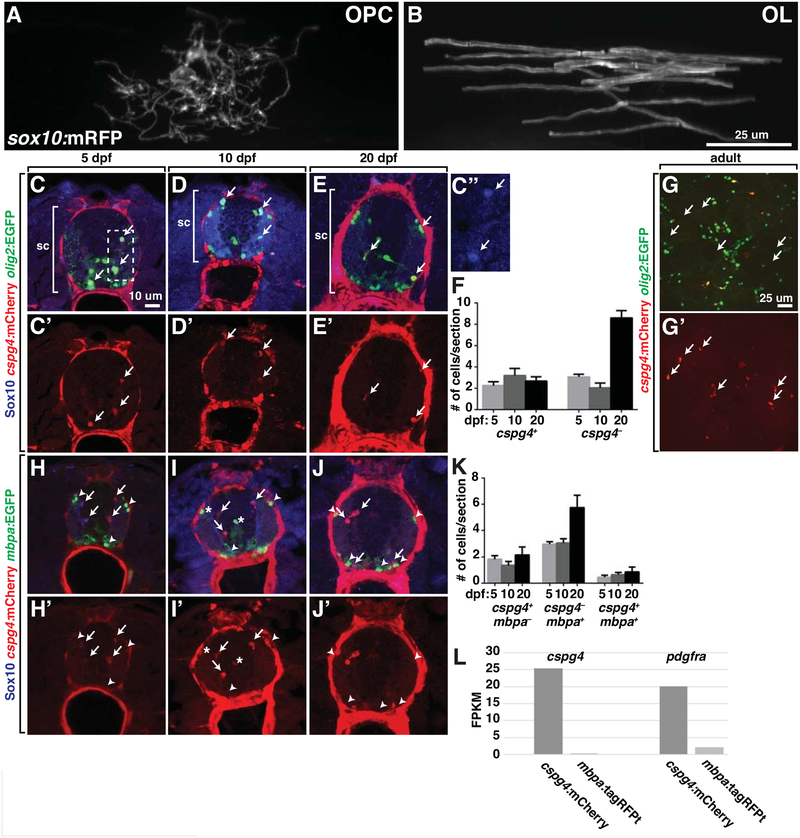
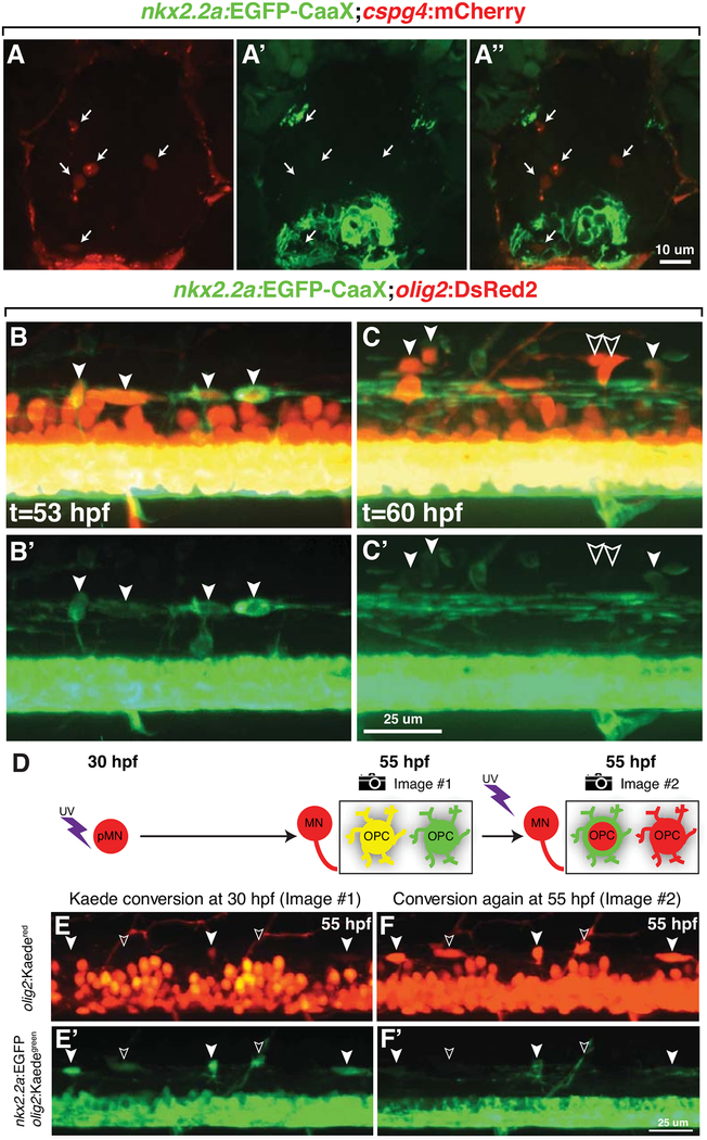
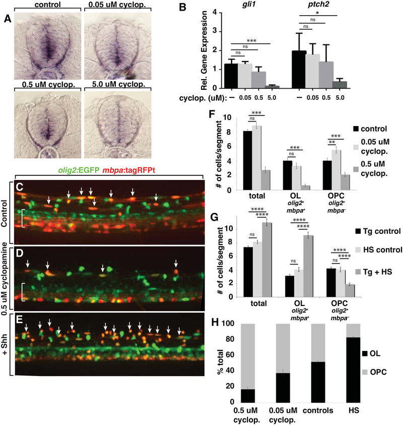
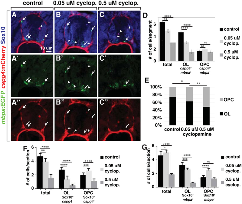
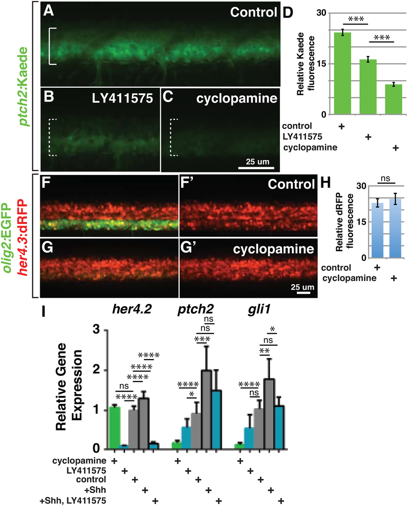
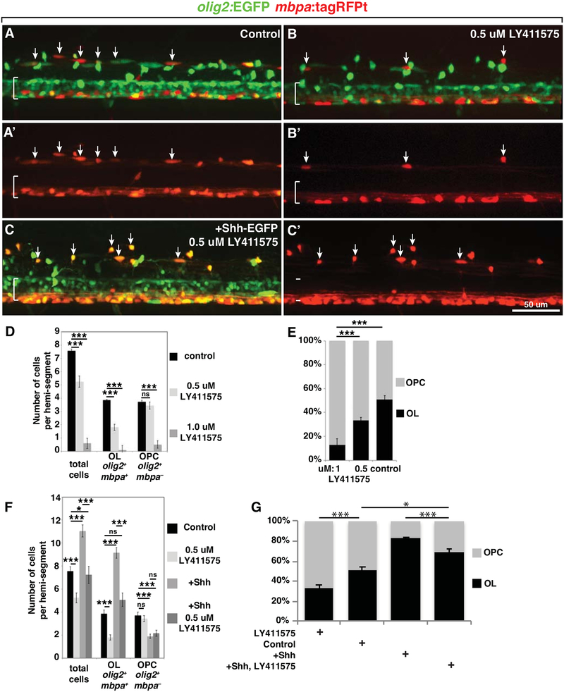
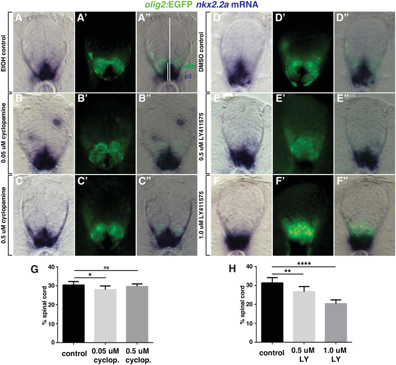
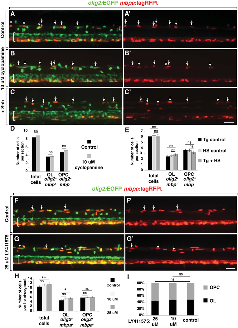
During development of the central nervous system oligodendrocyte precursor cells (OPCs) give rise to both myelinating oligodendrocytes and NG2 glia, which are the most proliferative cells in the adult mammalian brain. NG2 glia retain characteristics of OPCs, and some NG2 glia produce oligodendrocytes, but many others persist throughout adulthood. Why some OPCs differentiate as oligodendrocytes during development whereas others persist as OPCs and acquire characteristics of NG2 glia is not known. Using zebrafish spinal cord as a model, we found that OPCs that differentiate rapidly as oligodendrocytes and others that remain as OPCs arise in sequential waves from distinct neural progenitors. Additionally, oligodendrocyte and persistent OPC fates are specified during a defined critical period by small differences in Shh signaling and Notch activity, which modulates Shh signaling response. Thus, our data indicate that OPCs fated to produce oligodendrocytes or remain as OPCs during development are specified as distinct cell types, raising the possibility that the myelinating potential of OPCs is set by graded Shh signaling activity.


Figures

Figure 1.
Figure 1.. OPCs and Oligodendrocytes Occupy Larval Zebrafish Spinal Cord.
(A) sox10:mRFP+ cell in a 7 dpf larvae with morphology characteristic of an OPC. (B) sox10:mRFP+ oligodendrocyte (OL) at 7 dpf. (C-E) Representative confocal images of transverse sections at the level of trunk spinal cord (sc, brackets) of 5, 10 and 20 dpf larvae showing cspg4:mCherry and olig2:EGFP expression combined with Sox10 immunohistochemistry. Examples of triple-labeled cells are indicated by arrows. C’-E’ show cspg4:mCherry alone. C” is an enlargement of the boxed area in panel C to show Sox10+ cells. (F) Number of OPCs (cspg4:mCherry+ olig2:EGFP+ Sox10+) and oligodendrocytes (cspg4:mCherry olig2:EGFP+ Sox10+) per transverse section for 5, 10 and 20 dpf larval stages. n=10 larvae for each stage. (G) Transverse section of adult spinal cord showing cspg4:mCherry and olig2:EGFP expression. Arrows indicate examples of double-labeled cells. G’ shows cspg4:mCherry alone. (H-J) Representative confocal images of transverse sections at the level of trunk spinal cord of 5, 10 and 20 dpf larvae showing cspg4:mCherry and mbpa:EGFP expression combined with Sox10 immunohistochemistry. Arrows indicate cspg4:mCherry+ mbpa:EGFP Sox10+ OPCs, arrowheads mark cspg4:mCherry mbpa:EGFP+ Sox10+ oligodendrocytes and asterisks indicate rare cspg4:mCherry+ mbpa:EGFP+ Sox10+ cells. (K) Number of OPCs (cspg4:mCherry+ mbpa:EGFP Sox10+) and oligodendrocytes (cspg4:mCherry mbpa:EGFP+ Sox10+ and cspg4:mCherry+ mbpa:EGFP+ Sox10+) per transverse section for 5, 10 and 20 dpf larval stages. n=10 larvae for each stage. (L) Expression (FPKM) of cspg4 and pdgfra in cspg4:mCherry+ and mbpa:tagRFPt+ cells isolated from 7 dpf larvae.
Figure 2.
Figure 2.. Distinct OPCs Arise Sequentially from Progenitors that Initiate olig2 Expression at Different Times.
(A-A”) Representative transverse section of 5 dpf Tg(nkx2.2a:EGFP-CaaX);Tg(cspg4:mCherry) larva showing red, green and combined channels. Arrows indicate cspg4:mCherry+ OPCs. (B-C’). Images captured from time-lapse videos of Tg(olig2:DsRed2);Tg(nkx2.2a:EGFP-CaaX) embryos at the level of the trunk spinal cord, with dorsal up. (A,A’) At 53 hpf, dorsally migrated olig2+ OPCs are nkx2.2a+ (filled arrowheads), indicating oligodendrocyte fate. (B,B’) At 60 hpf, some dorsally migrated olig2+ OPCs are nkx2.2a (outlined arrowheads). (D) Schematic representation of photoconversion experiment and result. Exposure of Tg(olig2:Kaede);Tg(nkx2.2a:EGFP-CaaX) to uv wavelength light at 30 hpf converts all olig2:Kaede+ cells from green to red. Motor neurons remain red because they no longer transcribe olig2 whereas OPCs continue to express olig2, forming newly synthesized green Kaede. OPCs that initiated olig2 expression prior to photoconversion are yellow whereas those that initiate olig2 expression after photoconversion are green. To determine subtype identity, Kaede was photoconverted again at 55 hpf, revealing nkx2.2a:EGFP-CaaX expression marking OPCs fated for oligodendrocyte development. (E,E’) Projection of confocal image stack of 55 hpf embryo that had been photoconverted at 55 hpf. OPCs that are both green and red (filled arrowheads) and only green (outlined arrowheads) are evident. (F,F’) Image of same embryo shown in D following second photoconversion at 55 hpf. OPCs that were green and red are nkx2.2a+, indicating oligodendrocyte fate, whereas OPCs that were only green are nkx2.2a.
Figure 3.
Figure 3.. Different Levels of Shh Signaling Specify OPC and Oligodendrocyte Fates.
(A) Representative images of transverse sections of trunk spinal cord obtained from 48 hpf embryos processed, in parallel, for in situ RNA hybridization to detect ptch2 expression. Embryos were treated beginning at 24 hpf with control solution or 0.05, 0.5 or 5.0 μM cyclopamine (cyclop.). (B) Relative levels of gli1 and ptch2 transcripts detected by semi-quantitative RT-PCR. n=5 biological replicates with 20 larvae pooled per sample. (C-E) Representative images of confocal stacks of living 5 dpf Tg(olig2:EGFP);Tg(mbpa:tagRFPt) larvae at the level of the trunk spinal cord. Dorsal spinal cord is toward the top and brackets indicate the pMN domain in ventral spinal cord. Arrows mark olig2+ mbpa+ oligodendrocytes. (C) Larva treated with control solution. (D) Larva treated with 0.5 M cyclopamine. (E) Heat shocked larva carrying the Tg(hsp70l:shha-EGFP) transgene in addition to the reporter transgenes. (F) Average number of dorsal olig2+ cells, olig2+ mbp+ oligodendrocytes (OL) and olig2+ mbp OPCs in control (n=50 embryos, 2676 cells), 0.5 μM cyclopamine-treated (n=17 embryos, 285 cells) and 0.05 μM cyclopamine-treated (n=7 embryos, 624 cells) larvae. (G) Average number of dorsal olig2+ cells, olig2+ mbp+ oligodendrocytes and olig2+ mbp OPCs in Tg control (heat-shocked larvae lacking hsp70l:shha-EGFP transgene; n=19 larvae, 842 cells), HS control (hsp70l:shha-EGFP larvae not subjected to heat-shock; n=10 larvae, 490 cells) and Tg + HS (heat-shocked hsp70l:shha-EGFP larvae; n=10 larvae, 663 cells). (H) Proportion of all dorsal olig2+ cells consisting of olig2+ mbp+ oligodendrocytes and olig2+ mbp OPCs from cyclopamine treatment and heat-shock experiments. * < 0.05, ** < 0.01, *** < 0.001, **** < 0.0001. ns, not significant, where p > 0.05. Data are presented as mean ± SEM. qPCR data evaluated using unpaired t test with Welsh correction. Cell count data evaluated using unpaired, two tailed t test.
Figure 4.
Figure 4.. Changing the level of Hedgehog signaling changes the proportions of spinal cord oligodendrocytes and OPCs.
(A-C”) Representative images of transverse sections through the trunk spinal cords of 5 dpf Tg(cspg4:mCherry);Tg(mbpa:EGFP) larvae and processed to detect Sox10 expression (blue). Arrows indicate mbpa:EGFP+ oligodendrocytes and arrowheads mark cspg4:mCherry+ OPCs. (D) Average number of total cspg4+ and mbpa+ cells, cspg4+ mbp OPCs and cspg4 mbp+ oligodendrocytes in control (n=10 larvae, 643 cells), 0.5 μM cyclopamine-treated (n=10 larvae, 307 cells) and 0.05 μM cyclopamine-treated (n=10 larvae, 502 cells). (E) Proportions of cspg4+ mbp OPCs and cspg4 mbp+ oligodendrocytes. (F) Average number of total Sox10+ cspg4+ and Sox10+ cspg4 cells, Sox10+ cspg4+ OPCs and Sox10+ cspg4 oligodendrocytes in control (n=10 larvae), 0.5 μM cyclopamine-treated (n=10 larvae) and 0.05 μM cyclopamine-treated (n=10 larvae). p values are indicated by asterisks. * < 0.05, ** < 0.01, *** < 0.001, **** < 0.0001. ns, not significant, where p > 0.05. Data are presented as mean ± SEM. Cell count data evaluated using unpaired, two tailed t test.
Figure 5.
Figure 5.. Notch Activity Enhances Shh Signaling.
(A-C) Representative confocal images of living 48 hpf Tg(ptch2:Kaede) embryos at the level of the trunk spinal cord with dorsal up. Brackets mark ventral spinal cord. (A) Embryo treated with control solution. (B) Embryo treated with 2.5 μM LY411575 to inhibit Notch signaling. (C) Embryo treated with 5 μM cyclopamine. (D) Relative Kaede fluorescence intensity measurements for control (n=5), LY411575 treated (n=6) and cyclopamine treated (n=5) embryos. (F-G’) Representative confocal images of living 48 hpf Tg(olig2:EGFP);Tg(her4.3:dRFP) embryos at the level of the trunk spinal cord. (F) Embryo treated with control solution. (G) Embryo treated with 5 μM cyclopamine. (H) Relative dRFP fluorescence intensity measurements for control (n=18) and cyclopamine-treated (n=11) Tg(olig2:EGFP);Tg(her4.3:dRFP) embryos. (I) Relative levels of her4.2, ptch2 and gli1 transcripts. Cyclopamine and LY411575 concentrations were 5.0 and 2.5 μM, respectively. +Shh indicates heat shocked Tg(hsp70l:shha-EGFP) embryos. p values are indicated by asterisks. * < 0.05, ** < 0.01, *** < 0.001, **** < 0.0001. ns, not significant, where p > 0.05. Data are presented as mean ± SEM. Fluorescence intensity data evaluated using two-tailed t test. qPCR data evaluated using unpaired t test with Welsh correction.
Figure 6.
Figure 6.. Notch Activity Modulates Shh Signaling to Specify Oligodendrocytes and OPCs.
(A-C’) Representative projections of confocal stack images of living 5 dpf Tg(olig2:EGFP);Tg(mbpa:tagRFPt) larvae. Dorsal is up and brackets mark the pMN domain of ventral spinal cord. Arrows indicate olig2+ mbpa+ oligodendrocytes. Panel pairs show olig2:EGFP and mbpa:tagRFPt expression combined and mbpa:tagRFPt expression alone. (A,A’) Larva treated with control solution. (B,B’) Larvae treated with 0.5 μM LY411575. (C,C’) Heat shocked larva carrying the Tg(hsp70l:shha-EGFP) transgene in addition to the reporter transgenes and treated with 0.5 μM LY411575. (D) Average number of total dorsal spinal cord olig2+ cells, olig2+ mbp+ oligodendrocytes and olig2+ mbpa OPCs in control (n=10, 454 cells), 0.5 μM LY411575 treated (n=11, 346 cells) and 1.0 μM LY411575 treated (n=17, 60 cells) larvae. (E) The effect of Notch inhibition on the proportions of olig2+ mbpa+ oligodendrocytes and olig2+ mbpa OPCs. (F) Average number of total number of dorsal spinal cord olig2+ cells, olig2+ mbpa+ oligodendrocytes and olig2+ mbpa OPCs in control, 0.5 μM LY411575 treated, Shh overexpressing and Shh overexpressing + 0.5 μM LY411575 treated (n=21, 760 cells) larvae. For this graph the control data were combined from Figures 3F and 5D. The LY411575 data are from Figure 5D and the +Shh data are from Figure 3F, shown here for comparison. (G) Proportion of all dorsal olig2+ cells consisting of olig2+ mbpa+ oligodendrocytes and olig2+ mbpa OPCs following Notch inhibition and Shh overexpression in the presence and absence of Notch inhibitor. p values are indicated by asterisks. * < 0.05, ** < 0.01, *** < 0.001, **** < 0.0001. ns, not significant, where p > 0.05. Data are presented as mean ± SEM. Data were evaluated using unpaired, two-tailed t test.
Figure 7.
Figure 7.. Effects of Shh and Notch inhibitors on nkx2.2a expression dynamics.
(A-F) Representative examples of differential interference contrast (DIC), epifluorescent and combined images of transverse sections of Tg(olig2:EGFP) embryos treated from 24–48 hpf with control solutions (A,D), cyclopamine (B,C) or LY411575 (E,F) and processed to detect nkx2.2a mRNA expression. nkx2.2a expression marks the p3 domain whereas olig2:EGFP expression marks the pMN domain. White lines on panel A” indicate height measurements for the nkx2.2a expression domain and spinal cord. (G) Averaged percent height of the spinal cord occupied by nkx2.2a expression for EtOH treated control (n=8), 0.05 μM cyclopamine treated (n=7) and 0.5 μM cyclopamine treated (n=7) embryos. (H) Averaged percent height of the spinal cord occupied by nkx2.2a expression for DMSO treated control (n=8), 0.5 μM LY411575 treated (n=8) or 0.5 μM LY411575 treated (n=8) embryos. p values are indicated by asterisks. * < 0.05, ** < 0.01, **** < 0.0001. ns, not significant, where p > 0.05. Data are presented as mean ± SEM. Data were evaluated using unpaired, two-tailed t test.
Figure 8.
Figure 8.. Manipulation of Shh and Notch Signaling Following Specification Does Not Alter Oligodendrocyte and OPC Formation.
(A-C’, F-G’) Representative projections of confocal stack images of living 5 dpf Tg(olig2:EGFP);Tg(mbpa:tagRFPt) larvae. Dorsal is up and brackets mark the pMN domain of ventral spinal cord. Arrows indicate olig2+ mbpa+ oligodendrocytes. Panel pairs show olig2:EGFP and mbpa:tagRFPt expression combined and mbpa:tagRFPt expression alone. (A,A’) Larva treated with control solution. (B,B’) Larva treated with 10 μM cyclopamine beginning at 72 hpf. (C,C’) hsp70l:Shh-GFP; olig2:EGFP; mbpa:tagRFPt larva heat-shocked at 72hpf. (D) Average number of dorsally migrated olig2+ mbpa+ oligodendrocytes and olig2+ mbpa OPCs for control (n=24 larvae,1042 cells) and 10 μM cyclopamine (n=11 larvae, 596 cells) treatments. (E) Average number of dorsally migrated olig2+ mbpa+ oligodendrocytes and olig2+ mbpa OPCs for transgenic (Tg) control (n=8 larvae), heat-shock (HS) control (n=5 larvae) and +Shh (n=16 larvae, 590 cells) treatments. (F,F’) Larva treated with control solution. (G,G’) Larva treated with 25 μM Notch inhibitor LY411575 beginning at 72 hpf. (H) Average number of dorsally migrated olig2+ mbpa+ oligodendrocytes and olig2+ mbpa OPCs in control (n=9 larvae, 571 cells),10 μM LY411575 (n=15 larvae, 1021 cells), and 25 μM LY411575 (n=15 larvae, 1064 cells) treatments. p values are indicated by asterisks. * < 0.05, ** < 0.01. ns, not significant, where p > 0.05. Data are presented as mean ± SEM. Data were evaluated using unpaired, two-tailed t test. All scale bars represent 25 um.

Similar articles

Cited by

References

    1. Agius E, Soukkarieh C, Danesin C, Kan P, Takebayashi H, Soula C, and Cochard P (2004). Converse control of oligodendrocyte and astrocyte lineage development by Sonic hedgehog in the chick spinal cord. Dev. Biol 270, 308–321. - PubMed
    1. Barth KA, and Wilson SW (1995). Expression of zebrafish nk2.2 is influenced by sonic hedgehog/vertebrate hedgehog-1 and demarcates a zone of neuronal differentiation in the embryonic forebrain. Development 121, 1755–1768. - PubMed
    1. Birey F, Kloc M, Chavali M, Hussein I, Wilson M, Christoffel DJ, Chen T, Frohman MA, Robinson JK, Russo SJ, et al. (2015). Genetic and Stress-Induced Loss of NG2 Glia Triggers Emergence of Depressive-like Behaviors through Reduced Secretion of FGF2. Neuron 88, 941–956. - PMC - PubMed
    1. Bussmann J, and Schulte-Merker S (2011a). Rapid BAC selection for tol2-mediated transgenesis in zebrafish. Development 138, 4327–4332. - PubMed
    1. Bussmann J, and Schulte-Merker S (2011b). Rapid BAC selection for tol2-mediated transgenesis in zebrafish. Development 138, 4327–4332. - PubMed

Publication types

MeSH terms