Review
. 2018 Oct 19;6(4):25.
doi: 10.3390/jdb6040025.

Drosophila as a Model for Developmental Biology: Stem Cell-Fate Decisions in the Developing Nervous System

Affiliations
Review

Drosophila as a Model for Developmental Biology: Stem Cell-Fate Decisions in the Developing Nervous System

Katherine Harding et al. J Dev Biol. .

Abstract

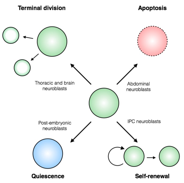
Stem cells face a diversity of choices throughout their lives. At specific times, they may decide to initiate cell division, terminal differentiation, or apoptosis, or they may enter a quiescent non-proliferative state. Neural stem cells in the Drosophila central nervous system do all of these, at stereotypical times and anatomical positions during development. Distinct populations of neural stem cells offer a unique system to investigate the regulation of a particular stem cell behavior, while comparisons between populations can lead us to a broader understanding of stem cell identity. Drosophila is a well-described and genetically tractable model for studying fundamental stem cell behavior and the mechanisms that underlie cell-fate decisions. This review will focus on recent advances in our understanding of the factors that contribute to distinct stem cell-fate decisions within the context of the Drosophila nervous system.

Keywords: Drosophila; apoptosis; cell cycle; development; neuroblast; stem cell biology.


Conflict of interest statement

The authors declare no conflict of interest.

Figures

Figure 1
Figure 1
Stem cell-fate decisions in the Drosophila central nervous system.Drosophila neural stem cells, called neuroblasts, exhibit diverse stem cell behaviors. Thoracic and brain neuroblasts are eliminated by terminal division (top left), while abdominal neuroblasts undergo apoptosis (top right). Temporal controls regulate the period of neuroblast quiescence between embryonic and larval stages of development (bottom left). Certain larval neuroblasts within the inner proliferating center (IPC) of the optic lobe switch from renewing asymmetric to amplifying symmetric divisions (bottom right). Execution of these cell-fate decisions is intimately tied to neuroblast identity in time and space.
Figure 2
Figure 2
Neuroblast populations within the Drosophila central nervous system. (A) Cell-fate decisions are mapped onto anatomical populations of neuroblasts during embryonic and larval development. The majority of abdominal neuroblasts undergo apoptosis during embryogenesis, leaving 3 neuroblasts per hemisegment in the larval CNS. Neuroblasts stop proliferating between embryonic and larval development, except for 4 mushroom body neuroblasts. Following reactivation, neuroblasts in the thorax and brain undergo terminal division, while the remaining abdominal neuroblasts die through apoptosis. (B) Proliferation patterns of neuroblasts throughout the larval CNS. Most neuroblasts use the Type I division pattern, except a small population of Type II neuroblasts in the brain. The neuroblasts in the inner proliferating center of the optic lobe have been observed to divide symmetrically.
Figure 3
Figure 3
Spatial patterning of the Drosophila ventral nerve cord. (A) A single hemisegment containing approximately 30 neuroblasts is shown in the inset. Gene expression is patterned within the hemisegment: two examples of spatially restricted molecular markers are shown (mirror and wingless; from the Hyper-Neuroblast Map, Doe laboratory). (B) Upon delamination from the neuroectoderm, neuroblast identity is specified within a hemisegment by a Cartesian grid generated by expression patterns of the columnar and segment polarity genes (reviewed in [16]). (C) Along the anterior-posterior axis of the embryo, hemisegment identity is also subject to spatial regulation by the Hox gene family. All of these spatial patterns are superimposed in vivo, providing each neuroblast with a unique spatial identity.
Figure 4
Figure 4
The temporal series and division patterns of Drosophila neuroblasts. (A) The temporal transcription factor (TTF) sequence expressed in embryonic Type I neuroblasts. The last member, grainyhead, has been shown to regulate both proliferation and apoptosis of neuroblasts, suggesting that context-dependent co-factors may dictate the cell-fate outcome for a given neuroblast. (B) The TTF sequence expressed in larval Type II neuroblasts. Progression to the late factors is dependent on the steroid hormone ecdysone, and leads to a terminal division [26,32]. (C) Three types of asymmetric neuroblast proliferation patterns observed in the nervous system. Type 0 divisions have only been observed following a period of Type I divisions in embryonic neuroblasts.
Figure 5
Figure 5
Localization of proteins during asymmetric and symmetric neuroblast divisions. (A) A typical asymmetric neuroblast division involves segregation of apical and basal cortex complex members to opposite sides of the dividing neuroblast. Proteins within the apical cortex complex are retained in the stem cell, while factors associated with the basal cortex complex are transmitted to the daughter GMC. (B) Symmetric divisions of the IPC neuroblasts display low Prospero and Miranda protein levels, but Brat and Numb proteins are divided equally between the two new stem cells [51].
Figure 6
Figure 6
Regulation of neuroblast quiescence and reactivation. (A) The majority of post-embryonic neuroblasts exit the cell cycle at G2 phase (G2Q), while the remaining ~25% display a canonical G0 arrest [73]. Tribbles promotes and maintains quiescence through degradation of Cdc25/Stg and inhibition of Akt signaling (not shown). (B) Prospero regulates neural differentiation and is required for neuroblast quiescence, but the mechanism underlying its ability to induce two different cell fates is unclear. The connections between G2Q and the Prospero regulatory network are also unknown. (C) Secreted factors from the larval fatbody are sensed by the brain, leading to secretion of insulin-like peptides (dILPs). dILPs are also expressed by stellate surface glia, and these act on the neuroblast to promote growth and proliferation.
Figure 7
Figure 7
Regulation of neuroblast apoptosis. (A) The RHG genes grim, reaper and sickle are inactive in non-apoptotic cells, allowing DIAP1 to sequester and inhibit caspases. Apoptosis is induced following transcriptional activation of the RHG genes. RHG binding to DIAP1 promotes its turnover and releases caspases to allow their activation. (B) The intergenic enhancer NBRR is required for neuroblast apoptosis and for expression of the surrounding RHG genes. (C) A proposed model for regulation of RHG genes by long-range DNA interactions between the NBRR and RHG promoters mediated by the cohesin complex.
Figure 8
Figure 8
Regulation of neuroblast terminal division. (A) The RNA-binding proteins Imp and Syp have opposing effects on Prospero-mediated terminal division. Imp is sufficient to prevent cell cycle exit, but Syp may play an indirect permissive role for Prospero function. (B) Imp and Syp expression correlates to progression of the larval temporal series. Imp expression is down-regulated following the pulse of ecdysone, but Syp expression begins prior to this pulse.
Figure 9
Figure 9
Misregulation of stem cell and differentiated cell identity. (A) Notch activity is detected equally in the stem cell and GMC following neuroblast division, but quickly becomes restricted to the neuroblast [128]. The super elongation complex (SEC) promotes a positive feedback loop with Notch signaling in the neuroblast, while Numb inhibits Notch specifically in the GMC. (B) Loss of differentiated cell identity can lead to unchecked proliferation in the nervous system. Two classes of dedifferentiation mutants have been identified: pros and brat mutants fail to restrict the GMC to a single terminal division, while mdlc, lola or nerfin-1 mutants display ectopic expression of neuroblast markers in neurons. lola and nerfin-1 mutants also result in ectopic proliferation of neuronal cells.

Similar articles

Cited by

References

    1. Truman J.W., Bate M. Spatial and Temporal Patterns of Neurogenesis in the Central Nervous System of Drosophila melanogaster. Dev. Biol. 1988;125:145–157. doi: 10.1016/0012-1606(88)90067-X. - DOI - PubMed
    1. White K., Grether M.E., Abrams J.M., Young L., Farrell K., Steller H. Genetic Control of Programmed Cell Death in Drosophila. Science. 1994;264:677–683. doi: 10.1126/science.8171319. - DOI - PubMed
    1. Bello B.C., Hirth F., Gould A.P. A Pulse of the Drosophila Hox Protein Abdominal-A Schedules the End of Neural Proliferation via Neuroblast Apoptosis. Neuron. 2003;37:209–219. doi: 10.1016/S0896-6273(02)01181-9. - DOI - PubMed
    1. Maurange C., Cheng L., Gould A.P. Temporal Transcription Factors and Their Targets Schedule the End of Neural Proliferation in Drosophila. Cell. 2008;133:891–902. doi: 10.1016/j.cell.2008.03.034. - DOI - PubMed
    1. Abrams J.M., White K., Fessier L.I., Steller H. Programmed cell death during Drosophila embryogenesis. Development. 1993;117:29–43. - PubMed

LinkOut - more resources