Review
. 2018 Oct 19;19(10):3244.
doi: 10.3390/ijms19103244.

The Proposal of Molecular Mechanisms of Weak Organic Acids Intake-Induced Improvement of Insulin Resistance in Diabetes Mellitus via Elevation of Interstitial Fluid pH

Affiliations
Review

The Proposal of Molecular Mechanisms of Weak Organic Acids Intake-Induced Improvement of Insulin Resistance in Diabetes Mellitus via Elevation of Interstitial Fluid pH

Yoshinori Marunaka. Int J Mol Sci. .

Abstract

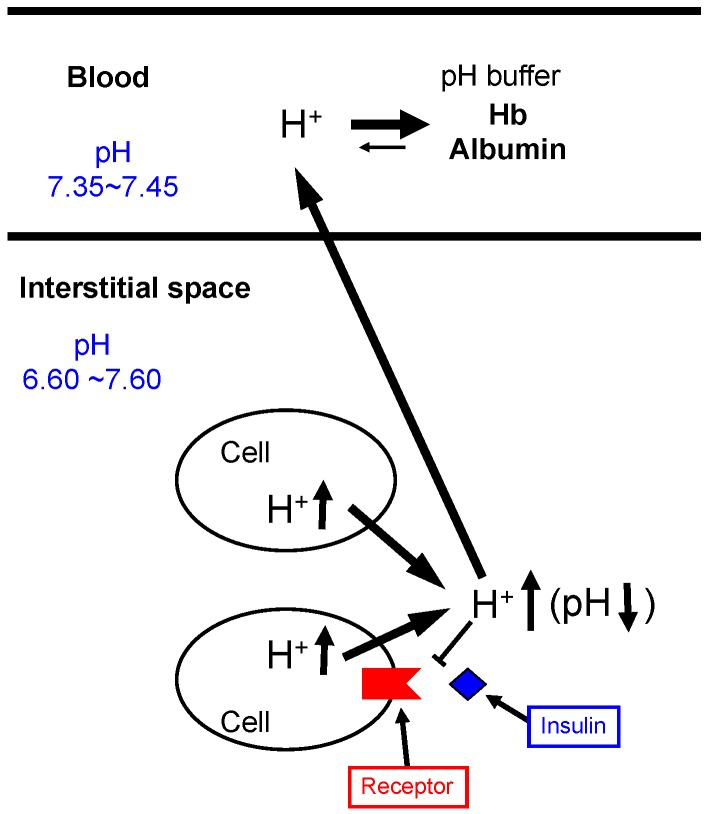
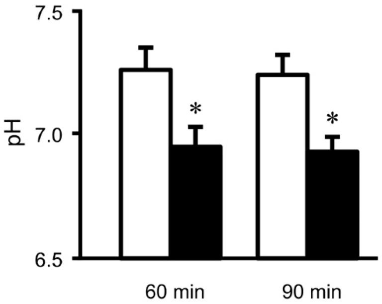
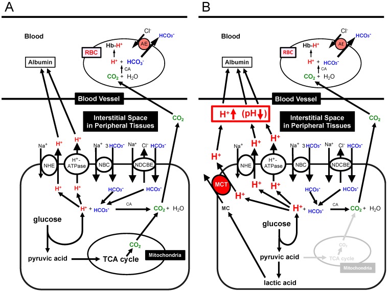
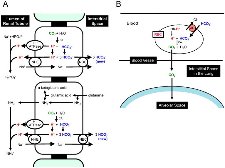
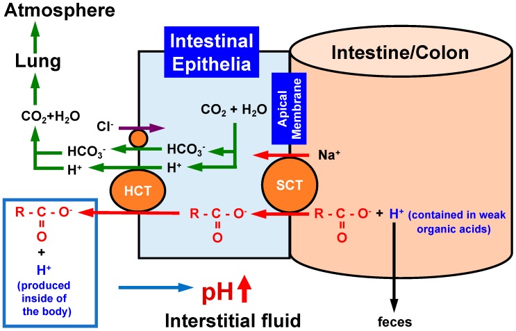
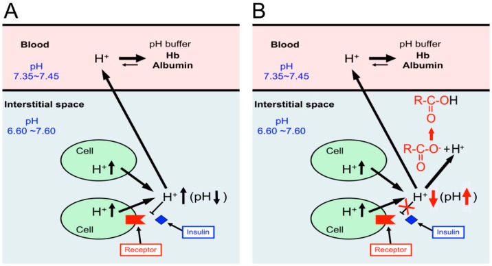
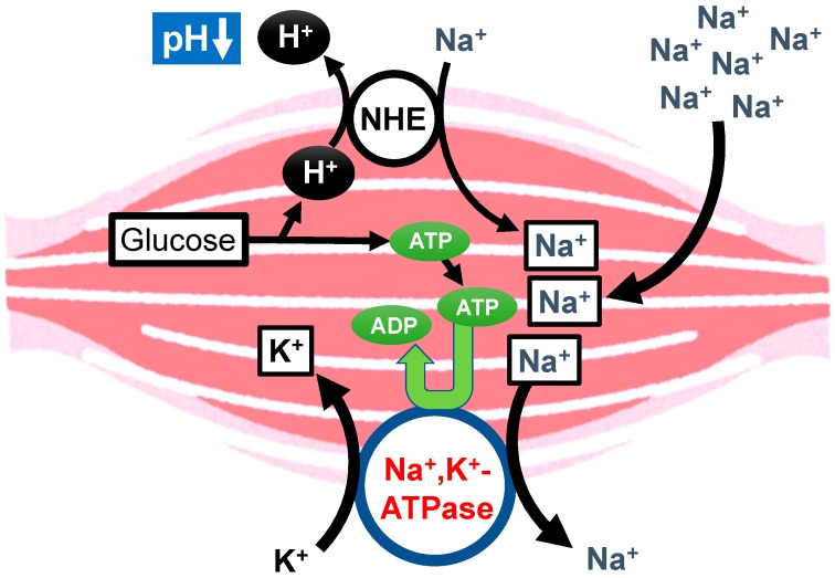
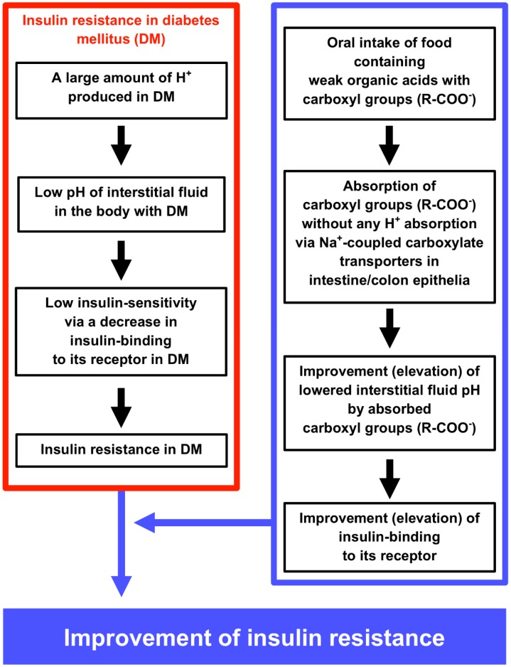
Blood contains powerful pH-buffering molecules such as hemoglobin (Hb) and albumin, while interstitial fluids have little pH-buffering molecules. Thus, even under metabolic disorder conditions except severe cases, arterial blood pH is kept constant within the normal range (7.35~7.45), but the interstitial fluid pH under metabolic disorder conditions becomes lower than the normal level. Insulin resistance is one of the most important key factors in pathogenesis of diabetes mellitus, nevertheless the molecular mechanism of insulin resistance occurrence is still unclear. Our studies indicate that lowered interstitial fluid pH occurs in diabetes mellitus, causing insulin resistance via reduction of the binding affinity of insulin to its receptor. Therefore, the key point for improvement of insulin resistance occurring in diabetes mellitus is development of methods or techniques elevating the lowered interstitial fluid pH. Intake of weak organic acids is found to improve the insulin resistance by elevating the lowered interstitial fluid pH in diabetes mellitus. One of the molecular mechanisms of the pH elevation is that: (1) the carboxyl group (R-COO-) but not H⁺ composing weak organic acids in foods is absorbed into the body, and (2) the absorbed the carboxyl group (R-COO-) behaves as a pH buffer material, elevating the interstitial fluid pH. On the other hand, high salt intake has been suggested to cause diabetes mellitus; however, the molecular mechanism is unclear. A possible mechanism of high salt intake-caused diabetes mellitus is proposed from a viewpoint of regulation of the interstitial fluid pH: high salt intake lowers the interstitial fluid pH via high production of H⁺ associated with ATP synthesis required for the Na⁺,K⁺-ATPase to extrude the high leveled intracellular Na⁺ caused by high salt intake. This review article introduces the molecular mechanism causing the lowered interstitial fluid pH and insulin resistance in diabetes mellitus, the improvement of insulin resistance via intake of weak organic acid-containing foods, and a proposal mechanism of high salt intake-caused diabetes mellitus.

Keywords: alkalization; binding affinity; food; insulin; interstitial fluid; pH; weak organic acid.


Conflict of interest statement

The author declares no conflict of interest.

Figures

Figure 1
Figure 1
pH of interstitial fluids and blood, and binding affinity of insulin to its receptor. Blood contains very strong, powerful pH-buffering molecules such as hemoglobin (Hb) and albumin, while interstitial fluids have little pH-buffering molecules. Thus, even under metabolic disorder conditions except severe disorders, arterial blood pH is kept constant within the normal range (7.35–7.45), but pH of interstitial fluids becomes lower than the normal level. Modified from Figure 1 in World J Diabetes 6(1): 125–135, 2015 [1].
Figure 2
Figure 2
pH of interstitial (extracellular) fluids around the hippocampus of Otsuka Long-Evans Tokushima Fatty (OLETF: a model rat of type 2 diabetes mellitus) and normal (Wistar) rats. The pH value is shown as the mean ± SEM (n = 4). The pH values shown in Figure 2 were measured at 60 and 90 min after antimony pH electrodes reached interstitial (extracellular) fluids around the brain hippocampus of OLETF rats (closed columns) and normal (Wistar) rats (open columns). *, p < 0.05 compared with that in normal (Wistar) rats at each measured time. Modified from Mol Cell Therapies 2:6, 2014 [2].
Figure 3
Figure 3
Production of H+ and CO2 and transporting systems of H+ and CO2 in peripheral tissues. (A) Production of H+ and CO2 and transporting systems of H+ and CO2 in peripheral tissues with ‘normal’ mitochondrial function: the glycolysis process produces H+ and TCA cycle generates CO2. (B) Production of H+ and CO2 and transporting systems of H+ and CO2 in peripheral tissues with ‘dysfunction’ of mitochondria. Much more amounts of H+ are produced via glycolysis in a case of mitochondrial dysfunction in order to produce the same amount of ATP as that with normal mitochondrial function. In a case of mitochondrial dysfunction, TCA cycle has no or little function, thus the required amount of ATP is mainly generated via glycolysis, leading to production of much more amounts of H+ and lactic acid than the normal case. Modified from Figure 2 in World J Diabetes 6(1): 125–135, 2015 [1].
Figure 4
Figure 4
Production of HCO3 in the kidney (A) and transporting systems of H+ and CO2 in the lung (B). (A) The H+ extrusion system into urine and new synthesis of HCO3 in the kidney. CO2 is converted into H+ and HCO3 via a CA-facilitated process. The H+ generated from CO2 is extruded into urine as a form of NH4+ or HPO4 by binding to NH3 or HPO42− (NH3 + H+→NH4+: HPO42− + H+→H2PO4). If CO2 is supplied, HCO3 is newly generated and functions as a pH-buffering material coupled with a process of H+ extrusion into urine. (B) The extrusion system of CO2 in the lung. CO2 is converted from H+ and HCO3 produced in the peripheral tissues. In the lung, the process (H+ + HCO3→CO2 + H2O), which is the reversible process occurring in peripheral tissue (refer to the process in RBC described in Figure 2), occurs due to low CO2 circumstances in the lung. HCO3 generated from CO2 in peripheral tissues is consumed to produce CO2, which is released to atmosphere. This means that HCO3 generated from CO2 in peripheral tissues is not a net source of HCO3 when CO2 is released to atmosphere in the lung. Modified from Figure 2 in World J Diabetes 6(1): 125–135, 2015 [1].
Figure 5
Figure 5
Action of oral intake of foods containing weak organic acids with carboxyl groups on pH regulation of the interstitial fluid. When we intake the weak organic ‘acid’ containing a carboxyl part (R-COO), only the carboxyl part (R-COO) is absorbed via sodium-coupled carboxylate transporters (SCT) expressed in the apical membrane of the intestine. Incorporated carboxyl groups are transported from the intracellular space to the extracellular space (the interstitial space) of epithelial cells via H+-coupled carboxylate transporters (HCT). H+ contained in weak organic acids is not absorbed in the intestine, but is excreted into feces. Thus, weak organic acids behave as ‘bases’ by combing with H+ produced in the body, elevating pH. This means that weak organic acids play a role as pH buffers in the interstitial fluid.
Figure 6
Figure 6
Insulin resistance caused by lowered interstitial fluid pH in diabetes mellitus (A), and elevation of interstitial fluid pH and improvement of insulin resistance by intake of foods containing weak organic acids with carboxylic groups (R-COO) (B). (A) The lowered value of interstitial fluid pH diminishes the insulin binding affinity to its receptor, causing the insulin resistance in diabetes mellitus. (B) Elevation of interstitial fluid pH by intake of foods containing weak organic acids with carboxylic groups (R-COO) increases the binding affinity of insulin to its receptor, improving the insulin resistance.
Figure 7
Figure 7
Proposal of the molecular mechanism of the high salt intake-induced development of diabetes mellitus. High salt intake produces a lot of H+, since high salt intake leads to consumption of ATP required for the Na+, K+-ATPase to extrude the high leveled intracellular Na+ caused by high salt intake in cells such as muscles.
Figure 8
Figure 8
The molecular mechanism causing low interstitial fluid pH and insulin resistance in diabetes mellitus (DM) (left column), and the improvement of insulin resistance by oral intake of foods containing weak organic acids with carboxyl groups (right column). The left column inside of the red square indicates the molecular mechanism causing DM and insulin resistance via lowering interstitial fluid pH by producing a lot of H+. The right column inside of the blue square indicates the molecular mechanism of oral intake of foods containing weak organic acids with carboxyl groups improving lowered pH of interstitial fluids, insulin binding and insulin resistance under the DM condition.

References

    1. Marunaka Y. Roles of interstitial fluid pH in diabetes mellitus: Glycolysis and mitochondrial function. World J. Diabetes. 2015;6:125–135. doi: 10.4239/wjd.v6.i1.125. - DOI - PMC - PubMed
    1. Marunaka Y., Yoshimoto K., Aoi W., Hosogi S., Ikegaya H. Low pH of interstitial fluid around hippocampus of the brain in diabetic OLETF rats. Mol. Cell. Ther. 2014;2:6. doi: 10.1186/2052-8426-2-6. - DOI - PMC - PubMed
    1. Fujita T. The metabolic syndrome in Japan. Nat. Clin. Pract. Cardiovasc. Med. 2008;5(Suppl. 1):S15–S18. doi: 10.1038/ncpcardio0808. - DOI - PubMed
    1. Marunaka Y., Hagiwara N., Tohda H. Insulin activates single amiloride-blockable Na channels in a distal nephron cell line (A6) Am. J. Physiol. Renal Physiol. 1992;263:F392–F400. doi: 10.1152/ajprenal.1992.263.3.F392. - DOI - PubMed
    1. Mills E., Kuhn C.M., Feinglos M.N., Surwit R. Hypertension in CB57BL/6J mouse model of non-insulin-dependent diabetes mellitus. Am. J. Physiol. 1993;264:R73–R78. doi: 10.1152/ajpregu.1993.264.1.R73. - DOI - PubMed