. 2018 Oct 21;16(10):395.
doi: 10.3390/md16100395.

A High-Content Screening Assay for the Discovery of Novel Proteasome Inhibitors from Formosan Soft Corals

Affiliations

A High-Content Screening Assay for the Discovery of Novel Proteasome Inhibitors from Formosan Soft Corals

Xue-Hua Ling et al. Mar Drugs. .

Abstract

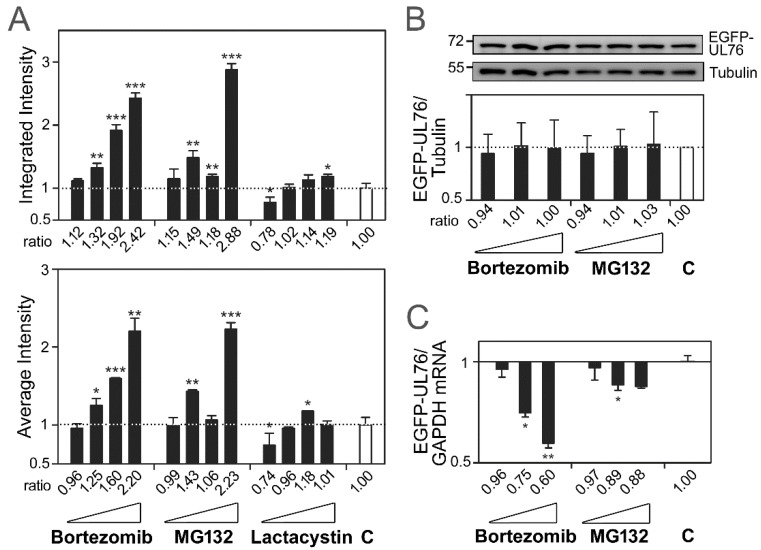
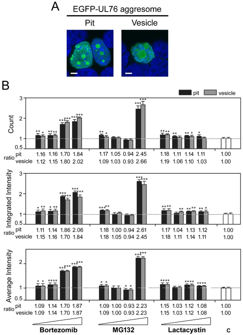
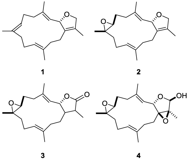
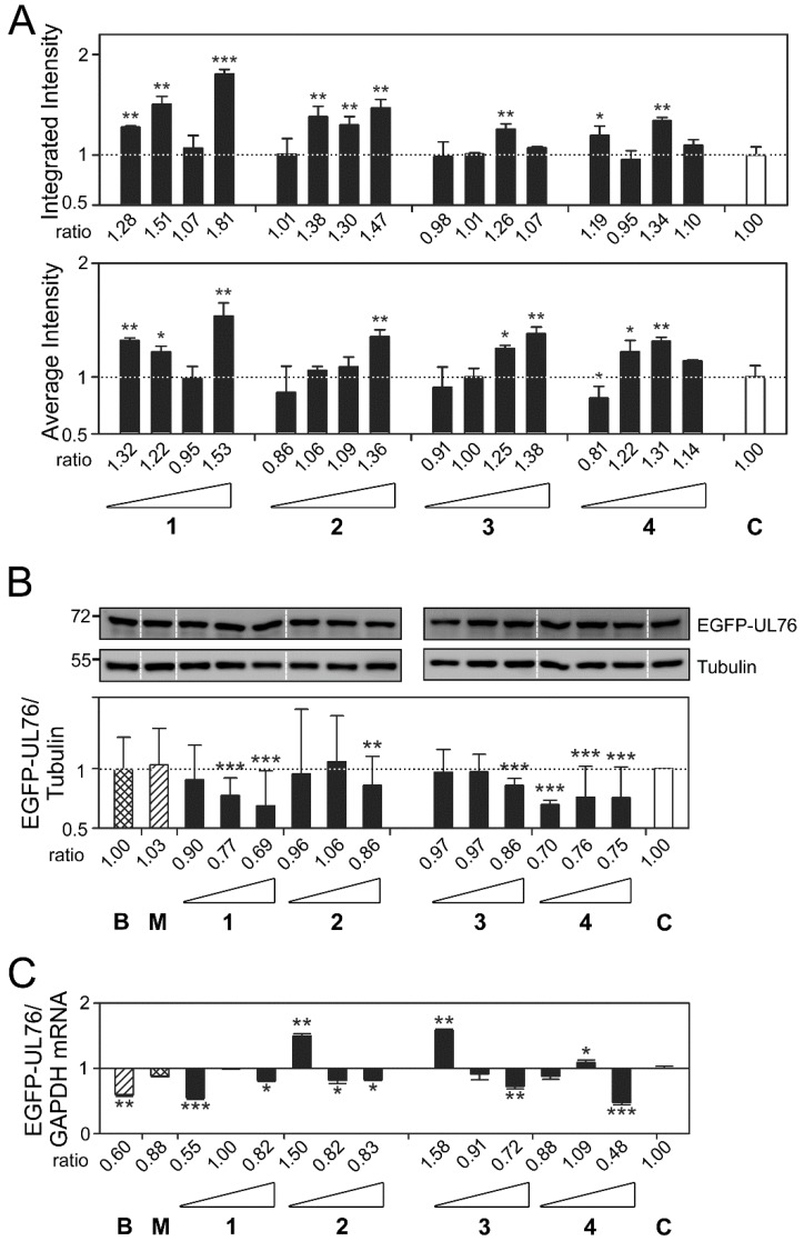
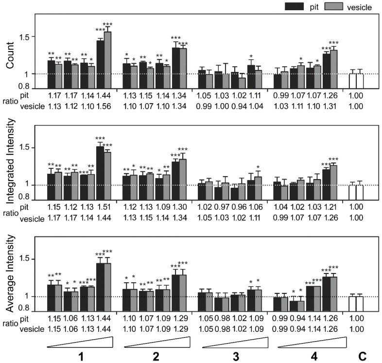
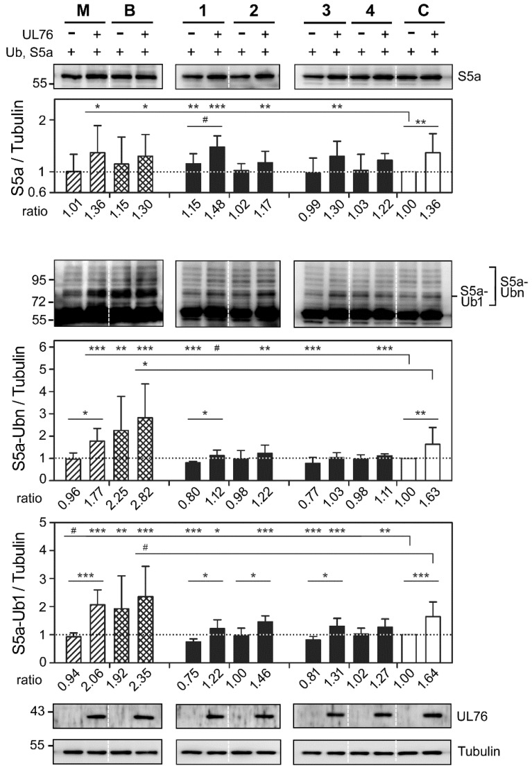
The ubiquitin-proteasome system (UPS) is a major proteolytic pathway that safeguards protein homeostasis. The main 26S proteasome consists of a 20S catalytic core proteasome and a 19S substrate recognition proteasome. UPS dysfunction underlies many important clinical diseases involving inflammation, tumors, and neurodegeneration. Currently, three 20S proteasome inhibitors, bortezomib, carfilzomib, and ixazomib, have been approved for the treatment of multiple myeloma. We aim to screen UPS inhibitors for biomedical purposes. The protein interaction network of human cytomegalovirus UL76 targets UPS, resulting in aggregations of ubiquitinated proteins termed aggresomes. In this study, we demonstrated that cell-based high-content measurements of EGFP-UL76 aggresomes responded to bortezomib and MG132 treatment in a dose-dependent manner. Employing this high-content screening (HCS) assay, we screened natural compounds purified from Formosan soft corals. Four cembrane-based compounds, sarcophytonin A (1), sarcophytoxide (2), sarcophine (3), and laevigatol A (4), were found to enhance the high-content profiles of EGFP-UL76 aggresomes with relative ratios of 0.2. By comparison to the mechanistic action of proteasome inhibitors, compounds 1 and 3 modulated the accumulation of ubiquitinated proteins, with a unique pattern likely targeting 19S proteasome. We confirmed that the EGFP-UL76 aggresome-based HCS system greatly improves the efficacy and sensitivity of the identification of proteasome inhibitors.

Keywords: cembrane; high-content screening; proteasome inhibitor; soft coral.


Conflict of interest statement

The authors declare no conflict of interest.

Figures

Figure 1
Figure 1
(A) The assessment of high-content measurements of EGFP-UL76 aggresome upon treatment with the proteasome inhibitors bortezomib (0.2, 1, 5, and 25 nM), MG132 (0.125, 0.25, 0.5, and 1 µM), and clasto-lactacystin β-lactone (0.08, 0.4, 2, and 10 µM). The integrated (top panel) and average (bottom panel) intensities per cell were measured, and the ratios were obtained by normalization to the control without proteasome inhibitor treatment, which is denoted by C throughout the text. Validation of the protein and transcript levels of EGFP-UL76 upon the addition of the proteasome inhibitors, bortezomib (1, 5, and 25 nM) and MG132 (0.25, 0.5, and 1 µM). (B) Western blot imaging and densitometric analyses were performed to quantitate the relative ratio of protein expression. The molecular mass markers are shown on the left in kDa. An equal level of α-tubulin was used as an internal loading control in each sample. Protein ratios of EGFP-UL76/tubulin were obtained by normalization to the control value. (C) Quantitative PCR was performed to assess the ratio of EGFP-UL76 mRNA expression. The transcript ratios of EGFP-UL76/GAPDH were obtained by normalization to the control value. All data points are the averages of at least three repetitive experiments. The error bars indicate standard deviations. The following symbols are used to indicate statistical significance throughout the text: * 0.01 < p < 0.05; ** 0.001 < p < 0.01; *** p < 0.001.
Figure 2
Figure 2
The high-content measurements of EGFP-UL76 aggresomes classified by diameter under proteasome inhibitor treatment. (A) Representative confocal images of EGFP-UL76 aggresomes expressed HEK293T cells transfected with pEGFP-UL76. Nuclei were counterstained with DAPI. Pit and vesicle denote aggresomes 1 to 5 µm and 5 to 20 µm in diameter, respectively. Scale bar: 5 µm. (B) Measurements of pit and vesicle aggresomes per cell were as follows: Count number, integrated intensity, and average intensities for pit and vesicle aggresomes, respectively. The relative ratio was normalized to the control values without inhibitor treatment. All data points are the averages of at least three repetitive experiments. The error bars indicate standard deviations. The following symbols are used to indicate statistical significance throughout the text: * 0.01 < p < 0.05; ** 0.001 < p < 0.01; *** p < 0.001.
Figure 3
Figure 3
Marine natural products exhibit proteasome inhibition by high-content assays. Sarcophytonin A is (1), sarcophytoxide (2), sarcophine (3), and laevigatol A (4).
Figure 4
Figure 4
Marine compounds modulated high-content measurements of EGFP-UL76 aggresomes. (A) Assessment of the integrated and average intensities of EGFP-UL76 aggresomes (1 to 50 µm) per cell. The tested concentrations were 0.2, 1, 5, and 25 µg/mL for compounds 1, 2, 3, and 4, respectively. (B) Western blot imaging and densitometric analyses were performed to quantitate the EGFP-UL76/tubulin protein ratio with the addition of marine compound treatment at 1, 5, and 25 µg/mL. The molecular mass markers are shown on the left in kDa. (C) Quantitative PCR was conducted to assess the transcript ratio of EGFP-UL76/GAPDH in HEK293T cells treated with marine compounds at 1, 5, and 25 µg/mL. The high-content measurements of EGFP-UL76 with the addition of the proteasome inhibitors, bortezomib (25 nM, denoted B) and MG132 (1 µM, denoted M), were used as positive controls. All data points are the averages of at least three repetitive experiments. The error bars indicate standard deviations. The following symbols are used to indicate statistical significance throughout the text: * 0.01 < p < 0.05; ** 0.001 < p < 0.01; *** p < 0.001.
Figure 5
Figure 5
Classification of the high-content measurements of EGFP-UL76 aggresomes by size with marine compound treatment at 0.2, 1, 5, and 25 µg/mL. Pit and vesicle denote aggresomes 1 to 5 µm and 5 to 20 µm in diameter, respectively. The following pit and vesicle measurements per cell were determined: Count number, integrated intensity, and average intensity. The relative ratio was normalized to control values without marine compound treatment. All data points are the averages of at least three repetitive experiments. The error bars indicate standard deviations. The following symbols are used to indicate statistical significance throughout the text: * 0.01 < p < 0.05; ** 0.001 < p < 0.01; *** p < 0.001.
Figure 6
Figure 6
Marine compounds promote the accumulation of cellular ubiquitin-conjugated proteins. HEK293T cells transiently expressed S5a and ubiquitin proteins by transfection with pcDNA3-HA-S5a and pcDNA3-FLAG-Ub (denoted +). Cells expressing UL76 were transfected with pEF-UL76 (+), and cells not expressing UL76 were instead transfected with the cloning vector, pEF1/Myc-His C (−). Antibodies that recognized the UL76 and FLAG epitopes detected UL76 and ubiquitin-conjugated proteins, respectively. The cellular proteins were resolved in 8.5% SDS-PAGE. An equal level of α-tubulin was used as an internal loading control in each sample. The molecular mass markers are shown on the left in kDa. Cells were treated with the following drug concentrations: MG132 (M), 1 µM; bortezomib (B), 25 nM; compounds 1, 2, 3, and 4, 25 µg/mL. The intensities of the indicated polyubiquitinated area (Ubn, marked by brackets) were quantitated by densitometric analysis. Data were obtained from the average of at least three repeats. The relative ratios of ubiquitinated proteins were normalized to the values with neither UL76 production nor drug treatment. All data points are the averages of at least three repetitive experiments. The error bars indicate standard deviations. The following symbols are used to indicate statistical significance throughout the text: # 0.05 < p < 0.1; * 0.01 < p < 0.05; ** 0.001 < p < 0.01; *** p < 0.001.
Figure 7
Figure 7
Marine compounds modulated the accumulation of cellular S5a, polyubiquitinated S5a, and monoubiquitinated S5a. HEK293T cells were transfected and exposed to the drug as described in Figure 7. S5a was detected by antibody against the HA epitope. The intensity of ubiquitinated S5a was detected by prolonging the acquisition time in the imaging detection system. The intensities of the indicated S5a, polyubiquitinated S5a (S5a-Ubn, marked by brackets), and monoubiquitinated S5a (S5a-Ub1, marked by flat bar) areas were quantitated by densitometric analyses. The ratios were normalized to the values with neither UL76 production nor drug treatment. All data points are the averages of at least three repetitive experiments. The error bars indicate standard deviations. The following symbols are used to indicate statistical significance throughout the text: # 0.05 < p < 0.1; * 0.01 < p < 0.05; ** 0.001 < p < 0.01; *** p < 0.001.

Similar articles

Cited by

References

    1. Wegley Kelly L., Haas A.F., Nelson C.E. Ecosystem microbiology of coral reefs: Linking genomic, metabolomic, and miogeochemical dynamics from animal symbioses to reefscape processes. mSystems. 2018;3:e00162-17. doi: 10.1128/mSystems.00162-17. - DOI - PMC - PubMed
    1. Quinn R.A., Vermeij M.J., Hartmann A.C., Galtier d’Auriac I., Benler S., Haas A., Quistad S.D., Lim Y.W., Little M., Sandin S., et al. Metabolomics of reef benthic interactions reveals a bioactive lipid involved in coral defence. Proc. Biol. Sci. 2016;283:20160469. doi: 10.1098/rspb.2016.0469. - DOI - PMC - PubMed
    1. Aratake S., Tomura T., Saitoh S., Yokokura R., Kawanishi Y., Shinjo R., Reimer J.D., Tanaka J., Maekawa H. Soft coral Sarcophyton (Cnidaria: Anthozoa: Octocorallia) species diversity and chemotypes. PLoS ONE. 2012;7:e30410. doi: 10.1371/journal.pone.0030410. - DOI - PMC - PubMed
    1. Blunt J.W., Carroll A.R., Copp B.R., Davis R.A., Keyzers R.A., Prinsep M.R. Marine natural products. Nat. Prod. Rep. 2018;35:8–53. doi: 10.1039/C7NP00052A. - DOI - PubMed
    1. Lehmann G., Udasin R.G., Livneh I., Ciechanover A. Identification of UBact, a ubiquitin-like protein, along with other homologous components of a conjugation system and the proteasome in different gram-negative bacteria. Biochem. Biophys. Res. Commun. 2017;483:946–950. doi: 10.1016/j.bbrc.2017.01.037. - DOI - PubMed

MeSH terms

LinkOut - more resources