. 2018 Oct 22;18(1):1016.
doi: 10.1186/s12885-018-4950-0.

Recombinant PAPP-A resistant insulin-like growth factor binding protein 4 (dBP4) inhibits angiogenesis and metastasis in a murine model of breast cancer

Affiliations

Recombinant PAPP-A resistant insulin-like growth factor binding protein 4 (dBP4) inhibits angiogenesis and metastasis in a murine model of breast cancer

Y E Smith et al. BMC Cancer. .

Abstract

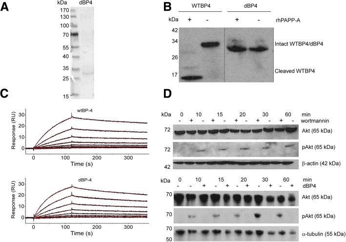
Background: The Insulin-like growth factor (IGF) pathway plays a role in tumour development and progression. In vivo, IGF1 activity is regulated by the IGF binding proteins (IGFBPs). IGFBP4 inhibits the activity of IGF1 but proteolytic cleavage by pregnancy-associated plasma protein-A (PAPP-A) releases active IGF1. A modified IGFBP4, dBP4, which was resistant to PAPP-A cleavage but retained IGF1 binding capacity, was engineered, expressed in Human Embryonic Kidney (HEK) 293 cells and purified. This study examined the effects of dBP4 on IGF1-induced cell migration, invasion and angiogenesis in vitro. The effect of intra-tumour injections of dBP4 on tumour angiogenesis and metastasis was examined using the 4T1.2luc orthotopic model of breast cancer.

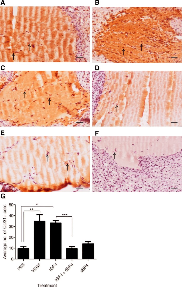
Methods: PAPP-A resistance and IGF binding capacity of dBP4 were characterized by Western blot and surface plasmon resonance, respectively. 4T1.2luc are mouse mammary adenocarcinoma cells transfected with luciferase to allow in vivo imaging. The effect of dBP4 on IGF1-induced Akt activation in 4T1.2luc cells was assessed by Western blot. Cell migration and invasion assays were performed using 4T1.2luc cells. Angiokit™ assays and Matrigel® implants were used to assess the effects of dBP4 on angiogenesis in vitro and in vivo, respectively. An orthotopic breast cancer model - 4T1.2luc cells implanted in the mammary fat pad of BALB/c mice - was used to assess the effect of intra tumour injection of purified dBP4 on tumour angiogenesis and metastasis. Tumour growth and lung metastasis were examined by in vivo imaging and tumour angiogenesis was evaluated by CD31 immunohistochemistry.

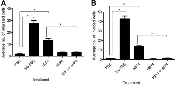
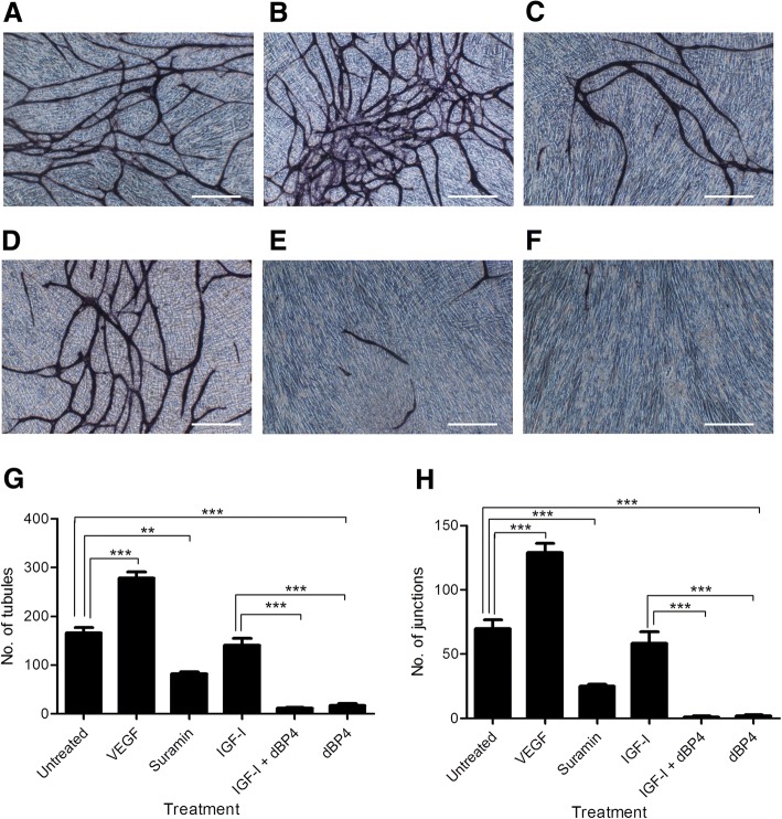
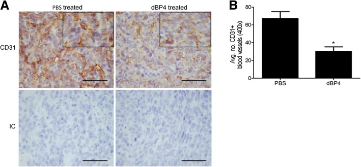
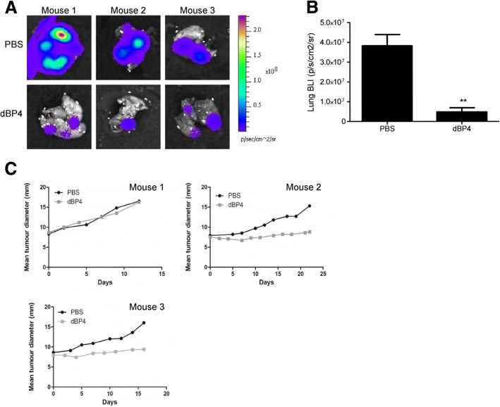
Results: Our engineered, PAPP-A resistant IGFBP4 (dBP4) retained IGF1 binding capacity and inhibited IGF1 activation of Akt as well as IGF1-induced migration and invasion by 4T1.2 mammary adenocarcinoma cells. dBP4 inhibited IGF1-induced angiogenesis in vitro and in Matrigel implants in vivo. Direct intra-tumour injection of soluble dBP4 reduced angiogenesis in 4T1.2 luc mammary tumours tumour and reduced lung metastasis.

Conclusion: A PAPP-A resistant IGFBP4, dBP4, inhibits angiogenesis and metastasis in 4T1.2 mammary fat pad tumours. This study highlights the therapeutic potential of dBP4 as an approach to block the tumour-promoting actions of IGF1.

Keywords: Angiogenesis; IGFBP4/dBP4; Insulin-like growth factor (IGF); Pregnancy associated plasma protein A/PAPP-A.


Conflict of interest statement

Ethics approval

Animal studies were approved by the Ethics Committee of Royal College of Surgeons in Ireland, carried out under animal license guidelines of the Department of Health, Ireland and in accordance with the UK Co-ordinating Committee on Cancer Research (UKCCCR) Guidelines for the Welfare of Animals in Experimental Neoplasia.

Consent for publication

Not applicable.

Competing interests

The authors declare that they have no competing interests.

Publisher’s Note

Springer Nature remains neutral with regard to jurisdictional claims in published maps and institutional affiliations.

Figures

Fig. 1
Fig. 1
dBP4 binds IGF1, is resistant to PAPP-A cleavage and inhibits IGF-induced Akt phosphorylation. a SDS-PAGE analysis of purified dBP4 protein. b dBP4 or wtIGFBP4 (WTBP4) were treated with recombinant PAPP-A in the presence of IGF1 for 24 h. Intact IGFBP4 and cleavage fragments were identified by western blot with anti-IGFBP4 antibody. Position of molecular weight (kDa) markers is indicated. Empty lanes on the blot were removed between WTBP4 and dBP4 for clarity. c Surface plasmon resonance. Twofold serial dilutions of purified HIS-tagged mouse wtIGFBP4 or dBP4 (50 nM to 0.4 nM) were injected over a surface with immobilized mouse IGF1. Recorded binding curves are shown in black and a global 1:1 L fit is shown in red. d 25 μg of cell lysate was fractionated by SDS-PAGE, and total Akt or pAkt identified by western blot. (upper panel) 4T1.2luc cells were pre-treated with the PI3K inhibitor, wortmannin, followed by IGF1 (100 ng/ml). β-actin is shown as loading control. (lower panel) 4T1.2luc cells were treated with IGF1 (100 ng/ml) or IGF1 pre-incubated with 2.5 μg/ml dBP4. α-tubulin is shown as loading control. Blots are representative of three independent experiments
Fig. 2
Fig. 2
dBP4 inhibits migration and invasion of 4T1.2luc cells. a Chemotaxis and b invasion of 4T1.2luc cells towards human IGF1 (100 ng/ml), dBP4 (2.5 μg/ml) or IGF1 (100 ng/ml) with dBP4 (2.5 μg/ml). PBS and 5% (v/v) FBS were used as negative and positive controls, respectively. Data (n = 3 independent experiments) are expressed as mean ± SEM of migrated (a) or invaded (b) cells from three fields of view (original magnification 200×) (*P < 0.001, one-way ANOVA with Tukey post hoc test)
Fig. 3
Fig. 3
dBP4 inhibits angiogenesis in vitro. Representative images of human microvascular endothelial cells stained with CD31. a Untreated control, b VEGF positive control, c suramin negative control, d IGF1, e IGF1 plus dBP4, f dBP4 (scale bar 500 μm). g number of tubules and (h) number of junctions. Data (n = 3) are expressed as mean ± SEM. (**P < 0.01, *** P < 0.001 one-way ANOVA with Tukey post hoc test)
Fig. 4
Fig. 4
dBP4 inhibits IGF1-induced angiogenesis in vivo. 12 week old female BALB/c mice (n = 5/group) were injected subcutaneously with Matrigel®. After 7 days implants were excised and stained for CD31 (pointed arrow indicates CD31+ cells). Representative images of a PBS negative control, b VEGF positive control, c IGF1, d IGF1 and dBP4, e dBP4. f shows negative isotype control (scale bars 200 μm). g Mean ± SEM CD31+ cells in three fields of view/implant (n = 5). (*P < 0.05), **P < 0.01, ***P < 0.001) one-way ANOVA with Tukey post hoc test)
Fig. 5
Fig. 5
dBP4 inhibits angiogenesis in 4T1.2luc tumours. 4T1.2luc cells (5 × 104) were implanted into the mammary fat pad of female BALB/c mice (n = 3/group). Once tumours reached a MTD of 8–8.5 mm, mice received an intra-tumour injection of 50 μg dBP4 or PBS every 2–3 days. Once tumours reached a MTD of 17 mm, mice were sacrificed. Primary tumours were excised and blood vessels visualized by staining for CD31 a shows representative images of CD31+ stained tumours from PBS or dBP4 treated mice. IC: Negative isotype control. (scale bar 50 μm). Enlarged area shows vessel morphology. b Mean ± SEM CD31+ vessels in 5 fields of view/tumour (n = 3/group). (*P < 0.05, unpaired Students t-test)
Fig. 6
Fig. 6
a Bioluminescence (BLI) images of lungs from dBP4 or PBS treated mice (n = 3). b Metastatic lung burden following treatment expressed as BLI. Data are expressed as mean ± SEM (*P < 0.05, **P < 0.01, unpaired Students t test). c Primary tumour growth curves of individual mice

References

    1. Pollak M. Insulin and insulin-like growth factor signalling in neoplasia. Nat Rev Cancer. 2008;8:915–928. doi: 10.1038/nrc2536. - DOI - PubMed
    1. Leroith D, Werner H, Beitner-Johnson D, Roberts CT., Jr Molecular and cellular aspects of the insulin-like growth factor I receptor. Endocr Rev. 1995;16:143–163. doi: 10.1210/edrv-16-2-143. - DOI - PubMed
    1. Petley T, Graff K, Jiang W, Yang H, Florini J. Variation among cell types in the signaling pathways by which IGF1 stimulates specific cellular responses. Horm Metab Res. 1999;31:70–76. doi: 10.1055/s-2007-978701. - DOI - PubMed
    1. Hermanto U, Zong CS, Wang LH. Inhibition of mitogen-activated protein kinase kinase selectively inhibits cell proliferation in human breast cancer cells displaying enhanced insulin-like growth factor I-mediated mitogen-activated protein kinase activation. Cell Growth Differ. 2000;11:655–664. - PubMed
    1. Grey A, Chen Q, Xu X, Callon K, Cornish J. Parallel phosphatidylinositol-3 kinase and p42/44 mitogen-activated protein kinase signaling pathways subserve the mitogenic and antiapoptotic actions of insulin-like growth factor I in osteoblastic cells. Endocrinology. 2003;144:4886–4893. doi: 10.1210/en.2003-0350. - DOI - PubMed

MeSH terms

Substances