

Anatomical study of jugular foramen in the neck
- PMID: 30348503
- PMCID: PMC9422587
- DOI: 10.1016/j.bjorl.2018.09.004
Anatomical study of jugular foramen in the neck
Abstract
Introduction: The anatomical complexity of the jugular foramen makes surgical procedures in this region delicate and difficult. Due to the advances in surgical techniques, approaches to the jugular foramen became more frequent, requiring improvement of the knowledge of this region anatomy.
Objective: To study the anatomy of the jugular foramen, internal jugular vein and glossopharyngeal, vagus and accessory nerves, and to identify the anatomical relationships among these structures in the jugular foramen region and lateral-pharyngeal space.
Methods: A total of 60 sides of 30 non-embalmed cadavers were examined few hours after death. The diameters of the jugular foramen and its anatomical relationships were analyzed.
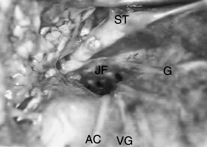
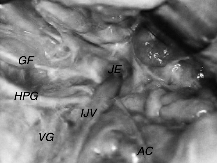
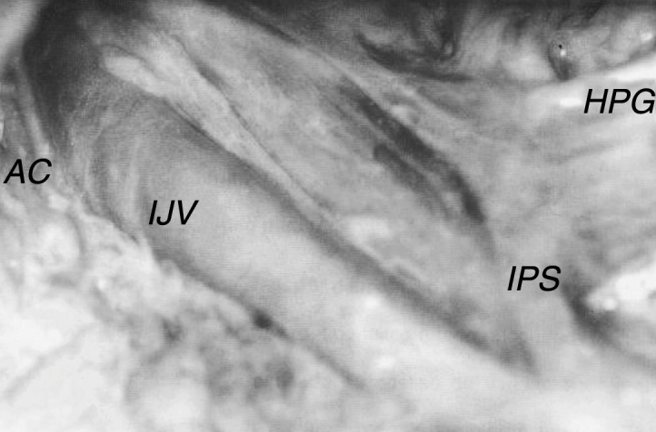
Results: The diameters of the jugular foramen and internal jugular vein were greater on the right side in most studied specimens. The inferior petrosal sinus ended in the internal jugular vein up to 40mm below the jugular foramen; in 5% of cases. The glossopharyngeal nerve exhibited an intimate anatomical relationship with the styloglossus muscle after exiting the skull, and the vagal nerve had a similar relationship with the hypoglossal nerve. The accessory nerve passed around the internal jugular vein via its anterior wall in 71.7% of cadavers.
Conclusion: Anatomical variations were found in the dimensions of the jugular foramen and the internal jugular vein, which were larger in size on the right side of most studied bodies; variations also occurred in the trajectory and anatomical relationships of the nerves. The petrosal sinus can join the internal jugular vein below the foramen.
Introdução: A complexidade anatômica do forame jugular torna a realização de procedimentos cirúrgicos nessa região delicada e difícil. Devido aos avanços obtidos nas técnicas cirúrgicas, as abordagens do forame jugular têm sido feitas com maior frequência, o que requer uma melhoria correspondente no conhecimento de sua anatomia.
Objetivo: Estudar a anatomia do forame jugular, da veia jugular interna e dos nervos glossofaríngeo, vago e acessório, assim como as relações anatômicas entre estas estruturas na região do forame jugular e no espaço parafaríngeo.
Método: Foram examinados 60 lados de 30 cadáveres frescos algumas horas após a morte. Os diâmetros e suas relações anatômicas foram analisados.
Resultados: Os diâmetros do forame jugular e da veia jugular interna foram maiores no lado direito na maioria dos espécimes estudados. O seio petroso inferior terminava na veia jugular interna até 40 mm abaixo do forame jugular, em 5% dos casos. O nervo glossofaríngeo exibiu uma relação íntima anatômica com o músculo estiloglosso após a sua saída do crânio e o nervo vago exibiu uma relação semelhante com o nervo hipoglosso. O nervo acessório passou em torno da veia jugular interna via sua parede anterior em 71,7% dos cadáveres.
Conclusão: Foram encontradas variações anatômicas nas dimensões do forame jugular e da veia jugular interna, que apresentaram tamanhos maiores à direita na maioria dos espécimes estudados; variações também ocorreram na trajetória e nas relações anatômicas dos nervos. O seio petroso pode se unir à veia jugular interna abaixo do forame.
Keywords: Accessory nerve; Base do crânio; Glossopharyngeal nerve; Jugular veins; Nervo acessório; Nervo glossofaríngeo; Nervo vago; Skull base; Vagus nerve; Veias jugulares.
Copyright © 2018 Associação Brasileira de Otorrinolaringologia e Cirurgia Cérvico-Facial. Published by Elsevier Editora Ltda. All rights reserved.
Figures
References
-
- Tummala R.P., Coscarela E., Morcos J.J. Surgical anatomy of the jugular foramen. Oper Tech Neurosurg. 2005;8:2–5.
-
- Dias G.J., Perumal V., Smith C., Cornwal J. The relationship between jugular foramen asymmetry and superior sagital venous sinus laterality. Anthropol Sci. 2014;22:115–120.
-
- Prades J.M., Martin C.H., Veyret C.H., Merzougui N., Chelikh L. Anatomic basis of the infratemporal approach of the jugular foramen. Surg Radiol Anat. 1994;16:11–20. - PubMed
-
- Katsuta T., Rhoton A.L., Jr., Matsushima T. The jugular foramen: microsurgical anatomy and operative approaches. Neurosurgery. 1997;41:149–201. - PubMed
-
- Keles B., Semaan M.T., Fayad J.N. The medial wall of the jugular foramen: a temporal bone anatomic study. Otolaryngol Head Neck Surg. 2009;141:401–407. - PubMed
MeSH terms
LinkOut - more resources
Full Text Sources
