. 2018 Dec 21;63(1):e01754-18.
doi: 10.1128/AAC.01754-18. Print 2019 Jan.

The Reaction Mechanism of Metallo-β-Lactamases Is Tuned by the Conformation of an Active-Site Mobile Loop

Affiliations

The Reaction Mechanism of Metallo-β-Lactamases Is Tuned by the Conformation of an Active-Site Mobile Loop

Antonela R Palacios et al. Antimicrob Agents Chemother. .

Abstract

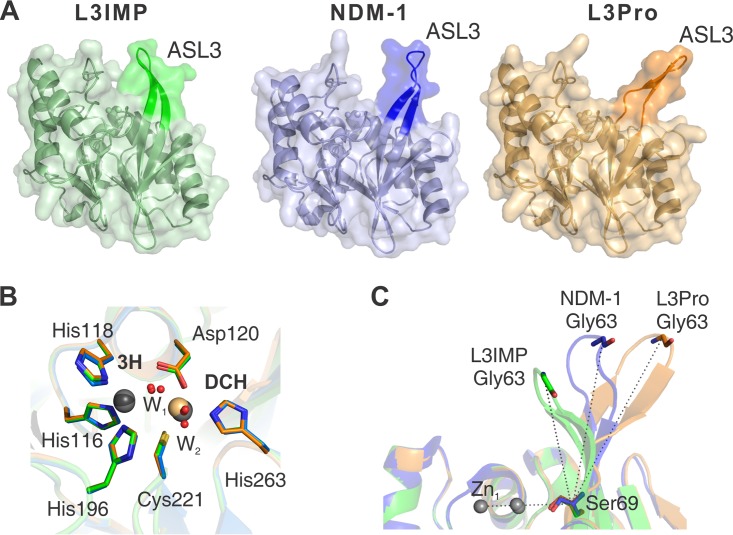
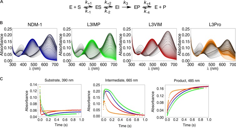
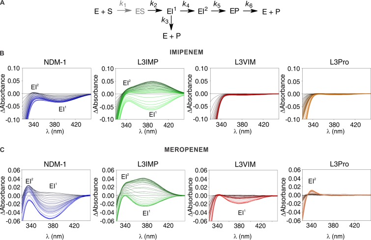
Carbapenems are "last resort" β-lactam antibiotics used to treat serious and life-threatening health care-associated infections caused by multidrug-resistant Gram-negative bacteria. Unfortunately, the worldwide spread of genes coding for carbapenemases among these bacteria is threatening these life-saving drugs. Metallo-β-lactamases (MβLs) are the largest family of carbapenemases. These are Zn(II)-dependent hydrolases that are active against almost all β-lactam antibiotics. Their catalytic mechanism and the features driving substrate specificity have been matter of intense debate. The active sites of MβLs are flanked by two loops, one of which, loop L3, was shown to adopt different conformations upon substrate or inhibitor binding, and thus are expected to play a role in substrate recognition. However, the sequence heterogeneity observed in this loop in different MβLs has limited the generalizations about its role. Here, we report the engineering of different loops within the scaffold of the clinically relevant carbapenemase NDM-1. We found that the loop sequence dictates its conformation in the unbound form of the enzyme, eliciting different degrees of active-site exposure. However, these structural changes have a minor impact on the substrate profile. Instead, we report that the loop conformation determines the protonation rate of key reaction intermediates accumulated during the hydrolysis of different β-lactams in all MβLs. This study demonstrates the existence of a direct link between the conformation of this loop and the mechanistic features of the enzyme, bringing to light an unexplored function of active-site loops on MβLs.

Keywords: New Delhi metallo-β-lactamase; antibiotic resistance; enzyme mechanism; enzyme structure; metallo-β-lactamase.


Figures

FIG 1
FIG 1
Engineered substitutions of loop L3 in NDM-1. Sequence alignment of the L3 variants, highlighting the differences at the loop L3 region, including the standard BBL numbering (28).
FIG 2
FIG 2
Expression levels and Zn(II) limitation susceptibility of NDM-1 and L3 variants. (A) Immunoblot demonstrating steady-state expression in E. coli DH5α. Proteins were detected from whole-cell lysates (lanes 2 to 5 and lane 14), spheroplasts (lanes 6 to 9), and periplasmic extracts (lanes 10 to 13). Wild-type NDM-1 (W) corresponds to lanes 2, 6, and 10; L3IMP (I) corresponds to lanes 3, 7, and 11; L3VIM (V) corresponds to lanes 4, 8, and 12; L3Pro (P) corresponds to lanes 5, 9, and 13. Empty plasmid (E) corresponds to lane 14. Lane 1 shows the protein ladder marker. The GroEL molecular weight is 60 kDa and that for MBP is 47 kDa. (B) Immunoblot demonstrating steady-state expression of wild-type NDM-1 and the L3 variants in E. coli DH5α treated with DPA. After induction, cells were incubated with (+) or without (–) DPA, and protein expression was detected from of whole-cell lysates. Wild-type NDM-1 (W) corresponds to lanes 2 and 3, L3IMP (I) corresponds to lanes 4 and 5, L3VIM (V) corresponds to lanes 6 and 7, L3Pro (P) corresponds to lanes 8 and 9, and empty plasmid (E) corresponds to lanes 10 and 11. Untreated cells were loaded before treated ones. Lane 1 shows the protein ladder marker. (C) Antimicrobial susceptibility profiles of E. coli DH5α/pMBLe producing β-lactamases against cefotaxime at increasing DPA concentrations. E. coli DH5α expressing NDM-1 is shown in blue, E. coli DH5α expressing L3IMP is shown in green, E. coli DH5α expressing L3VIM is shown in red, and E. coli DH5α expressing L3Pro is shown in orange.
FIG 3
FIG 3
Difference spectrum of Co(II)-substituted wild-type NDM-1 and L3 variants. The difference spectra of the Co(II)-substituted MβLs were obtained by subtraction of the spectrum of the nonmetallated β-lactamase from the one corresponding to the final bi-Co(II) substituted variant. The difference spectrum of the wild-type NDM-1 is shown in blue; that for L3IMP is shown in green, that for L3VIM is shown in red, and that for L3Pro is shown in orange.
FIG 4
FIG 4
X-ray crystal structures of the L3IMP and L3Pro variants compared to NDM-1. Crystal structures are indicated by color: NDM-1 (PDB code 3SPU, chain D) in blue, L3IMP in green (PDB code 6C6I, 1.65 Å), and L3Pro in orange (PDB code 6CAC, 1.80 Å). The images were generated after the complete alignment of NDM-1 and the two L3 variants. (A) The loop L3 position is highlighted in a darker color. (B) Relevant conserved amino acids from the active sites of NDM-1 (blue), L3IMP (green), and L3Pro (orange) are represented by sticks; metal ions (Zn(II) in gray and Cd(II) in light orange) and water molecules (red) are represented as spheres. The position and orientation of the metal ligands is conserved among the three structures. The distances between the ions in the two sites are very similar (≈3.8 Å) among the different proteins. The positions of the ions in the DCH site display a slight variability among the structures, while in the 3H site the position is unchanged. (C) Angle determined by the loop L3 and the plane of the active site of each mutant. Angles were calculated between Zn1, Cα of Ser69, and Cα of Gly63. The values obtained for each loop L3 were as follows: L3IMP, 68°; NDM-1, 88°; and L3Pro, 110°.
FIG 5
FIG 5
Photodiode array stopped-flow spectra and traces of nitrocefin hydrolysis by NDM-1 and its L3 variants. (A) Reaction mechanism for nitrocefin hydrolysis by NDM-1 (adapted from Yang et al. [32]). (B) Electronic absorption spectra upon the reaction of 10 μM nitrocefin and 10 μM enzyme in 100 mM HEPES (pH 7.5), 0.2 M NaCl, and 0.3 mM ZnSO4 at 6°C. The reaction progresses from black to color: NDM-1 in blue, L3VIM in red, L3IMP in green, and L3Pro in orange. Absorption bands with peaks at 390, 485, and 605 nm correspond to maximum absorption of the substrate, product, and the anionic intermediate of the reaction, respectively. (C) Temporary profiles of substrate (390 nm), intermediate (665 nm), and product (485 nm) during the reaction described in panel A. Traces: NDM-1, blue; L3VIM, red; L3IMP, green; L3Pro, yellow.
FIG 6
FIG 6
Photodiode array stopped-flow spectra of carbapenems hydrolysis by NDM-1 and the L3 variants. (A) General reaction mechanism for carbapenems hydrolysis by MβLs (adapted from Lisa et al. [33]). The ES complex does not accumulate and is thus depicted in a lighter color (gray). (B) Sequence of difference spectra collected upon the reaction of 100 μM imipenem and 100 μM β-lactamase. The reactions progress from black to color: NDM-1, blue; L3VIM, red; L3IMP, green; and L3Pro, orange. The ionic intermediates, EI1 and EI2, were detected as absorption bands with maxima at 390 and 343 nm. The time interval spans up to 0.2 s. (C) Sequence of difference spectra upon the reaction of 100 μM meropenem and 100 μM enzyme. The reactions progress from black to color: NDM-1, blue; L3VIM, red; L3IMP, green; and L3Pro, orange. The ionic intermediates, EI1 and EI2, were detected as absorption bands with maxima at 390 and 330 nm. The time interval spans up 0.2 s.

References

    1. Fisher JF, Meroueh SO, Mobashery S. 2005. Bacterial resistance to beta-lactam antibiotics: compelling opportunism, compelling opportunity. Chem Rev 105:395–424. doi:10.1021/cr030102i. - DOI - PubMed
    1. Bonomo RA, Tolmasky M. 2007. Enzyme-mediated resistance to antibiotics: mechanisms, dissemination, and prospects for inhibition, 1st ed, vol 83 ASM Press, Washington, DC.
    1. Bush K, Jacoby GA. 2010. Updated functional classification of β-lactamases. Antimicrob Agents Chemother 54:969–976. doi:10.1128/AAC.01009-09. - DOI - PMC - PubMed
    1. King AM, Reid-Yu SA, Wang W, King DT, De Pascale G, Strynadka NC, Walsh TR, Coombes BK, Wright GD. 2014. Aspergillomarasmine A overcomes metallo-beta-lactamase antibiotic resistance. Nature 510:503–506. doi:10.1038/nature13445. - DOI - PMC - PubMed
    1. Gonzalez MM, Kosmopoulou M, Mojica MF, Castillo V, Hinchliffe P, Pettinati I, Brem J, Schofield CJ, Mahler G, Bonomo RA, Llarrull LI, Spencer J, Vila AJ. 2015. Bisthiazolidines: a substrate-mimicking scaffold as an inhibitor of the NDM-1 carbapenemase. ACS Infect Dis 1:544–554. doi:10.1021/acsinfecdis.5b00046. - DOI - PMC - PubMed

Publication types

MeSH terms