. 2018 Dec 21;63(1):e01043-18.
doi: 10.1128/AAC.01043-18. Print 2019 Jan.

Identification of Functional MKK3/6 and MEK1/2 Homologs from Echinococcus granulosus and Investigation of Protoscolecidal Activity of Mitogen-Activated Protein Kinase Signaling Pathway Inhibitors In Vitro and In Vivo

Affiliations

Identification of Functional MKK3/6 and MEK1/2 Homologs from Echinococcus granulosus and Investigation of Protoscolecidal Activity of Mitogen-Activated Protein Kinase Signaling Pathway Inhibitors In Vitro and In Vivo

Chuanshan Zhang et al. Antimicrob Agents Chemother. .

Abstract

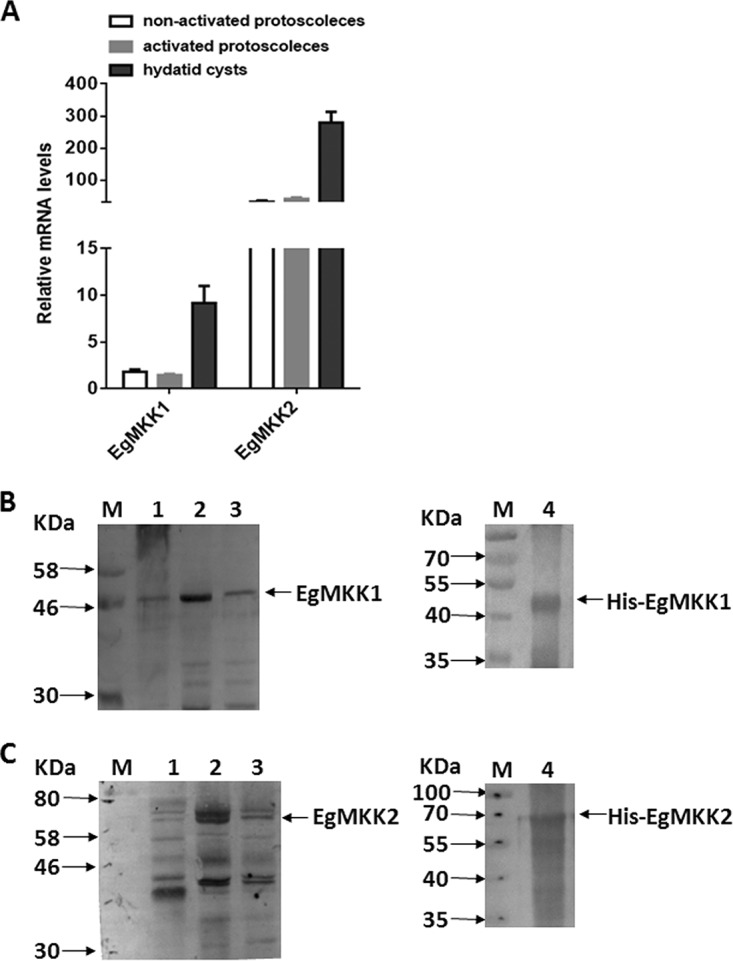
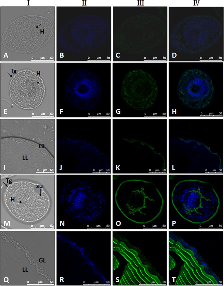
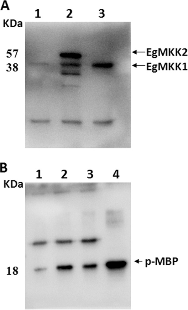
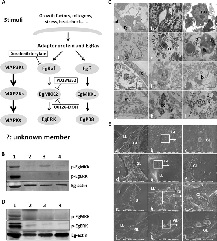
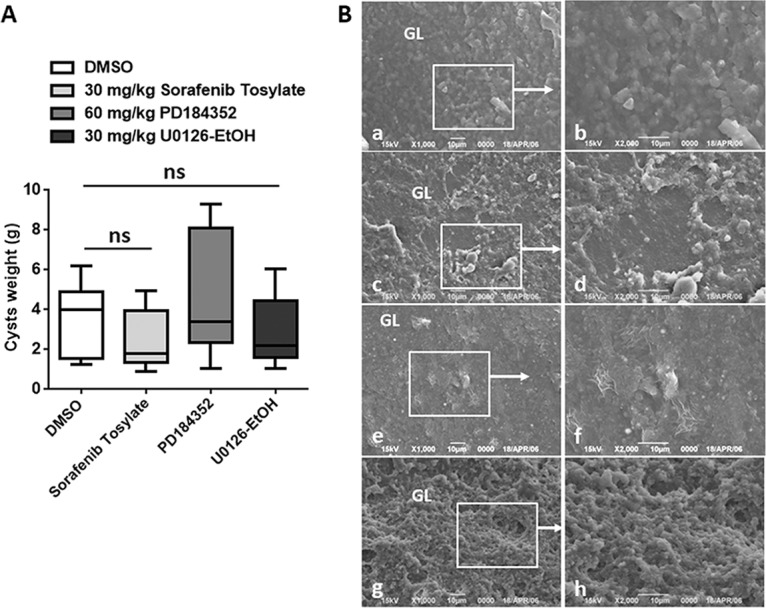
Cystic echinococcosis is a zoonosis caused by the larval stage of Echinococcus granulosussensu lato There is an urgent need to develop new drugs for the treatment of this disease. In this study, we identified two new members of mitogen-activated protein kinase (MAPK) cascades, MKK3/6 and MEK1/2 homologs (termed EgMKK1 and EgMKK2, respectively), from E. granulosussensu stricto Both EgMKK1 and EgMKK2 were expressed at the larval stages. As shown by yeast two-hybrid and coimmunoprecipitation analyses, EgMKK1 interacted with the previously identified Egp38 protein but not with EgERK. EgMKK2, on the other hand, interacted with EgERK. In addition, EgMKK1 and EgMKK2 displayed kinase activity toward the substrate myelin basic protein. When sorafenib tosylate, PD184352, or U0126-ethanol (EtOH) was added to the medium for in vitro culture of E. granulosus protoscoleces (PSCs) or cysts, an inhibitory and cytolytic effect was observed via suppressed phosphorylation of EgMKKs and EgERK. Nonviability of PSCs treated with sorafenib tosylate or U0126-EtOH, and not with PD184352, was confirmed through bioassays, i.e., inoculation of treated and untreated protoscoleces into mice. In vivo treatment of E. granulosussensu stricto-infected mice with sorafenib tosylate or U0126-EtOH for 4 weeks demonstrated a reduction in parasite weight, but the results did not show a significant difference. In conclusion, the MAPK cascades were identified as new targets for drug development, and E. granulosus was efficiently inhibited by their inhibitors in vitro The translation of these findings into in vivo efficacy requires further adjustment of treatment regimens using sorafenib tosylate or, possibly, other kinase inhibitors.

Keywords: Echinococcus granulosus; MAPK kinases; chemotherapy; cystic echinococcosis; inhibitor.


Figures

FIG 1
FIG 1
mRNA and protein levels of EgMKK1 and EgMKK2 expression in the larval stages. (A) Quantitative PCR analysis of EgMKK1 and EgMKK2 mRNA levels. Quantitative real-time PCR analysis was performed on total RNA isolated from nonactivated PSCs, activated PSCs, and hydatid cysts. Shown is a bar graph comparing the relative expression levels (means ± SD) of EgMKK1 and EgMKK2 normalized to the levels of Egelp. The relative expression value was averaged from triple samples. (B and C) Immunoblot detection of the EgMKK1 (B) and EgMKK2 (C) proteins. Cell lysates of nonactivated PSCs (lane 1), activated PSCs (lane 2), in vitro-cultivated cysts (lane 3), and Escherichia coli BL21 expressing a His-EgMKK1 fusion protein or a His-EgMKK2 fusion protein (lane 4) were separated on a 12% SDS-polyacrylamide gel. Protein marker sizes are indicated to the left (M).
FIG 2
FIG 2
Immunolocalization of the EgMKK1 and EgMKK2 proteins on parasite sections. Sections were labeled with purified EgMKK1-immunized rabbit sera (E to L), EgMKK2-immunized rabbit sera (M to T), and F(ab′)2 goat anti-rabbit antibody conjugated to Alexa 488 fluorochrome. Column I, phase-contrast images; column II, nuclear staining images (4ʹ,6-diamidino-2-phenylindole dihydrochloride [DAPI]); column III, anti-EgMKK1 or anti-EgMKK2 fluorescence images; column ІV, nuclear staining (blue) image merged with anti-EgMKK1 or anti-EgMKK2 fluorescence (green). Negative controls (panels A to D) were incubated with rabbit preimmune sera. Panels E to H and M to P represent invaginated PSCs. Panels I to L and Q to T represent in vitro-cultivated hydatid cysts. Tg, tegument; H, hooks; su, suckers; GL, germinal layer; LL, laminated layer.
FIG 3
FIG 3
Interaction of EgMKK1 and EgMKK2 proteins with downstream signaling partners. (A) Yeast two-hybrid analyses. Translational fusions were produced between the Gal4 activation domain (pGADT7 vector) or the Gal4 DNA binding domain (pGBKT7 vector) and EgMKK1, EgMKK2, EgERK, Egp38, or Eg14-3-3.2. Double transformants of yeast strain Y2HGold were assessed for colony growth after 3 to 5 days of incubation. Growth of a representative number of double transformants on selection plates is shown. As positive (pGBKT7-p53 × pGADT7-T) and negative (pGBKT7-Lam × pGADT7-T) controls, the recommended plasmids of the Matchmaker kit (Clontech, Japan) were used. (B to E) Co-IP experiments. (B and C) 293T cells were cotransfected with expression vectors for V5-tagged EgMKK1 and Myc-tagged Egp38 (lane 3) as well as V5-tagged EgMKK2 and Myc-tagged EgERK (lane 2) or Eg14-3-3.2 (lane 6), and whole-cell lysates were directly analyzed (input) (B) or precipitated with V5 antibody (co-IP) (C) and subjected to immunoblot analysis with the indicated antibody. Extracts from V5-EgMKK1 or EgMKK2 and pCMV-Myc-N coexpression in 293T cells were used as negative controls. (D and E) The immunoblot was probed with anti-Myc antibody to detect interacting proteins. (B, D, and E) Lanes 1 and 5, pCMV-Myc-N; lane 2, V5-tagged EgMKK2 plus Myc-tagged EgpERK; lane 3, V5-tagged EgMKK1 plus Myc-tagged Egp38; lanes 4 and 7, 293T cells; lane 6, V5-tagged EgMKK2 plus Myc-tagged Egp14-3-3.2. (C) Lane 1, V5-tagged EgMKK2 plus pCMV-Myc-N; lane 2, V5-tagged EgMKK2 plus Myc-tagged EgpERK; lane 3, V5-tagged EgMKK1 plus Myc-tagged Egp38; lane 4, V5-tagged EgMKK1 plus pCMV-Myc-N; lane 5, V5-tagged EgMKK2 plus Myc-tagged Egp14-3-3.2; lane 6, 293T cells. Protein marker sizes are indicated to the left (M).
FIG 4
FIG 4
Kinase activities of EgMKK1 and EgMKK2 proteins. (A) pcDNA3.1/V5-EgMKK1 (lane 2) and -EgMKK2 (lane 3) vectors were transfected into 293T cells and expressed as V5 fusion proteins. Whole-cell lysates were precipitated with V5 antibody (co-IP) and subjected to immunoblot analysis with anti-V5 antibody. Extracts from 293T cells transfected with the pcDNA3.1/V5 vector were used as negative controls (lane 1). (B) V5-tagged EgMKK1 (lane 3) or EgMKK2 (lane 2) was determined from extracts by immunoprecipitation with a V5 antibody followed by an in vitro kinase assay with MBP as a substrate. Extracts from NIH 3T3 cells were used as positive controls (lane 4). Protein marker sizes are indicated to the left (M).
FIG 5
FIG 5
Effects of MAPK pathway inhibitors on E. granulosus sensu stricto PSC viability in vitro. (A) Loss of viability of E. granulosus sensu stricto PSCs at 1, 25, 50, 100, 200, and 400 µM for sorafenib tosylate (a potent and selective inhibitor of Raf) over 4 days; (B) loss of viability of E. granulosus sensu stricto PSCs at 1, 25, 50, 100, 200, and 400 µM for PD184352 (a potent and selective inhibitor of MEK) over 4 days; (C) loss of viability of E. granulosus sensu stricto PSCs at 1, 25, 50, 100, 200, and 400 µM for U0126-EtOH (a specific inhibitor of MEK) over 8 days. Albendazole at 1 and 400 µM was used as a reference anti-infective agent. PSCs incubated in the absence of an inhibitor (supplemented with 0.2% DMSO) were used as negative controls. Each point represents the mean percentage of viable PSCs from three different experiments. Asterisks indicate a statistically significant difference (P < 0.05) from the corresponding control.
FIG 6
FIG 6
In vitro effects of inhibitor treatments on E. granulosus sensu stricto PSCs and cysts by targeting MAPK pathway members for 30 days. (A) Model of the E. granulosus ERK/p38-like MAPK pathway. (B and D) Representative immunoblots of EgMKKs and EgERK revealed with heterologous antibodies against the phosphorylated forms of human MEK1/2 and ERK1/2. Shown are total protein extracts from PSCs (B) or hydatid cysts (D) cultured in the presence of medium containing DMSO (control) (lane 1), sorafenib tosylate (25 µM) (lane 2), PD184352 (100 µM) (lane 3), or U0126-EtOH (100 µM) (lane 4). Actin was used as a loading control. (C) Ultrastructural changes of PSCs detected by TEM after incubation with either DMSO (control) or inhibitors. (E) Morphology changes of germinal layers from hydatid cysts detected by SEM after incubation with either DMSO (control) or inhibitors. (a to c) Control PSCs or cysts; (d to f) 25 µM sorafenib tosylate treatment; (g to i) 100 µM PD184352 treatment; (k to m) 100 µM U0126-EtOH treatment. mt, microtriches; Tg, tegument; nu, nucleus; mu, muscle cells; cc, chromatin condensation; b, residual lamellar bodies; GL, germinal layer; LL, laminated layer. The white arrows point to local tissue magnification.
FIG 7
FIG 7
In vivo treatment of secondary E. granulosus sensu stricto-infected BALB/c mice with MAPK pathway inhibitors. (A) Box plots indicating the distribution of cyst weights (grams) in the different treatment groups. Reductions in the weights of recovered parasites in relation to those in the control groups were achieved by treatment with 30 mg/kg sorafenib tosylate and 30 mg/kg U0126-EtOH, but they were not statistically significant (ns) (P =0.1994 and P =0.5360, respectively). (B) Representative images obtained by SEM of hydatid cysts from inhibitor-treated mice. (a and b) control cysts; (c and d) mice treated with 30 mg/kg sorafenib tosylate; (e and f) mice treated with 60 mg/kg PD184352; (g and h) mice treated with 30 mg/kg U0126-EtOH. GL, germinal layer; LL, laminated layer. The white arrows point to local tissue magnification. Each treatment group comprised 10 animals.

Similar articles

Cited by

References

    1. Kern P. 2003. Echinococcus granulosus infection: clinical presentation, medical treatment and outcome. Langenbecks Arch Surg 388:413–420. doi:10.1007/s00423-003-0418-y. - DOI - PubMed
    1. Craig P, Mastin A, van Kesteren F, Boufana B. 2015. Echinococcus granulosus: epidemiology and state-of-the-art of diagnostics in animals. Vet Parasitol 213:132–148. doi:10.1016/j.vetpar.2015.07.028. - DOI - PubMed
    1. Nazligul Y, Kucukazman M, Akbulut S. 2015. Role of chemotherapeutic agents in the management of cystic echinococcosis. Int Surg 100:112–114. doi:10.9738/INTSURG-D-14-00068.1. - DOI - PMC - PubMed
    1. Mandal S, Mandal MD. 2012. Human cystic echinococcosis: epidemiologic, zoonotic, clinical, diagnostic and therapeutic aspects. Asian Pac J Trop Med 5:253–260. doi:10.1016/S1995-7645(12)60035-2. - DOI - PubMed
    1. Lu G, Li J, Zhang C, Li L, Bi X, Li C, Fan J, Lu X, Vuitton DA, Wen H, Lin R. 2016. Molecular cloning and characterization of a P38-like mitogen-activated protein kinase from Echinococcus granulosus. Korean J Parasitol 54:759–768. doi:10.3347/kjp.2016.54.6.759. - DOI - PMC - PubMed

Publication types

MeSH terms