. 2018 Dec 3;217(12):4141-4154.
doi: 10.1083/jcb.201804163. Epub 2018 Oct 22.

Ribosome biogenesis factor Ltv1 chaperones the assembly of the small subunit head

Affiliations

Ribosome biogenesis factor Ltv1 chaperones the assembly of the small subunit head

Jason C Collins et al. J Cell Biol. .

Abstract

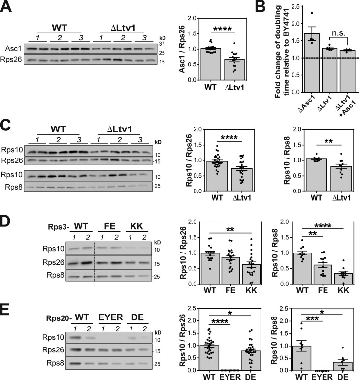
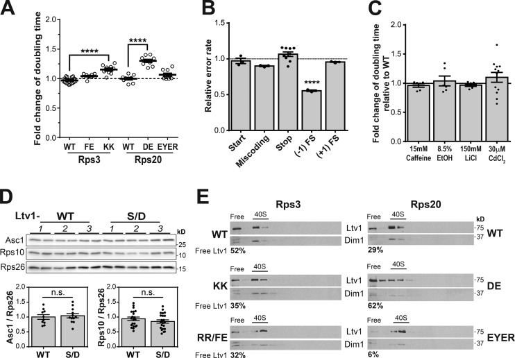
The correct assembly of ribosomes from ribosomal RNAs (rRNAs) and ribosomal proteins (RPs) is critical, as indicated by the diseases caused by RP haploinsufficiency and loss of RP stoichiometry in cancer cells. Nevertheless, how assembly of each RP is ensured remains poorly understood. We use yeast genetics, biochemistry, and structure probing to show that the assembly factor Ltv1 facilitates the incorporation of Rps3, Rps10, and Asc1/RACK1 into the small ribosomal subunit head. Ribosomes from Ltv1-deficient yeast have substoichiometric amounts of Rps10 and Asc1 and show defects in translational fidelity and ribosome-mediated RNA quality control. These defects provide a growth advantage under some conditions but sensitize the cells to oxidative stress. Intriguingly, relative to glioma cell lines, breast cancer cells have reduced levels of LTV1 and produce ribosomes lacking RPS3, RPS10, and RACK1. These data describe a mechanism to ensure RP assembly and demonstrate how cancer cells circumvent this mechanism to generate diverse ribosome populations that can promote survival under stress.


Figures

None
Graphical abstract
Figure 1.
Figure 1.
Structure probing and genetic data indicate repositioning of Rps3. (A) Primer extension pattern of ribosomes from n = 3 biological replicates of WT and ΔLtv1 cells in the presence (+) or absence (–) of DMS. Differentially accessible residues as quantified below are highlighted in red (exposed) or blue (protected). Gray bars show ΔLtv1, and black bars show WT. (B and C) Quantification was done using Image Lab 6.0 and normalized to band 1, which causes a stop in all lanes. Differentially accessible residues are shown in the secondary (B) and tertiary (C) structure of the head rRNA. Rps3 (red), Rps10 (blue), Rps20 (green), Rps29 (cyan), Asc1 (yellow), and Rps17 (brown) binding sites are indicated in the colored boxes (B) or in the structure of mature 40S ribosome (C; PDB ID: 4V88). Enp1 and a peptide of Ltv1 are shown in purple. Their position was derived from a superposition of 40S in 4V88 and pre-40S in 6FAI. (D) Genetic interactions between Ltv1 deletion and mutations at the interface between Rps3 and Rps17. (E) Genetic interactions between Ltv1 deletion and mutations in the Rps3 protein network. Fold-changes were determined by comparison of the effects from Rps3, Rps20, or Rps17 mutations in the presence or absence of Ltv1. Data in D and E are three to five technical repeats of 3–12 biological replicates. Error bars represent the SEM, and significance was determined using an unpaired t test. ****, P < 0.0001.
Figure 2.
Figure 2.
Recruitment of Asc1 requires the Rps3 tail. (A) Asc1 and the C-terminal tail of Rps3 are required for growth on glycerol, a nonfermentable carbon source. V.O., vector only. (B) Asc1 occupancy in ribosomes from WT or C-terminally truncated Rps3 (Rps3214) cells was investigated by Coomassie-stained SDS-PAGE of purified 40S ribosomes. (C) In vitro binding of Asc1 to Asc1-deficient ribosomes containing full-length Rps3 or Rps3214. Shown is the Coomassie-stained SDS–PAGE gel of the pellet fractions. The empty lanes on the right show that Asc1 alone does not pellet.
Figure 3.
Figure 3.
Ltv1 is required for stoichiometric assembly of Rps10 and Asc1. (A) The levels of Asc1 relative to Rps26 in ribosomes purified from WT or ΔLtv1 cells were determined by Western blotting. Data are averages from two technical repeats of three biological replicates (1–3). (B) Similar to ΔAsc1 cells, ΔLtv1 cells grow slowly in glycerol. Overexpression of Asc1 does not rescue this effect (n = 4). (C–E) Changes in the RP composition of the head structure were determined by Western blotting of ribosomes purified from WT and ΔLtv1 cells (C) or cells containing mutations in the Rps3 protein network (D and E). Note that the same gel is used and shown to evaluate Asc1, Rps10, and Rps26 levels in A and C. A separate gel is run to evaluate Rps8 relative to Rps10. The data are averages from four to six technical repeats from two biological replicates. In all cases, significance was determined using an unpaired t test, and the error bars represent the SEM. *, P < 0.05; **, P < 0.01; ***, P < 0.001; ****, P < 0.0001.
Figure 4.
Figure 4.
Ribosomes from ΔLtv1 yeast display functional differences. (A) Effects on translational fidelity in ΔLtv1, ΔAsc1, and Rps10-reduced cells. Three to six technical repeats of three to six biological replicates were obtained. (B) Increased stress resistance in ΔLtv1, ΔAsc1, and Rps10 reduced cells (n = 6–12). The change in doubling time from addition of stress media was determined in ΔLtv1 and ΔAsc1 cells and normalized to the effect in WT cells. Similarly, Rps10-reduced cells were normalized to Rps10-replete cells. (C) ΔLtv1 cells are deficient in NRD (n = 5–6). Significance was determined by a two-tailed t test. (D) ΔLtv1 cells are sensitive to H2O2 (n = 6, with three technical repeats). In all cases, error bars represent the SEM, and unless otherwise noted, significance was determined using an unpaired student t test. *, P < 0.05; **, P < 0.01; ***, P < 0.001; ****, P < 0.0001.
Figure 5.
Figure 5.
Ltv1 facilitates RP assembly. (A) Genetic interactions between Ltv1-S/D and mutations in the Rps3 protein network. Effect from Rps3 and Rps20 mutations in the Ltv1-S/D background was normalized to the effect in WT background n = 3–6 biological replicates with three to five technical repeats. (B) Translational fidelity in Ltv1-S/D cells (n = 3–9). (C) No stress resistance in Ltv1-S/D cells (n = 3–12). (D) Ribosomal protein occupancy in Ltv1-S/D cells. The data are averages from two technical repeats of three to five biological replicate experiments. In all cases, the error bars represent the SEM, and the significance was determined using an unpaired t test. ****, P < 0.0001. (E) Release of Ltv1 is promoted by the Rps3 interaction network. Sucrose-gradient and Western analysis of lysates from cells containing WT or mutant Rps3 (left) and Rps20 (right), respectively. The percentage of free Ltv1 (vs. ribosome-bound Ltv1) is given below each Western blot.
Figure 6.
Figure 6.
LTV1 deficiency in human cancer cells. (A) Western blot for LTV1 from total protein in SF268 glioma and MDA–MB-231 breast cancer cell lines and their LTV1+/− heterozygous deletion derivatives. n = 3. (B and C) Northern blots and their quantification relative to U2 and SRP of total RNA from glioma and cancer cells (n = 6). (D) Analysis of ribosome content from equally loaded sucrose gradients of cancer cell lines (n = 3). (E) LTV1 distribution probed by sucrose-gradient and Western blot analysis of cell lysates from glioma (left) and breast cancer (right) cells. (F) Quantification of data in E (n = 3). (G) Occupancy of RPS3, RPS10, and RACK1 in ribosomes purified from glioma and cancer cells (n = 9–12). (H) Translational fidelity of glioma and breast cancer cells (n = 12). Error bars represent the SEM, and significance was determined using an unpaired student t test. *, P < 0.05; **, P < 0.01; ***, P < 0.001; ****, P < 0.0001. (I) cBioPortal analysis of genomic alterations in LTV1, BYSL (hEnp1), and CSNK1D (hCK1δ) from two datasets of cancer cell lines. Studies cited in this figure are from cBioPortal (Cerami et al., 2012; Gao et al., 2013).
Figure 7.
Figure 7.
Model of Ltv1-dependent head structure assembly. (A) Model for the Ltv1-dependent assembly of the small subunit head structure. Ribosomal proteins are indicated by their corresponding number, and assembly factors are defined as: L, Ltv1; E, Enp1; Y, Yar1; and H, Hrr25/Ck1δ. (B) Model of small subunit head assembly in ΔLtv1 cells.

Similar articles

Cited by

References

    1. Acker M.G., Kolitz S.E., Mitchell S.F., Nanda J.S., and Lorsch J.R.. 2007. Reconstitution of yeast translation initiation. Methods Enzymol. 430:111–145. 10.1016/S0076-6879(07)30006-2 - DOI - PubMed
    1. Adilakshmi T., Bellur D.L., and Woodson S.A.. 2008. Concurrent nucleation of 16S folding and induced fit in 30S ribosome assembly. Nature. 455:1268–1272. 10.1038/nature07298 - DOI - PMC - PubMed
    1. Ajore R., Raiser D., McConkey M., Jöud M., Boidol B., Mar B., Saksena G., Weinstock D.M., Armstrong S., Ellis S.R., et al. . 2017. Deletion of ribosomal protein genes is a common vulnerability in human cancer, especially in concert with TP53 mutations. EMBO Mol. Med. 9:498–507. 10.15252/emmm.201606660 - DOI - PMC - PubMed
    1. Arthur L., Pavlovic-Djuranovic S., Smith-Koutmou K., Green R., Szczesny P., and Djuranovic S.. 2015. Translational control by lysine-encoding A-rich sequences. Sci. Adv. 1:e15000154 10.1126/sciadv.1500154 - DOI - PMC - PubMed
    1. Burwick N., Shimamura A., and Liu J.M.. 2011. Non-Diamond Blackfan anemia disorders of ribosome function: Shwachman Diamond syndrome and 5q- syndrome. Semin. Hematol. 48:136–143. 10.1053/j.seminhematol.2011.01.002 - DOI - PMC - PubMed

Publication types

MeSH terms

Substances