. 2018 Nov 6;115(45):E10548-E10555.
doi: 10.1073/pnas.1811167115. Epub 2018 Oct 22.

Structural and mechanistic insights into the function of the unconventional class XIV myosin MyoA from Toxoplasma gondii

Affiliations

Structural and mechanistic insights into the function of the unconventional class XIV myosin MyoA from Toxoplasma gondii

Cameron J Powell et al. Proc Natl Acad Sci U S A. .

Abstract

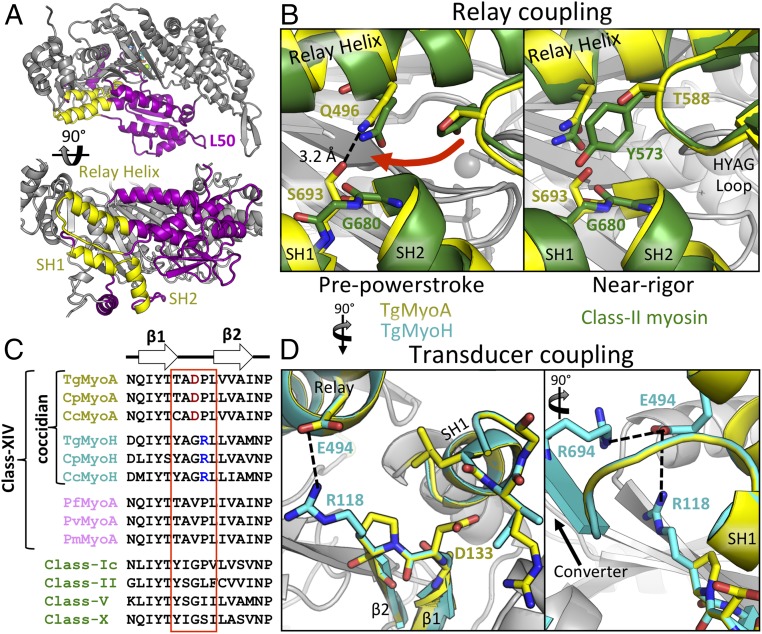
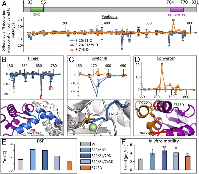
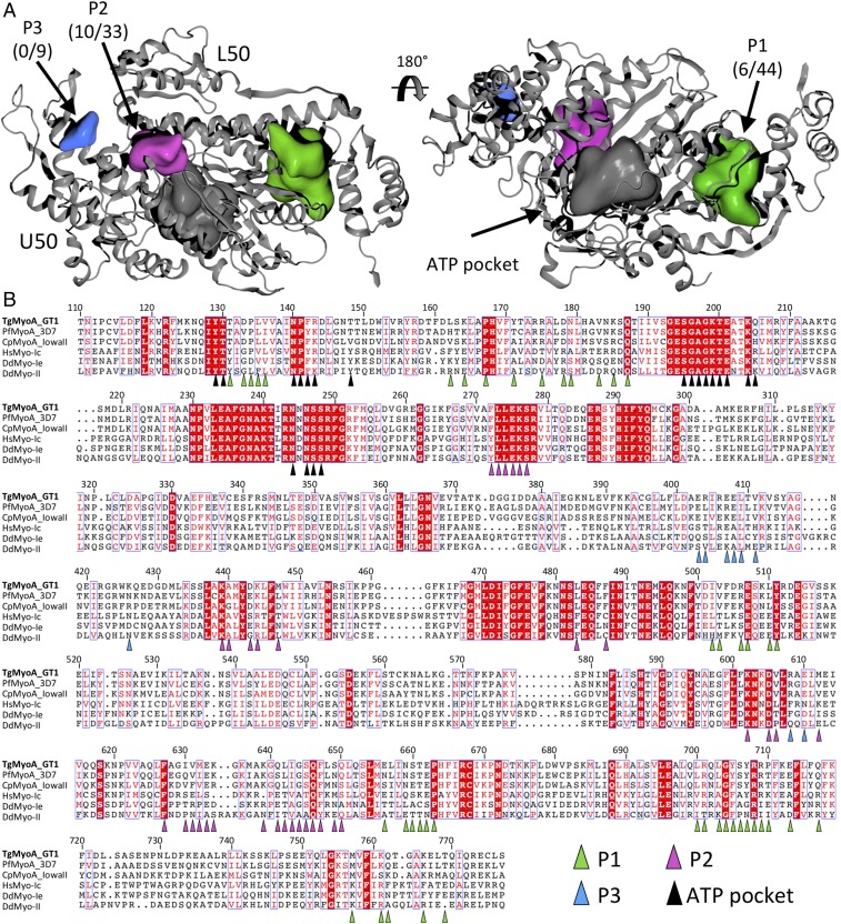
Parasites of the phylum Apicomplexa are responsible for significant morbidity and mortality on a global scale. Central to the virulence of these pathogens are the phylum-specific, unconventional class XIV myosins that power the essential processes of parasite motility and host cell invasion. Notably, class XIV myosins differ from human myosins in key functional regions, yet they are capable of fast movement along actin filaments with kinetics rivaling previously studied myosins. Toward establishing a detailed molecular mechanism of class XIV motility, we determined the 2.6-Å resolution crystal structure of the Toxoplasma gondii MyoA (TgMyoA) motor domain. Structural analysis reveals intriguing strategies for force transduction and chemomechanical coupling that rely on a divergent SH1/SH2 region, the class-defining "HYAG"-site polymorphism, and the actin-binding surface. In vitro motility assays and hydrogen-deuterium exchange coupled with MS further reveal the mechanistic underpinnings of phosphorylation-dependent modulation of TgMyoA motility whereby localized regions of increased stability and order correlate with enhanced motility. Analysis of solvent-accessible pockets reveals striking differences between apicomplexan class XIV and human myosins. Extending these analyses to high-confidence homology models of Plasmodium and Cryptosporidium MyoA motor domains supports the intriguing potential of designing class-specific, yet broadly active, apicomplexan myosin inhibitors. The successful expression of the functional TgMyoA complex combined with our crystal structure of the motor domain provides a strong foundation in support of detailed structure-function studies and enables the development of small-molecule inhibitors targeting these devastating global pathogens.

Keywords: Apicomplexa; Toxoplasma gondii; X-ray crystallography; motility; myosin.


Conflict of interest statement

The authors declare no conflict of interest.

Figures

Fig. 1.
Fig. 1.
Structural analysis of the class XIV TgMyoA. (A) Schematic representation of TgMyoA expression and crystallization (blue line) constructs. (B) Sx200 SEC profile and SDS/PAGE gel showing homogenous preparation of monomeric TgMyoA 1–778. (C) Schematic illustration of actin cosedimentation assay (Top) showing that TgMyoA 1–778 is able to bind actin and is responsive to ATP (Bottom). F-actin with bound TgMyoA was pelleted at 340,000 × g. Supernatant containing excess globular actin and non–actin-binding MyoA (lane 1). ATP was added to the resuspended pellet, dissociating functional TgMyoA from the actin, and the F-actin was pelleted again, leaving only functional myosin in the supernatant (lane 2). The pellet containing F-actin and ATP nonresponsive myosin was then resuspended (lane 3). (D) Overview of TgMyoA 1–778 structure, highlighting the upper (orange) and lower (purple) 50-kDa domains and the putative SH3 (green), transducer (blue), and converter (red) subdomains. The region encompassing 779–831 (hashed lines) is not present in the crystal construct. (E) Topology map of TgMyoA motor domain with subdomains colored as in D.
Fig. 2.
Fig. 2.
Interactions in the SH helices maintain chemomechanical coupling. (A) Overview showing the position of the SH helices and relay helix (yellow) within the TgMyoA L50 domain (purple). (B) SH helices of TgMyoA (yellow) and class II myosin (green) highlight unique interactions between the SH1/SH2 pivot point, the relay helix, and the HYAG site in the PPS (Left) and near-rigor (Right) states. (C) Sequence alignments of β1/2 linker regions from various coccidian myosins (yellow and cyan), Plasmodium MyoA (pink), and non-class XIV myosins (green). (D) Unique interactions between the β1/2 linker, SH helices, and relay helix in TgMyoA and TgMyoH colored as in C.
Fig. 3.
Fig. 3.
Phosphomimetic mutations affect motility of TgMyoA by stabilizing or destabilizing key functional regions. (A) Schematic representation of TgMyoA aligned with HDX difference map showing change in percent deuterium incorporation of full-length TgMyoA phosphomimetic mutants in the near-rigor state relative to WT over 300 s. Error bars (SD) represent independent triplicates. Peptide number corresponds to the centroid amino acid of the peptide from which a data point is obtained. (BD). Expanded views of HDX difference map showing change in percent deuterium incorporation in different TgMyoA subdomains by the phosphomimetic mutants (Top) with the stabilized (blue) and destabilized (orange) regions mapped onto the TgMyoA crystal structure (Bottom). The protected regions in the relay, SH1, and converter associated with the S20/21D and S20/21/29D mutations are indicated as I, II, and III, respectively, in B; change in percentage incorporation in the switch II loop associated with the S-20/21D, S20/21/29D, and S743D mutations are shown in C (AlF4 is omitted in figure to prevent obstruction of switch II); and change in percentage incorporation in the converter subdomain associated with the S743D mutation is shown in D. (E) DSF thermal shift assay of TgMyoA comparing WT vs. phosphomimetic mutants. Error bars (SD) represent independent triplicates. (F) Actin filament displacement velocities of WT and phosphomimetic mutants in in vitro motility assays. Error bars (SD) represent five independent experiments. Mutant speeds were compared with WT by one-way ANOVA and Šídák’s multiple comparison tests: *P < 0.05 and **P < 0.005; “ns” indicates P > 0.05.
Fig. 4.
Fig. 4.
Pocket analysis of TgMyoA supports potential for class XIV-specific inhibitors. (A) Surface pocket analysis of the TgMyoA crystal structure. Solvent-accessible pockets, shown in green (P1), purple (P2), and blue (P3), were identified by using default parameters in OEDocking software, allowing the shape of the pockets to extend toward the solvent by one solvent radius. Pocket labels include the number of identical residues between TgMyoA and sequence-aligned myosins (B) among the total pocket residues. (B) Structure-guided sequence alignment of the core motor domains from class XIV, class I, and class II myosins. Highly variable N- and C-terminal regions have been omitted for clarity. Residues belonging to each of the solvent-accessible pockets are indicated with colored triangles.

References

    1. Harker KS, Ueno N, Lodoen MB. Toxoplasma gondii dissemination: A parasite’s journey through the infected host. Parasite Immunol. 2015;37:141–149. - PubMed
    1. Frenal K, Dubremetz JF, Lebrun M, Soldati-Favre D. Gliding motility powers invasion and egress in Apicomplexa. Nat Rev Microbiol. 2017;15:645–660. - PubMed
    1. Boucher LE, Bosch J. The apicomplexan glideosome and adhesins–Structures and function. J Struct Biol. 2015;190:93–114. - PMC - PubMed
    1. Frenal K, Foth BJ, Soldati-Favre D. Myosin class XIV and other myosins inprotists. In: Coluccio LM, editor. Myosins: A Superfamily of Molecular Motors, Proteins and Cell Regulation. Vol 7. Springer; Dordrecht, The Netherlands: 2008. pp. 421–440.
    1. Mueller C, Graindorge A, Soldati-Favre D. Functions of myosin motors tailored for parasitism. Curr Opin Microbiol. 2017;40:113–122. - PubMed

Publication types

Substances

Associated data

LinkOut - more resources