. 2018 Nov 6;115(45):E10702-E10711.
doi: 10.1073/pnas.1810653115. Epub 2018 Oct 22.

Isolation and characterization of NY-ESO-1-specific T cell receptors restricted on various MHC molecules

Affiliations

Isolation and characterization of NY-ESO-1-specific T cell receptors restricted on various MHC molecules

Michael T Bethune et al. Proc Natl Acad Sci U S A. .

Abstract

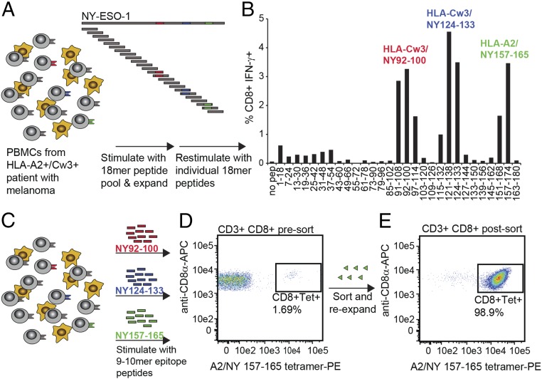
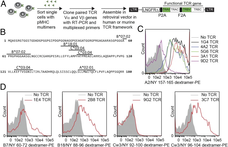
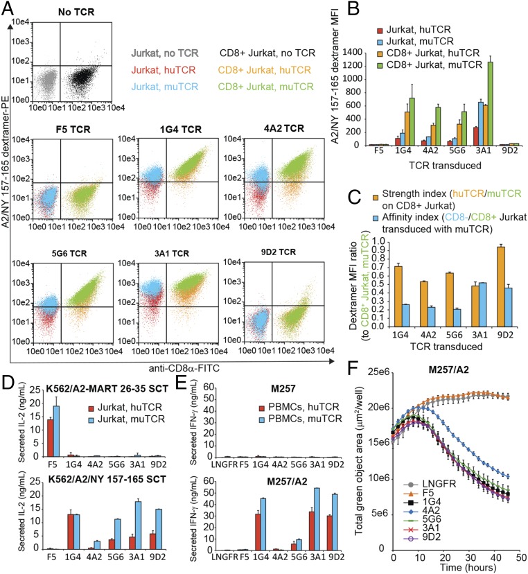
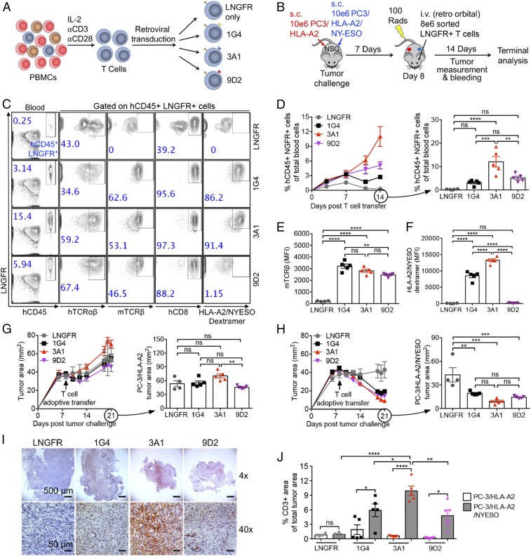
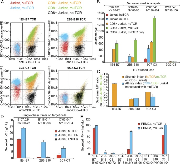
Tumor-specific T cell receptor (TCR) gene transfer enables specific and potent immune targeting of tumor antigens. Due to the prevalence of the HLA-A2 MHC class I supertype in most human populations, the majority of TCR gene therapy trials targeting public antigens have employed HLA-A2-restricted TCRs, limiting this approach to those patients expressing this allele. For these patients, TCR gene therapy trials have resulted in both tantalizing successes and lethal adverse events, underscoring the need for careful selection of antigenic targets. Broad and safe application of public antigen-targeted TCR gene therapies will require (i) selecting public antigens that are highly tumor-specific and (ii) targeting multiple epitopes derived from these antigens by obtaining an assortment of TCRs restricted by multiple common MHC alleles. The canonical cancer-testis antigen, NY-ESO-1, is not expressed in normal tissues but is aberrantly expressed across a broad array of cancer types. It has also been targeted with A2-restricted TCR gene therapy without adverse events or notable side effects. To enable the targeting of NY-ESO-1 in a broader array of HLA haplotypes, we isolated TCRs specific for NY-ESO-1 epitopes presented by four MHC molecules: HLA-A2, -B07, -B18, and -C03. Using these TCRs, we pilot an approach to extend TCR gene therapies targeting NY-ESO-1 to patient populations beyond those expressing HLA-A2.

Keywords: MHC; NY-ESO-1; T cell receptor gene therapy; TCR; immunotherapy.


Conflict of interest statement

Conflict of interest statement: A patent application has been filed (serial no. 62/727,485) entitled “Composition of NY-ESO-1-Specific T Cell Receptors Restricted on Multiple Major Histocompatibility Complex Molecules.”

Figures

Fig. 1.
Fig. 1.
Expansion and isolation of NY-ESO-1–specific T cell clones. PBMCs were obtained from patients with metastatic melanoma. T cell cloning strategy for a representative HLA-A2+, HLA-Cw3+ donor is shown. (A) Schematic outlining the expansion and testing strategy to identify NY-ESO-1–reactive T cell clones. PBMCs were incubated with 28 NY-ESO-1 18-mer peptides (overlapping by 12 aa) and then expanded for 10 d before restimulation with individual peptides in the presence of BFA. Epitopes presented by patient MHC alleles are colored red, blue, and green in those peptides containing the full epitope sequence. (B) Representative flow cytometry measurement of intracellular staining for IFN-γ in expanded PBMCs restimulated with individual NY-ESO-1–derived 18-mer peptides. (C) Schematic outlining the reexpansion strategy using individual 9–10-mer peptides verified to elicit a T cell response. (D) Representative flow cytometry data showing an NY-ESO-1–reactive subpopulation of CD3+CD8+ T cells before sorting. Sorted cells were expanded in the presence of IL-2 and irradiated autologous PBMCs. (E) Representative flow cytometry data showing an NY-ESO-1–reactive subpopulation of CD3+CD8+ T cells following sorting.
Fig. 2.
Fig. 2.
Cloning and functional screening of NY-ESO-1–specific TCRs. (A) Schematic of functional TCR cloning strategy. For each TCR, two constructs were prepared incorporating either human or murine TCR constant domains. (B) Protein sequence of NY-ESO-1 with epitopes relevant to this study delineated. (C) Flow cytometry histograms comparing HLA-A2/NY157–165 dextramer binding by HEK 293T cells transfected with vector backbone only, previously reported 1G4 TCR, and novel A2-restricted, NY-ESO-1–specific TCRs. (D) Flow cytometry histograms comparing indicated peptide–MHC dextramer binding by HEK 293T cells transfected with vector backbone only or the indicated novel NY-ESO-1-specificNY-ESO-1–specific TCR restricted on MHC alleles other than HLA-A2. Transfection experiments were performed twice, each in duplicate. Representative histograms are presented.
Fig. 3.
Fig. 3.
Function of A2-restricted, NY-ESO-1–specific TCRs. (A) Overlay of representative flow cytometry plots comparing A2/NY157–165 dextramer binding by Jurkat and CD8+ Jurkat cells expressing A2-restricted TCRs with human or murine constant domains. (B) Dextramer binding mean fluorescence intensity measurements from two independent experiments as in A. (C) Ratio of dextramer binding mean fluorescent intensity measurements from two independent experiments in B. (D) ELISA measuring secretion of IL-2 from TCR-transduced Jurkat cells following 48-h coincubation with K562 target cells expressing A2/MART26–35 or A2/NY157–165 single-chain trimer. Experiment was repeated three times, each with two technical replicates. Means ± SD for a representative experiment are shown. (E) ELISA measuring secretion of IFN-γ from TCR-transduced PBMCs following 48-h coincubation with the melanoma cell line M257 or an A2+ derivative. Experiment was repeated at least three times, each with two technical replicates. Means ± SD for a representative experiment are shown. (F) IncuCyte measurement of total green object area over time as a measurement of TCR-transduced T cell-mediated killing of GFP+ A2+ M257 cells. Means ± SD for four technical replicates are shown.
Fig. 4.
Fig. 4.
In vivo antitumor efficacy of NY-ESO-1 TCR–engineered human T cells. (A and B) Schematics of the experimental designs to (A) generate NY-ESO-1 TCR–engineered human T cells and to (B) study antitumor efficacy of these engineered T cells in an NSG mouse human prostate tumor xenograft model. NSG, immunodeficient NOD/SCID/γc−/− mice. (C) Representative flow cytometry plots characterizing engineered human T cells present in the peripheral blood of experimental mice on day 14 after adoptive T cell transfer. (D) Time course showing persistence of engineered human T cells (gated as LNGFR+ hCD45+) in the peripheral blood of experimental mice. (E and F) Mean fluorescence intensity measurements for (E) murine TCR and (F) HLA-A2/NYESO dextramer for engineered human T cells in the peripheral blood of experimental mice on day 14 after adoptive T cell transfer. (G and H) Measurements of cross-sectional area for (G) PC-3/HLA-A2 and (H) PC-3/HLA-A2/NYESO tumors. (I) Immunohistology images showing representative tumor sections. CD3+ cells are stained in red. (Scale bars: Upper, 500 μm; Lower, 50 μm.) (J) Percentage of CD3+ cell area over whole tumor section area. Representative of two experiments. Data are presented as the mean ± SEM (n = 4–5). ns, not significant; *P < 0.05, **P < 0.01, ***P < 0.001, ****P < 0.0001, by one-way ANOVA.
Fig. 5.
Fig. 5.
Function of NY-ESO-1–specific TCRs restricted on MHC alleles other than HLA-A2. (A) Overlay of representative flow cytometry plots comparing specified dextramer binding by Jurkat and CD8+ Jurkat cells expressing novel TCRs with human or murine constant domains. (B) Indicated dextramer binding mean fluorescence intensity measurements from two independent experiments as in A. (C) Ratio of respective dextramer binding mean fluorescent intensity measurements from two independent experiments in B. (D and E) ELISA measuring (D) secretion of IL-2 from TCR-transduced Jurkat cells or (E) secretion of IFN-γ from TCR-transduced PBMCs following 48-h coincubation with K562 target cells expressing indicated single-chain trimer. Experiments were repeated three times, each with two technical replicates. Means ± SD for a representative experiment are shown.
Fig. 6.
Fig. 6.
Targeting NY-ESO-1 epitopes restricted on multiple MHC alleles broadens the application of TCR gene therapy and makes it robust toward loss of heterozygosity at the MHC locus. (AD) T cells transduced with LNGFR only, A2-restricted 3A1 TCR, or B7-restricted 1E4 TCR—or a 1:1 mixture of 3A1-transduced and 1E4-transduced T cells—were coincubated for 48 h with HLA-A2+eGFP+ target cells, HLA-B7+eGFP+ target cells, or a 1:1 target cell mixture. (A and B) ELISA measuring secretion of IFN-γ from TCR-transduced PBMCs following 48-h coincubation with (A) M257 or (B) PC-3 tumor cell lines engineered to express eGFP and HLA-A*02:01 or HLA-B*07:02. PC-3 lines were additionally engineered to express NY-ESO-1. M257 lines express endogenous NY-ESO-1. Experiments were repeated three times, each with four or eight replicates. Means ± SD for a representative experiment are shown. (C and D) T cell-mediated killing of (C) M257 and (D) PC-3 tumor cell line derivatives measured over time using IncuCyte live-cell analysis. Total green object area (indicative of tumor cell density) at each time point measured over 48 h was normalized for each treatment relative to treatment with LNGFR-transduced T cells. Experiments were repeated three times, each with four or eight replicates. Results from a representative eight-replicate experiment are shown.

References

    1. Robinson J, et al. The IPD and IMGT/HLA database: Allele variant databases. Nucleic Acids Res. 2015;43:D423–D431. - PMC - PubMed
    1. González-Galarza FF, et al. Allele frequency net 2015 update: New features for HLA epitopes, KIR and disease and HLA adverse drug reaction associations. Nucleic Acids Res. 2015;43:D784–D788. - PMC - PubMed
    1. Johnson LA, et al. Gene transfer of tumor-reactive TCR confers both high avidity and tumor reactivity to nonreactive peripheral blood mononuclear cells and tumor-infiltrating lymphocytes. J Immunol. 2006;177:6548–6559. - PMC - PubMed
    1. Schumacher TN, Schreiber RD. Neoantigens in cancer immunotherapy. Science. 2015;348:69–74. - PubMed
    1. Bethune MT, Joglekar AV. Personalized T cell-mediated cancer immunotherapy: Progress and challenges. Curr Opin Biotechnol. 2017;48:142–152. - PubMed

Publication types

MeSH terms