

Lon Protease Has Multifaceted Biological Functions in Acinetobacter baumannii
- PMID: 30348832
- PMCID: PMC6304660
- DOI: 10.1128/JB.00536-18
Lon Protease Has Multifaceted Biological Functions in Acinetobacter baumannii
Abstract
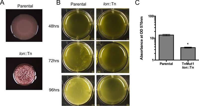
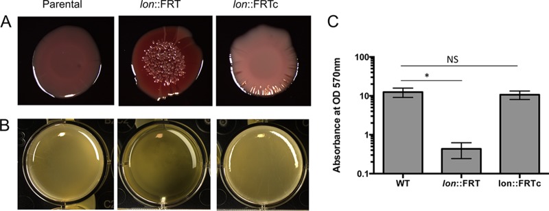
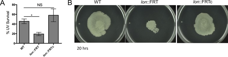
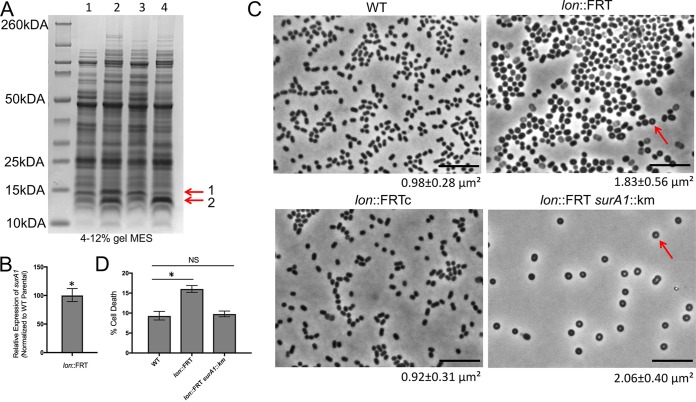
Acinetobacter baumannii is a Gram-negative opportunistic pathogen that is known to survive harsh environmental conditions and is a leading cause of hospital-acquired infections. Specifically, multicellular communities (known as biofilms) of A. baumannii can withstand desiccation and survive on hospital surfaces and equipment. Biofilms are bacteria embedded in a self-produced extracellular matrix composed of proteins, sugars, and/or DNA. Bacteria in a biofilm are protected from environmental stresses, including antibiotics, which provides the bacteria with selective advantage for survival. Although some gene products are known to play roles in this developmental process in A. baumannii, mechanisms and signaling remain mostly unknown. Here, we find that Lon protease in A. baumannii affects biofilm development and has other important physiological roles, including motility and the cell envelope. Lon proteases are found in all domains of life, participating in regulatory processes and maintaining cellular homeostasis. These data reveal the importance of Lon protease in influencing key A. baumannii processes to survive stress and to maintain viability.IMPORTANCEAcinetobacter baumannii is an opportunistic pathogen and is a leading cause of hospital-acquired infections. A. baumannii is difficult to eradicate and to manage, because this bacterium is known to robustly survive desiccation and to quickly gain antibiotic resistance. We sought to investigate biofilm formation in A. baumannii, since much remains unknown about biofilm formation in this bacterium. Biofilms, which are multicellular communities of bacteria, are surface attached and difficult to eliminate from hospital equipment and implanted devices. Our research identifies multifaceted physiological roles for the conserved bacterial protease Lon in A. baumannii These roles include biofilm formation, motility, and viability. This work broadly affects and expands understanding of the biology of A. baumannii, which will permit us to find effective ways to eliminate the bacterium.
Keywords: Acinetobacter; Lon protease; biofilms.
Copyright © 2018 American Society for Microbiology.
Figures
Similar articles
-
The LonA Protease Regulates Biofilm Formation, Motility, Virulence, and the Type VI Secretion System in Vibrio cholerae.J Bacteriol. 2016 Jan 11;198(6):973-85. doi: 10.1128/JB.00741-15. J Bacteriol. 2016. PMID: 26755629 Free PMC article.
-
Regulation of Acinetobacter baumannii biofilm formation.Future Microbiol. 2009 Apr;4(3):273-8. doi: 10.2217/fmb.09.5. Future Microbiol. 2009. PMID: 19327114 Free PMC article.
-
Effect of biofilm formation on the survival of Acinetobacter baumannii on dry surfaces.J Hosp Infect. 2012 Jan;80(1):56-60. doi: 10.1016/j.jhin.2011.08.013. Epub 2011 Oct 4. J Hosp Infect. 2012. PMID: 21975219
-
Biofilm formation in Acinetobacter baumannii.New Microbiol. 2014 Apr;37(2):119-27. Epub 2014 Apr 1. New Microbiol. 2014. PMID: 24858639 Review.
-
Acinetobacter baumannii Biofilm Formation and Its Role in Disease Pathogenesis: A Review.Infect Drug Resist. 2021 Sep 10;14:3711-3719. doi: 10.2147/IDR.S332051. eCollection 2021. Infect Drug Resist. 2021. PMID: 34531666 Free PMC article. Review.
Cited by
-
Structure, Substrate Specificity and Role of Lon Protease in Bacterial Pathogenesis and Survival.Int J Mol Sci. 2023 Feb 8;24(4):3422. doi: 10.3390/ijms24043422. Int J Mol Sci. 2023. PMID: 36834832 Free PMC article. Review.
-
c-di-GMP inhibits LonA-dependent proteolysis of TfoY in Vibrio cholerae.PLoS Genet. 2020 Jun 26;16(6):e1008897. doi: 10.1371/journal.pgen.1008897. eCollection 2020 Jun. PLoS Genet. 2020. PMID: 32589664 Free PMC article.
-
Mechanisms Protecting Acinetobacter baumannii against Multiple Stresses Triggered by the Host Immune Response, Antibiotics and Outside-Host Environment.Int J Mol Sci. 2020 Jul 31;21(15):5498. doi: 10.3390/ijms21155498. Int J Mol Sci. 2020. PMID: 32752093 Free PMC article. Review.
-
Proteomic survey of the DNA damage response in Caulobacter crescentus.bioRxiv [Preprint]. 2023 Jun 30:2023.03.24.534141. doi: 10.1101/2023.03.24.534141. bioRxiv. 2023. Update in: J Bacteriol. 2023 Oct 26;205(10):e0020623. doi: 10.1128/jb.00206-23. PMID: 36993274 Free PMC article. Updated. Preprint.
-
Essential roles of Lon protease in the morpho-physiological traits of the rice pathogen Burkholderia glumae.PLoS One. 2021 Sep 15;16(9):e0257257. doi: 10.1371/journal.pone.0257257. eCollection 2021. PLoS One. 2021. PMID: 34525127 Free PMC article.
References
-
- Valencia R, Arroyo LA, Conde M, Aldana JM, Torres M-J, Fernández-Cuenca F, Garnacho-Montero J, Cisneros JM, Ortiz C, Pachón J, Aznar J. 2009. Nosocomial outbreak of infection with pan–drug‐resistant Acinetobacter baumannii in a tertiary care university hospital. Infect Control Hosp Epidemiol 30:257–263. doi:10.1086/595977. - DOI - PubMed
Publication types
MeSH terms
Substances
Grants and funding
LinkOut - more resources
Full Text Sources
Molecular Biology Databases
