Comparative Study
. 2018 Dec 21;293(51):19686-19698.
doi: 10.1074/jbc.RA118.004373. Epub 2018 Oct 22.

Biochemical and structural studies reveal differences and commonalities among cap-snatching endonucleases from segmented negative-strand RNA viruses

Affiliations
Comparative Study

Biochemical and structural studies reveal differences and commonalities among cap-snatching endonucleases from segmented negative-strand RNA viruses

Tobias Holm et al. J Biol Chem. .

Abstract

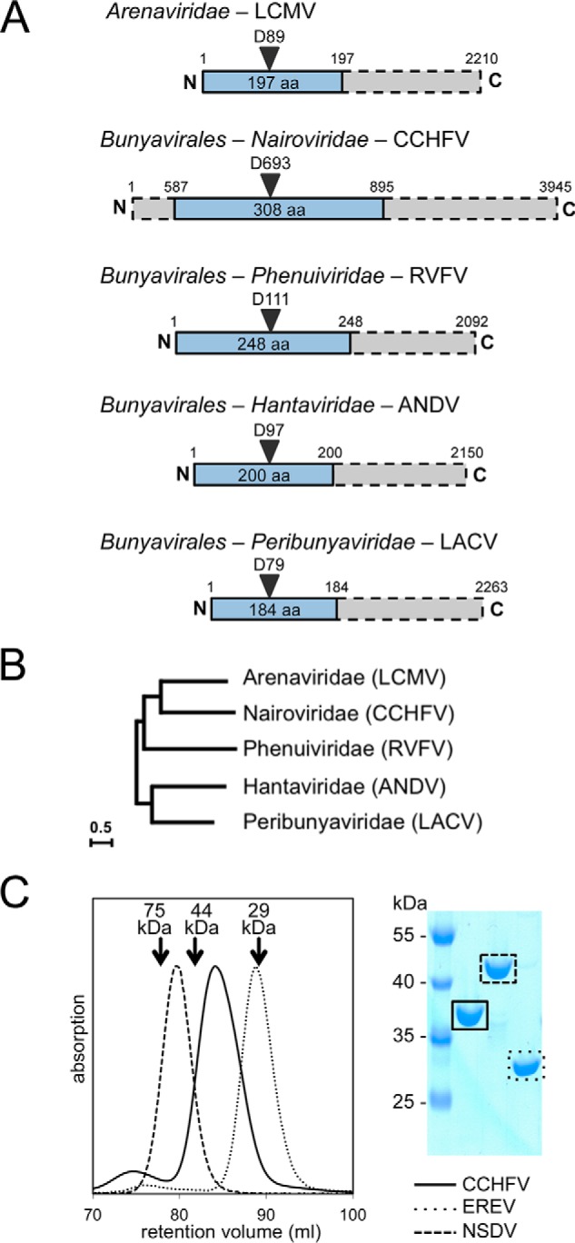
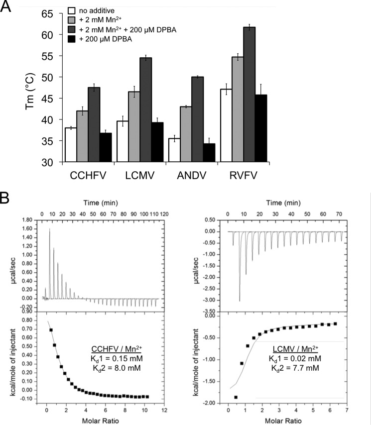
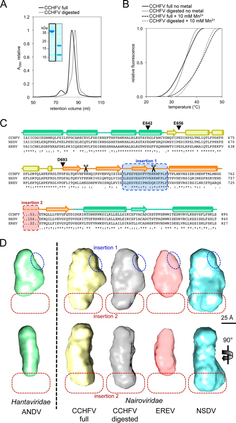
Viruses rely on many host cell processes, including the cellular transcription machinery. Segmented negative-strand RNA viruses (sNSV) in particular cannot synthesize the 5'-cap structure for their mRNA but cleave off cellular caps and use the resulting oligonucleotides as primers for their transcription. This cap-snatching mechanism, involving a viral cap-binding site and RNA endonuclease, is both virus-specific and essential for viral proliferation and therefore represents an attractive drug target. Here, we present biochemical and structural results on the putative cap-snatching endonuclease of Crimean-Congo hemorrhagic fever virus (CCHFV), a highly pathogenic bunyavirus belonging to the Nairoviridae family, and of two additional nairoviruses, Erve virus (EREV) and Nairobi sheep disease virus (NSDV). Our findings are presented in the context of other cap-snatching endonucleases, such as the enzymatically active endonuclease from Rift Valley fever virus (RVFV), from Arenaviridae and Bunyavirales, belonging to the His- and His+ endonucleases, respectively, according to the absence or presence of a metal ion-coordinating histidine in the active site. Mutational and metal-binding experiments revealed the presence of only acidic metal-coordinating residues in the active site of the CCHFV domain and a unique active-site conformation that was intermediate between those of His+ and His- endonucleases. On the basis of small-angle X-ray scattering (SAXS) and homology modeling results, we propose a protein topology for the CCHFV domain that, despite its larger size, has a structure overall similar to those of related endonucleases. These results suggest structural and functional conservation of the cap-snatching mechanism among sNSVs.

Keywords: cap-snatching; endonuclease; enzyme structure; metal ion–protein interaction; nairovirus; negative-strand RNA virus; recombinant protein expression; small-angle X-ray scattering (SAXS); structure-function; viral transcription.


Conflict of interest statement

The authors declare that they have no conflicts of interest with the contents of this article

Figures

Figure 1.
Figure 1.
Domain boundaries of the CCHFV cap-snatching endonuclease. A, schematic diagram of cap-snatching endonuclease domains (blue) within L proteins (gray bars) of sNSVs. The order/family, an example species, and residue numbers for the example species are indicated. B, evolutionary relationship between sNSVs based on the RdRp sequences, displayed in an unrooted maximum likelihood tree (adapted from Ref. 15), shows close relationship between Nairoviridae and Arenaviridae. C, size-exclusion chromatography and Coomassie-stained denaturing gel show that the three nairovirus endonuclease domains are pure, monomeric, and of different sizes. The molecular masses and retention volumes of size standard proteins are indicated above the chromatogram.
Figure 2.
Figure 2.
Nuclease activity of different cap-snatching endonucleases. Enzymes were incubated with a radioactively labeled nucleic acid substrate in the presence or absence of divalent metal ions, as indicated. Substrate and reaction products were separated on a denaturing polyacrylamide gel and visualized by autoradiography. A, His+ endonucleases show activity on ssRNA in the presence of manganese and calcium, whereas their catalytic site mutants are inactive. A and B, all three nairovirus endonucleases, as well as the His− endonuclease from LCMV, do not show significant activity on any of the tested RNA substrates. C, all tested His+ endonucleases (ANDV, LACV, and RVFV) do not cleave dsRNA, ssDNA, or dsDNA, but only ssRNA as shown exemplarily for the RVFV endonuclease.
Figure 3.
Figure 3.
Stabilization of cap-snatching endonucleases by a known endonuclease inhibitor and manganese binding. A, thermal stability of His+ and His− endonucleases in the presence of 2 mm MnCl2 and 200 μm DPBA measured in a thermal shift assay (Tm, melting temperature). Thermal stabilization by DPBA is only observed in the presence of manganese ions. B, manganese binding to 160 μm CCHFV (left panel) or 140 μm LCMV (right panel) endonuclease protein was measured at 20 °C by isothermal titration calorimetry. The upper plot shows the binding isotherm, and the lower plot shows the integrated values. The dissociation constants from fitting a two-site sequential binding mode (Kd1 and Kd2) to one representative experiment are indicated.
Figure 4.
Figure 4.
CCHFV endonuclease active site conformation. A, active site architecture of His+ (left panel) and His− (right panel) endonucleases. Superposition of relevant side chains (residue numbers are given in the table below) and the divalent metal ions of IAV, LACV, and HNTV are opposed to LASV, LCMV, and CASV. B, sequence alignment of the relevant area in the endonuclease domain of nairoviruses and potential His or Glu/Asp residues chosen for mutational studies. The alignment was generated using ClustalΩ (43). Conservation between residues is indicated as follows: *, identical; :, highly similar; ., similar. The predicted secondary structure elements are shown as cylinders for α-helices and arrows for β-sheets. Residues tested in mutagenesis studies are marked in blue (His) and red (Glu/Asp). The PD motif of the active site is shown at the far right. C, a Glu side chain but none of the His are involved in metal ion coordination, as shown by thermal stability testing of different CCHFV single-site mutants in presence of increasing amounts of Mn2+. As in B, His mutants are highlighted with a blue frame, and Glu/Asp mutants are highlighted with a red frame. IAV, influenza A virus; LASV, Lassa virus; LCMV, lymphocytic choriomeningitis virus; CASV, California Academy of Sciences virus.
Figure 5.
Figure 5.
Structural analysis of the CCHFV endonuclease and identification of two insertions. A, elution profiles from the size-exclusion column show a smaller size for the CCHFV endonuclease domain after trypsin treatment (gray) compared with the nontreated domain (black). Inset, the peak fractions are analyzed on a Coomassie-stained denaturing gel. B, melting curves of digested (gray) and full (black) CCHFV endonuclease domains are shown in absence (solid line) or in presence of 10 mm Mn2+ (dashed line). C, the structure-based sequence alignment of the three analyzed nairoviruses CCHFV, NSDV, and EREV was generated by PRALINE (16). Predicted and conserved secondary structure elements are shown as cylinders for α-helices and arrows for β-sheets. The color coding is as in Fig. 6. Insertion 1 (blue) is missing in the digested CCHFV domain (trypsin cleavage sites as determined by MS are indicated with scissors), and insertion 2 (red) is of variable size in the three different proteins. D, ab initio shapes of three nairovirus endonucleases (CCHFV, EREV, and NSDV) and the digested CCHFV domain compared with the ANDV endonuclease domain (9) as example for a non-nairovirus homolog are shown in front view (top panel) and side view (bottom panel). As in C, the proposed locations of the two insertions are marked with blue and red dashed lines.
Figure 6.
Figure 6.
Proposed structural topology of the putative CCHFV cap-snatching endonuclease. A, the sequence of the CCHFV endonuclease was manually aligned with His+ and His− endonucleases for which atomic structures are available. Secondary structure elements are shown as cylinders for α-helices and arrows for β-sheets and were taken from secondary structure prediction for CCHFV and from the crystal structures for the other proteins. Residues with a role in metal ion coordination are highlighted and align with the corresponding residues in the related proteins. Insertions 1 and 2, which are only present in nairoviruses, are highlighted in blue and red, respectively. B, structural topology of two His+ and a His− endonuclease (based on their crystal structures) and predicted topology of the CCHFV endonuclease, which takes an intermediate position. Important metal ion–coordinating side chains in the active site are shown as red circles (His) and blue circles (Glu/Asp). Related structural elements are colored as in A.

References

    1. Lukashev A. N., Klimentov A. S., Smirnova S. E., Dzagurova T. K., Drexler J. F., and Gmyl A. P. (2016) Phylogeography of Crimean Congo hemorrhagic fever virus. PLoS One 11, e0166744 10.1371/journal.pone.0166744 - DOI - PMC - PubMed
    1. Ergönül O. (2006) Crimean–Congo haemorrhagic fever. Lancet Infect. Dis. 6, 203–214 10.1016/S1473-3099(06)70435-2 - DOI - PMC - PubMed
    1. Zivcec M., Scholte F. E., Spiropoulou C. F., Spengler J. R., and Bergeron É. (2016) Molecular insights into Crimean–Congo hemorrhagic fever virus. Viruses 8, 106 10.3390/v8040106 - DOI - PMC - PubMed
    1. Ferron F., Weber F., de la Torre J. C., and Reguera J. (2017) Transcription and replication mechanisms of Bunyaviridae and Arenaviridae L proteins. Virus Res. 234, 118–134 10.1016/j.virusres.2017.01.018 - DOI - PMC - PubMed
    1. Reguera J., Gerlach P., and Cusack S. (2016) Towards a structural understanding of RNA synthesis by negative strand RNA viral polymerases. Curr. Opin. Struct. Biol. 36, 75–84 10.1016/j.sbi.2016.01.002 - DOI - PubMed

Publication types

LinkOut - more resources