. 2018 Dec 21;293(51):19511-19521.
doi: 10.1074/jbc.RA118.005421. Epub 2018 Oct 22.

It takes a dimer to tango: Oligomeric small heat shock proteins dissociate to capture substrate

Affiliations

It takes a dimer to tango: Oligomeric small heat shock proteins dissociate to capture substrate

Indu Santhanagopalan et al. J Biol Chem. .

Abstract

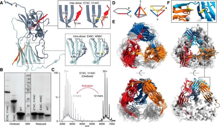
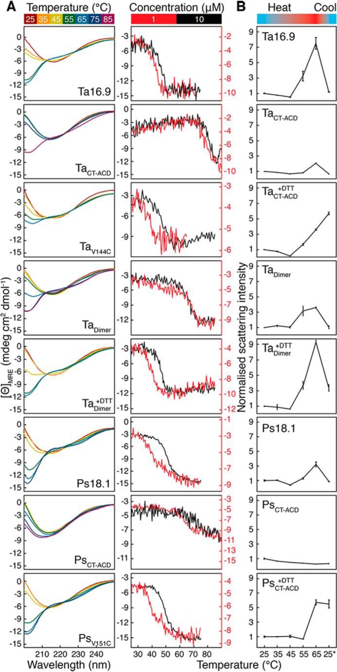
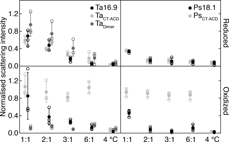
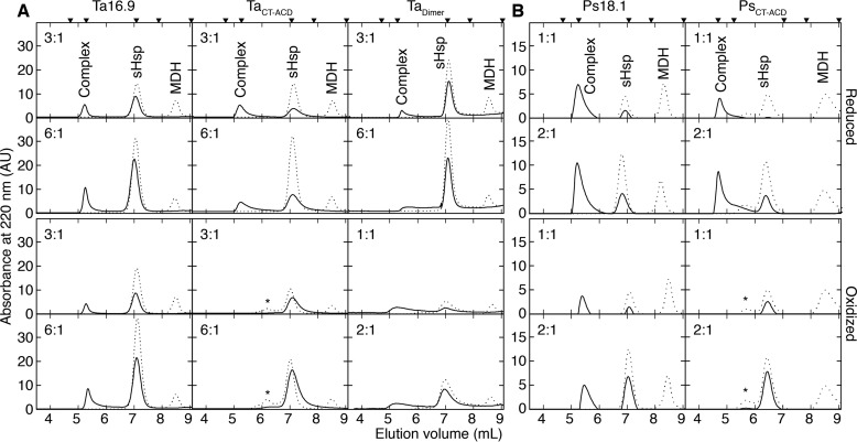
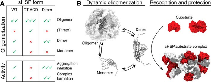
Small heat-shock proteins (sHsps) are ubiquitous molecular chaperones, and sHsp mutations or altered expression are linked to multiple human disease states. sHsp monomers assemble into large oligomers with dimeric substructure, and the dynamics of sHsp oligomers has led to major questions about the form that captures substrate, a critical aspect of their mechanism of action. We show here that substructural dimers of two plant dodecameric sHsps, Ta16.9 and homologous Ps18.1, are functional units in the initial encounter with unfolding substrate. We introduced inter-polypeptide disulfide bonds at the two dodecameric interfaces, dimeric and nondimeric, to restrict how their assemblies can dissociate. When disulfide-bonded at the nondimeric interface, mutants of Ta16.9 and Ps18.1 (TaCT-ACD and PsCT-ACD) were inactive but, when reduced, had WT-like chaperone activity, demonstrating that dissociation at nondimeric interfaces is essential for sHsp activity. Moreover, the size of the TaCT-ACD and PsCT-ACD covalent unit defined a new tetrahedral geometry for these sHsps, different from that observed in the Ta16.9 X-ray structure. Importantly, oxidized Tadimer (disulfide bonded at the dimeric interface) exhibited greatly enhanced ability to protect substrate, indicating that strengthening the dimeric interface increases chaperone efficiency. Temperature-induced size and secondary structure changes revealed that folded sHsp dimers interact with substrate and that dimer stability affects chaperone efficiency. These results yield a model in which sHsp dimers capture substrate before assembly into larger, heterogeneous sHsp-substrate complexes for substrate refolding or degradation, and suggest that tuning the strength of the dimer interface can be used to engineer sHsp chaperone efficiency.

Keywords: chaperone; chaperone efficiency; disulfides; dynamic light scattering (DLS); native mass spectrometry; oligomerization; protein design; protein engineering; protein folding; protein stability; small heat shock protein (sHsp); small-angle X-ray scattering (SAXS); stress response; substrate recognition; thermal stability.


Conflict of interest statement

The authors declare that they have no conflicts of interest with the contents of this article

Figures

Figure 1.
Figure 1.
Disulfide bonds introduced within the sHsp dimer or between sHsp dimers, the latter of which defines a new geometry of the sHsp dodecamer. A, positions of the Cys substitutions linking dimers (Inter-dimer; top inset) with the CTD of one dimer shown in red, and a segment of the β-sandwich of the other dimer in gray. Positions of the Cys substitutions linking monomers within a dimer (Intra-dimer; bottom inset) with segments of one monomer in dark gray and the other monomer in light gray. B, SDS-PAGE separation of the indicated wheat WT or mutant proteins either oxidized (left) or reduced (right). C, mass spectra of E74C V144C (TaCT-ACD) in the oxidized state before (black) and after activation (MS2) of the 32+ ion (gray), showing dissociation to covalent trimers. D, the possible theoretical geometries of dodecamers comprising dimers with each edge corresponding to a dimer. The only geometry consistent with a three-point linkage is a tetrahedron (middle). E, tetrahedral models of Ta16.9 (see text and supporting figures and methods for details). Three dimers are colored (blue, orange, red) and three are rendered in gray. F, the best-fit model highlighting positions of the disulfides in the dodecamer. Enlargement shows detail of an interdimer disulfide (between the blue and orange dimers) (left) and the intradimer disulfide (between the light blue and dark blue monomers within the dimer) (right).
Figure 2.
Figure 2.
Secondary structure, thermal stability, and size of the sHsps. A, left, far-UV CD spectra for Ta16.9, oxidized TaCT-ACD, TaV144C, oxidized Tadimer, reduced Tadimer, Ps18.1, oxidized PsCT-ACD, and PsV151C (all 10 μm) collected at temperatures from 25 to 65 °C. Spectra were also collected at 75 and 85 °C for oxidized TaCT-ACD, Tadimer, and PsCT-ACD. Right, mean residue ellipticity at 205 nm as a function of temperature for 10 μm (black) and 1 μm (red) sHsp. B, average values with standard deviations for the TSI of proteins from three experiments carried out at each temperature. Measurements were normalized with respect to 25 °C, which was assigned a value of 1.0.
Figure 3.
Figure 3.
Restricting dodecamer dissociation alters sHsp chaperone activity. Light scattering by aggregated MDH mixed with Ta16.9, TaCT-ACD, or Tadimer under reducing (top left) or oxidizing (bottom left) conditions, and Ps18.1 and PsCT-ACD under reducing (top right) or oxidizing (bottom right) conditions. The sHsp monomer: MDH monomer ratio is indicated on the x axis. Scattering values were normalized with respect to that for MDH heated alone, which was assigned a value of 1.0. Means (filled circles) from three replicate experiments (open circles) are plotted with standard deviation. Unheated MDH (4 °C) served as a control for absence of aggregation.
Figure 4.
Figure 4.
sHsp–MDH complex formation correlates with aggregation protection. A and B, SEC profiles were generated on a TSKgel SWxl Ultra SW column for soluble fractions of reaction mixtures of sHsp–MDH under reducing (top two rows) or oxidizing conditions (bottom two rows) for samples prepared as in Fig. 3 with (A) Ta16.9 and mutants or (B) Ps18.1 and PsCT-ACD. The dashed and solid lines represent unheated and heated reaction mixtures, respectively. The sHsp monomer: MDH monomer molar ratios are indicated at the top right corner of each plot. Calibration standards above the top panels correspond from left to right: Void volume, 670, 158, 44, and 17 kDa. Peaks seen at 7 ml and 8.5 ml correspond to sHsp and MDH, respectively. * indicates absorbance from protein with spurious disulfide linkages. Peaks between 4.7 and 6.5 ml correspond to sHsp–MDH complexes.
Figure 5.
Figure 5.
The sHsp dimer is the major substrate encounter species. A, table summarizing how the disulfide constrained sHsps (CT-ACD and Dimer) alter the population of dodecamer, dimer, or monomer compared with WT and resultant changes in chaperone activity. Check mark indicates presence and abundance of a specific protein form or extent of chaperone activity; × indicates absence of that protein form or activity. B, schematic showing how the dimer, which is the favored form in the dimer mutant and disfavored form in the CT-ACD mutants (as in A) is the major substrate capture form of the sHsp, followed by subsequent assembly of multiple dimers and substrates into heterogeneous complexes.

References

    1. Basha E., O'Neill H., and Vierling E. (2012) Small heat shock proteins and α-crystallins: Dynamic proteins with flexible functions. Trends Biochem. Sci. 37, 106–117 10.1016/j.tibs.2011.11.005 - DOI - PMC - PubMed
    1. Richter K., Haslbeck M., and Buchner J. (2010) The heat shock response: Life on the verge of death. Mol. Cell 40, 253–266 10.1016/j.molcel.2010.10.006 - DOI - PubMed
    1. Haslbeck M., and Vierling E. (2015) A first line of stress defense: Small heat shock proteins and their function in protein homeostasis. J. Mol. Biol. 427, 1537–1548 10.1016/j.jmb.2015.02.002 - DOI - PMC - PubMed
    1. Hilton G. R., Lioe H., Stengel F., Baldwin A. J., and Benesch J. L. P. (2013) Small heat-shock proteins: Paramedics of the cell. Top. Curr. Chem. 328, 69–98 10.1007/128_2012_324 - DOI - PubMed
    1. Santhanagopalan I., Basha E., Ballard K. N., Bopp N. E., and Vierling E. (2015) Model chaperones: Small heat shock proteins from plants. in The Big Book on Small Heat Shock Proteins (Tanguay R. M., and Hightower L. E., eds), pp. 119–153, Springer International Publishing, Cham, Switzerland: 10.1007/978-3-319-16077-1 - DOI

Publication types

LinkOut - more resources