. 2018 Oct 22;9(1):4394.
doi: 10.1038/s41467-018-06811-z.

Heterozygous deletion of chromosome 17p renders prostate cancer vulnerable to inhibition of RNA polymerase II

Affiliations

Heterozygous deletion of chromosome 17p renders prostate cancer vulnerable to inhibition of RNA polymerase II

Yujing Li et al. Nat Commun. .

Abstract

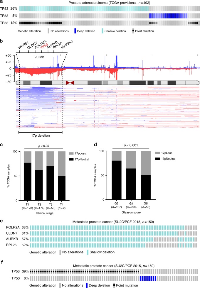
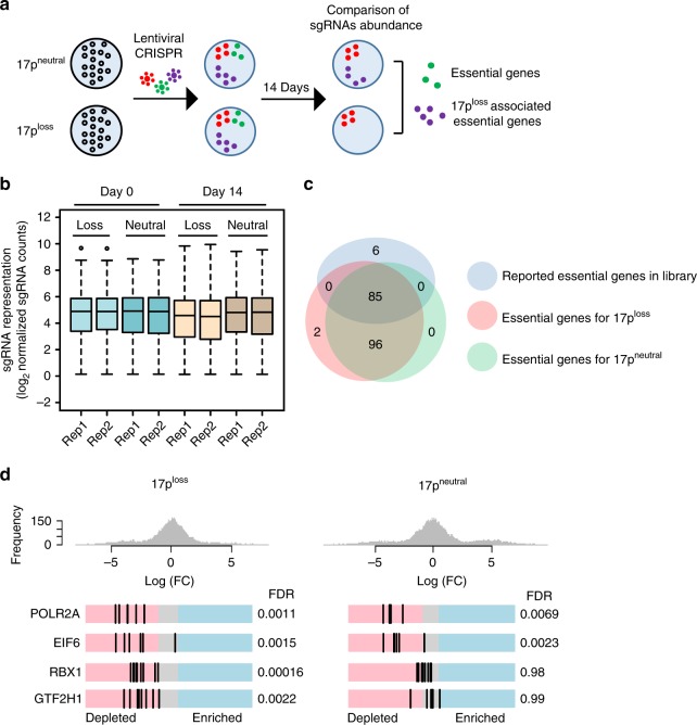
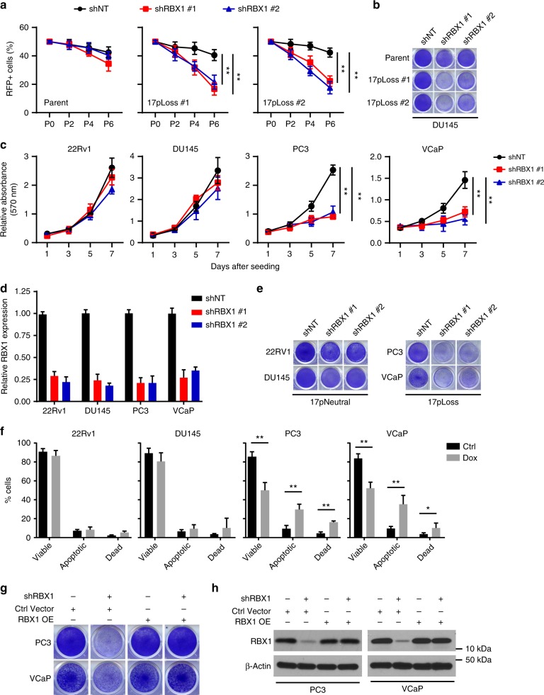
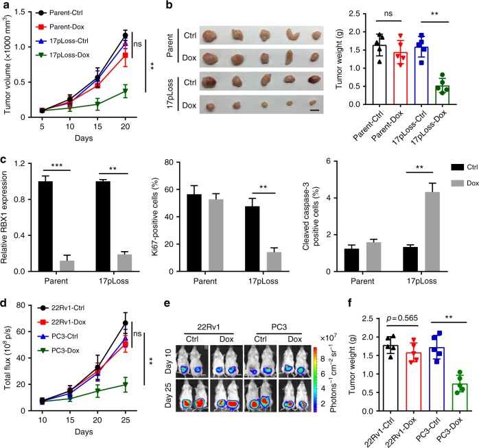
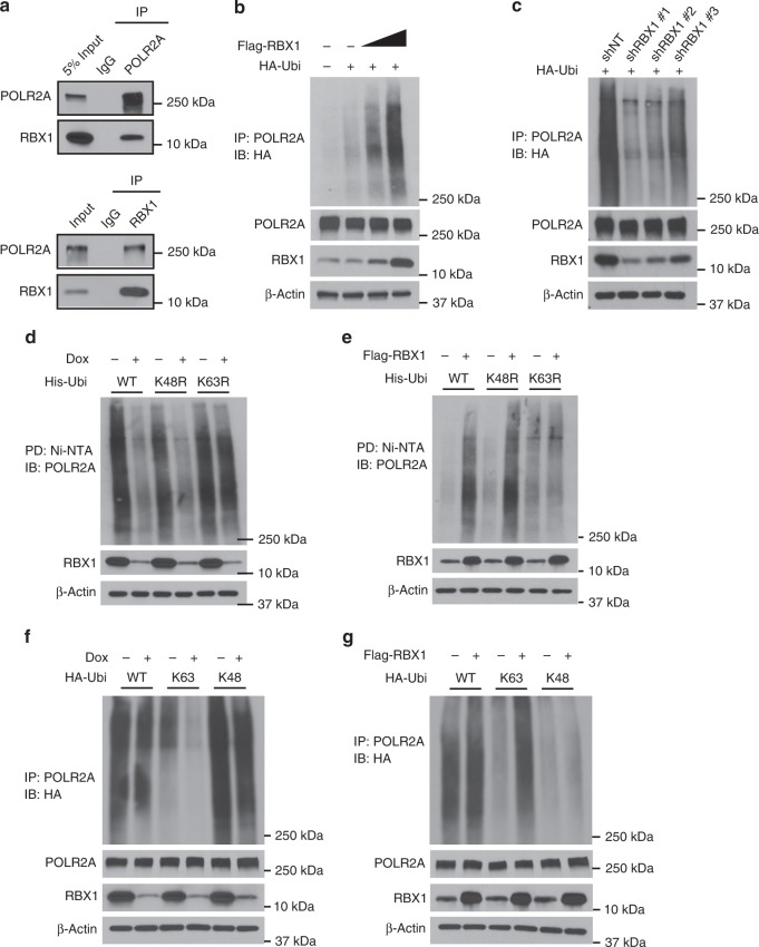
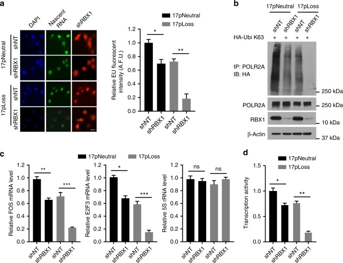
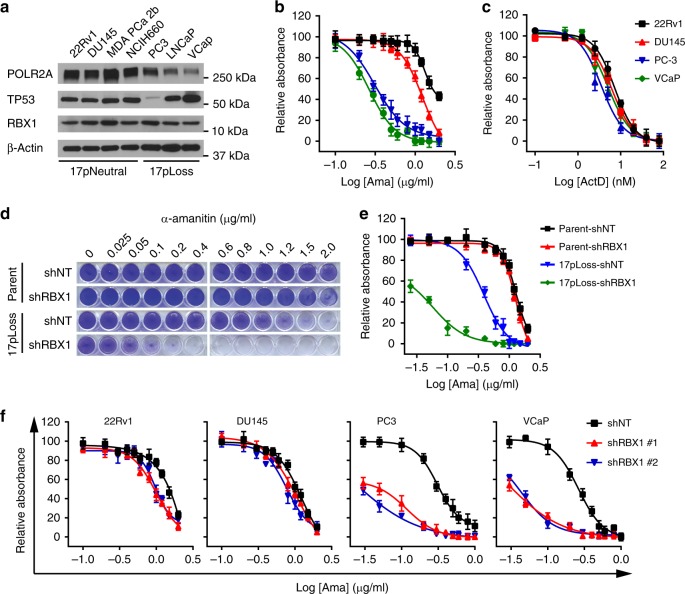
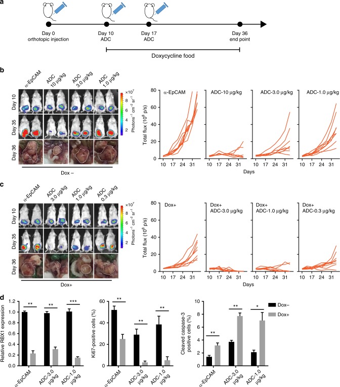
Heterozygous deletion of chromosome 17p (17p) is one of the most frequent genomic events in human cancers. Beyond the tumor suppressor TP53, the POLR2A gene encoding the catalytic subunit of RNA polymerase II (RNAP2) is also included in a ~20-megabase deletion region of 17p in 63% of metastatic castration-resistant prostate cancer (CRPC). Using a focused CRISPR-Cas9 screen, we discovered that heterozygous loss of 17p confers a selective dependence of CRPC cells on the ubiquitin E3 ligase Ring-Box 1 (RBX1). RBX1 activates POLR2A by the K63-linked ubiquitination and thus elevates the RNAP2-mediated mRNA synthesis. Combined inhibition of RNAP2 and RBX1 profoundly suppress the growth of CRPC in a synergistic manner, which potentiates the therapeutic effectivity of the RNAP2 inhibitor, α-amanitin-based antibody drug conjugate (ADC). Given the limited therapeutic options for CRPC, our findings identify RBX1 as a potentially therapeutic target for treating human CRPC harboring heterozygous deletion of 17p.


Conflict of interest statement

The authors declare no competing interests.

Figures

Fig. 1
Fig. 1
Chromosome 17p loss is a frequent genomic event in prostate cancer. a Genomic alterations of TP53 (point mutation, shallow deletion and deep deletion) in a TCGA prostate cancer dataset (TCGA provisional, n = 492) determined by cBioportal. Due to the intra-tumor heterogeneity, one tumor tissue may include tumor cells with homozygous deletion of TP53 or with mutant TP53, as shown in a few cases. b Integrated analysis of 17p deletion in 155 prostate cancer patient samples. Frequency plots of the copy number abnormalities indicate degree of copy number loss (red) or gain (blue). Representative genes in 17p deletion region are shown. c, d Distribution of heterozygous deletion of 17p among tumor clinical (c) and pathological (d) stages in the TCGA prostate cancer dataset (Fisher’s exact test). e, f Genomic alterations of 17p heterozygous deletion (e) and TP53 mutations (f) in the TCGA metastatic prostate cancer dataset (SU2C/PCF 2015, n = 150) determined by cBioportal
Fig. 2
Fig. 2
CRISPR-Cas9 screen identifies RBX1 as an essential gene for 17ploss prostate cancer cells. a Schematic illustration of CRISPR screening procedure in the isogenic pair of DU145 cells. b Box plots showing the distribution of sgRNA frequencies at different time points. c Overlapping of essential genes from this screen and from the previous reports. d Frequency histograms of enriched or depleted sgRNAs. POLR2A and EIF6 are two representatives of common essential genes. RBX1 and GTF2H1 are representatives of selective essential genes in the context of 17p loss
Fig. 3
Fig. 3
Prostate cancer cells with 17p deletion are highly sensitive to RBX1 depletion. a, b Effect of RBX1 knockdown on the proliferation of the parental and isogenic 17ploss DU145 cells, determined by direct competition assay. Cells expressing RFP and control nonspecific shRNA (shNT) or shRBX1 were sorted and mixed with control RFP-negative cells (1:1) and the RFP-positive cells were quantified at passages 2, 4, and 6 (a). Representative cell survival measured by staining with crystal violet was shown in b. ce Cell growth curves, based on crystal violet staining, of human prostate cancer cell lines expressing Dox-inducible shNT or RBX1-specific shRNA (shRBX1 #1, shRBX1 #2) (c). RBX1 knockdown efficiency and representative image were shown in d and e, respectively. f Fraction of apoptotic cells in the 17pneutral (22Rv1 and DU145) and 17ploss (PC3 and VCaP) cells expressing Dox-induced RBX1 shRNA at 4 days post Dox treatment. g, h Cell survival measured by crystal violet staining (g) and protein expression levels (h) of RBX1 in PC3 and VCaP cells expressing shRBX1, control or ectopic RBX1. Data are representative of three independent experiments and analyze by unpaired two-tailed t-test. Error bars denote SD. **, p < 0.01; ***, p < 0.001
Fig. 4
Fig. 4
Depletion of RBX1 inhibits the growth of 17ploss CRPC tumors in vivo. ac Tumor growth curves (a), gross tumor images and weights (b) of xenograft tumors derived from subcutaneously implanted parental and isogenic 17ploss DU145 cells expressing Dox-inducible RBX1 shRNA (n = 3). c RBX1 expression, cell proliferation and apoptosis in the above xenograft tumors were quantified. Scale bar, 10 mm. df Tumor growth curves (d), representative bioluminescent images (e), and gross tumor weights (f) of xenograft tumors derived from orthotopically implanted 22Rv1 and PC3 cells expressing Dox-inducible RBX1 shRNA (n = 5). Data are analyze by unpaired two-tailed t-test and are presented as the mean ± SD. ns not significant; **, p < 0.01
Fig. 5
Fig. 5
RBX1 modifies POLR2A by the K63-linked ubiquitination. a RBX1 physically interacts with POLR2A. Immunoprecipitation (IP) and western blot analyses were performed using indicated antibodies. Normal IgG was used as a negative control for IP. b, c RBX1 overexpression (b) or knockdown (c) increases or decreases the level of ubiquitinated POLR2A, respectively. DU145 cells transfected with indicated expression vectors were treated with MG132 and ubiquitinated POLR2A was pulled down (IP) and subject to immunoblotting (IB) analysis. d, e RBX1-mediated ubiquitination acts on the lysine-63 (K63) residue of POLR2A. DU145 cells were transfected with the indicated ubiquitin expression vectors and treated with MG132. Cells were also treated with Dox to induce RBX1 knockdown in d, or ectopic Flag-RBX1 was expressed in the cells in e. Ubiquitinated POLR2A was pulled down (PD) with Ni-NTA Agarose and subject to immunoblotting (IB) analysis. f, g RBX1 knockdown (f) or overexpression (g) decreases or increases the level of lysine-63 (K63) ubiquitination of POLR2A, respectively. Equal amounts of cell lysates were analyzed by immunoprecipitation and IB assays as described above. Data are representative of three independent experiments
Fig. 6
Fig. 6
RBX1 promotes the POLR2A-dependent RNA transcription. a Global RNA synthesis in the parental and isogenic 17ploss DU145 cells was evaluated by measurement of 5-EU incorporation after Dox-inducible RBX1 knockdown and the intensity was quantified with the CellProfiler Software. Scale bar, 25 µm. b Effect of RBX1 knockdown on the levels of lysine-63 (K63) ubiquitination of POLR2A in the parental and isogenic 17ploss DU145 cells. c qPCR analysis of the levels of short-lived mRNA (FOS and E2F3) compared to the control 5S rRNA in the isogenic DU145 cells with or without Dox-inducible RBX1 knockdown. d qPCR analysis of the in vitro transcription activity using HelaScribe DNA template. Data are representative of three independent experiments and analyze by unpaired two-tailed t-test. Error bars denote SD. ns not significant; **, p < 0.01; ***, p < 0.001
Fig. 7
Fig. 7
RBX1 depletion sensitized 17ploss prostate cancer cells to POLR2A inhibition. a Protein levels of POLR2A, p53, Rbx1, and β-Actin in human prostate cancer cell lines. b, c Cell proliferation of 17pneutral (22Rv1 and DU145) and 17ploss cells (PC3 and VCaP) treated with α-amanitin (b) or actinomycin D (c). d, e RBX1 depletion sensitizes the 17ploss DU145 cells to the treatment of the POLR2A inhibitor, α-amanitin. Representative images (d) and quantitative results (e) of cell survival are shown. f Cell proliferation of human prostate cancer cell lines under α-amanitin treatment. 17pneutral (22Rv1 and DU145) and 17ploss (PC3 and VCaP) cells, with or without Dox-induced RBX1 knockdown, were treated with increasing doses of α-amanitin. Data are representative of three independent experiments
Fig. 8
Fig. 8
Inhibition of RBX1 sensitizes 17ploss CRPC to the treatment of α-amanitin-based ADC. a Schematic illustration of orthotopic injection of 17ploss DU145 cells (1 × 106 cells) followed by twice i.p. injection of ADC and Dox food treatment. b, c Representative bioluminescent tumor images and individual tumor growth curves of xenograft tumors derived from orthotopically implanted 17ploss DU145 cells without (b) or with (c) Dox treatment. Once tumor was established, mice were randomly divided to 4 groups (n = 8) and treated with either free anti-EpCAM antibody or different doses of anti-EpCAM-amanitin conjugates (ADC). d Quantification of RBX1 knockdown efficiency, cell proliferation (Ki-67 staining) and apoptosis (cleaved caspase-3 staining) in the xenografted tumor tissues described above. Data are analyze by unpaired two-tailed t-test and are presented as the mean ± SD. **, p< 0.01; ***, p < 0.001

References

    1. Siegel RL, Miller KD, Jemal A. Cancer statistics, 2018. CA Cancer J. Clin. 2018;68:7–30. doi: 10.3322/caac.21442. - DOI - PubMed
    1. Chang AJ, Autio KA, Roach M, 3rd, Scher HI. High-risk prostate cancer-classification and therapy. Nat. Rev. Clin. Oncol. 2014;11:308–323. doi: 10.1038/nrclinonc.2014.68. - DOI - PMC - PubMed
    1. Huggins C, Hodges CV. Studies on prostatic cancer. I. The effect of castration, of estrogen and androgen injection on serum phosphatases in metastatic carcinoma of the prostate. CA Cancer J. Clin. 1972;22:232–240. doi: 10.3322/canjclin.22.4.232. - DOI - PubMed
    1. Attard G, et al. Prostate cancer. Lancet. 2016;387:70–82. doi: 10.1016/S0140-6736(14)61947-4. - DOI - PubMed
    1. Wong YN, Ferraldeschi R, Attard G, de Bono J. Evolution of androgen receptor targeted therapy for advanced prostate cancer. Nat. Rev. Clin. Oncol. 2014;11:365–376. doi: 10.1038/nrclinonc.2014.72. - DOI - PubMed

Publication types

MeSH terms