. 2018 Dec;50(12):1705-1715.
doi: 10.1038/s41588-018-0252-3. Epub 2018 Oct 22.

Long noncoding RNA MALAT1 suppresses breast cancer metastasis

Affiliations

Long noncoding RNA MALAT1 suppresses breast cancer metastasis

Jongchan Kim et al. Nat Genet. 2018 Dec.

Abstract

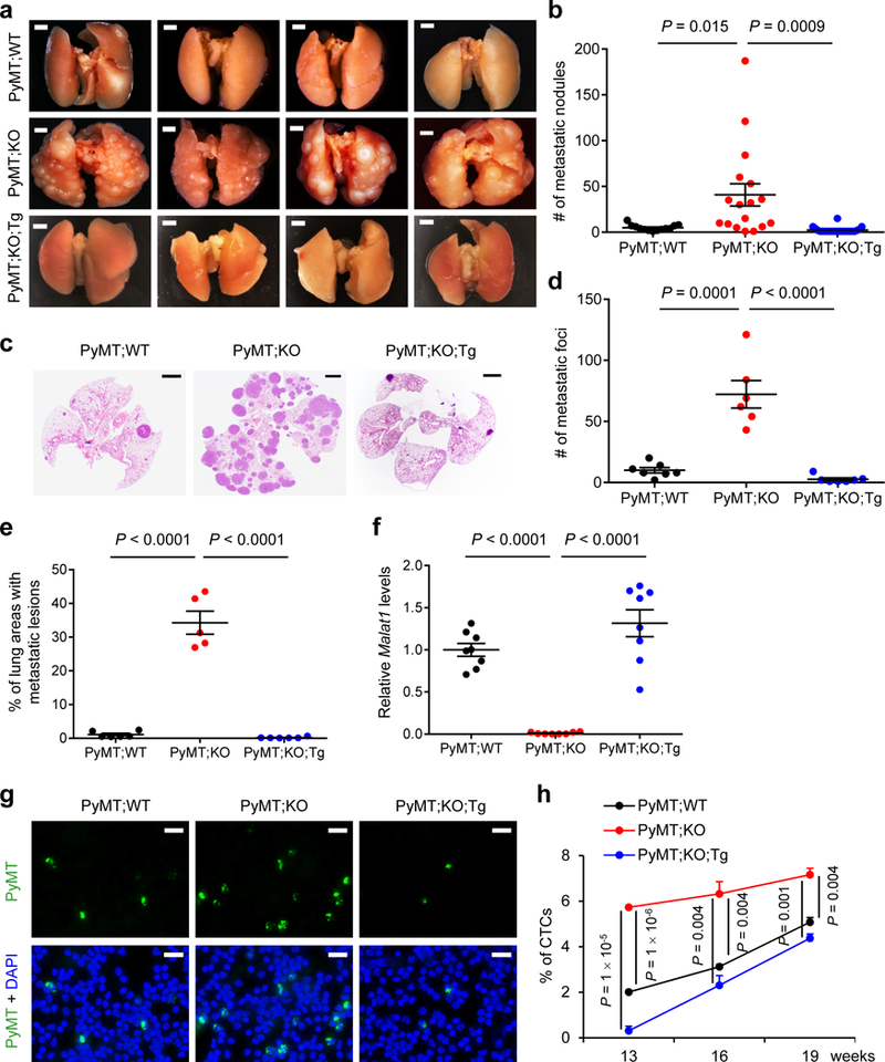
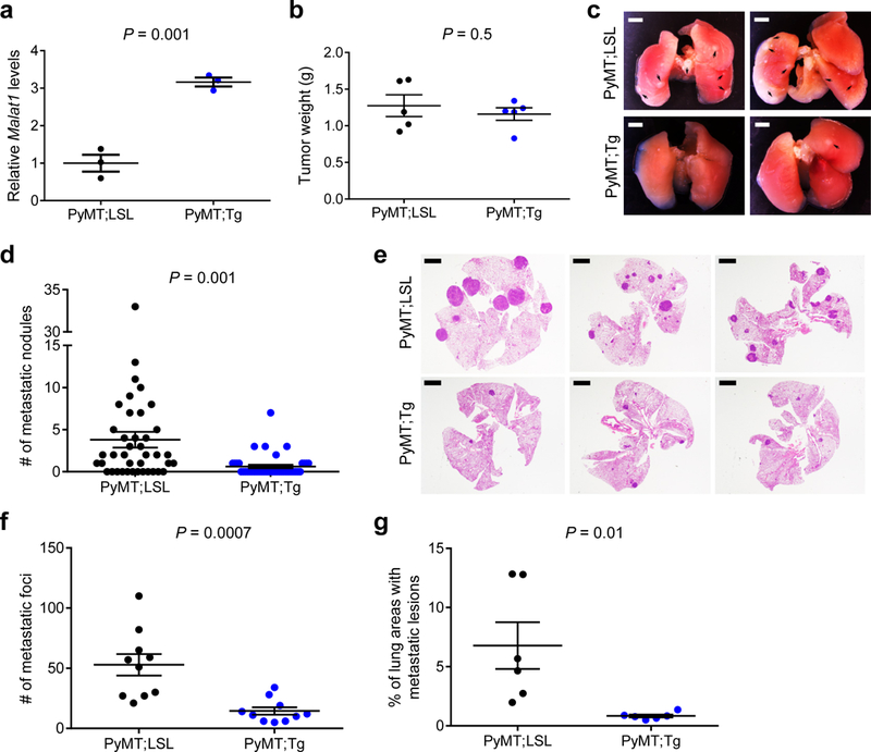
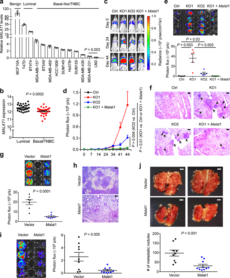
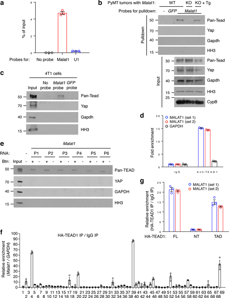
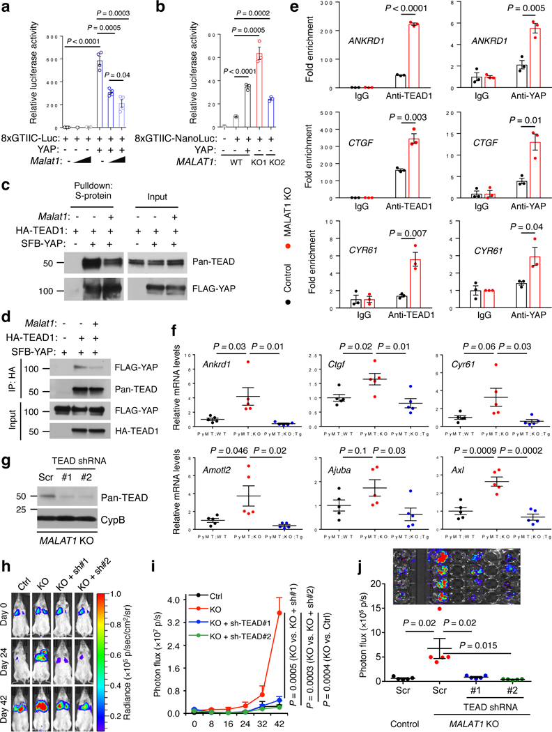
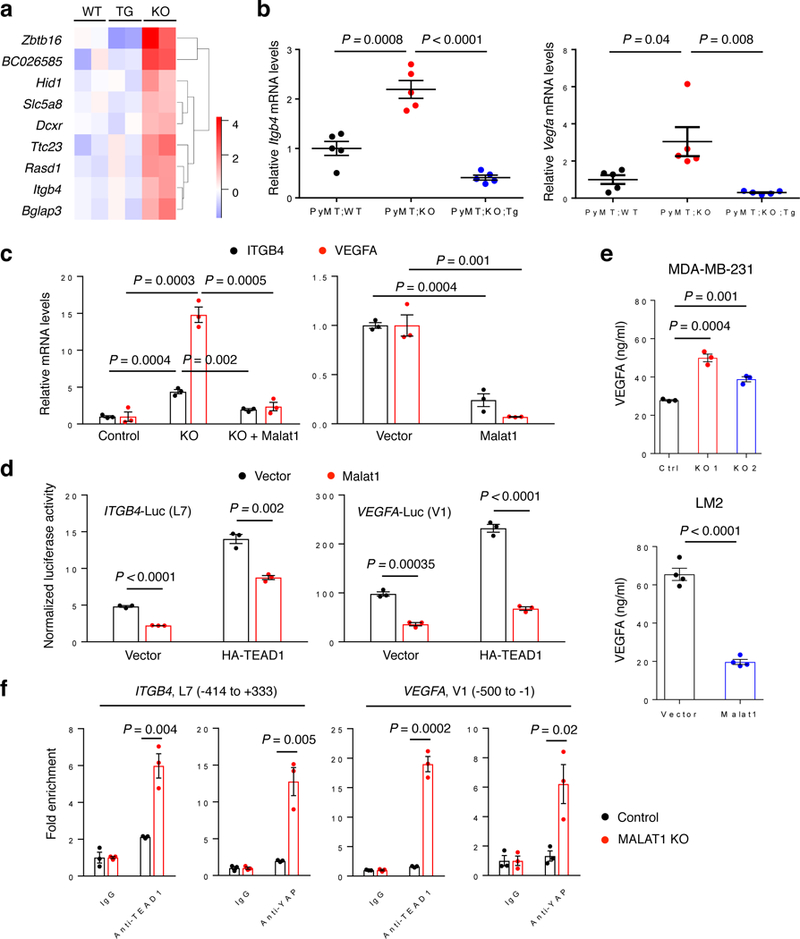
MALAT1 has previously been described as a metastasis-promoting long noncoding RNA (lncRNA). We show here, however, that targeted inactivation of the Malat1 gene in a transgenic mouse model of breast cancer, without altering the expression of its adjacent genes, promotes lung metastasis, and that this phenotype can be reversed by genetic add-back of Malat1. Similarly, knockout of MALAT1 in human breast cancer cells induces their metastatic ability, which is reversed by re-expression of Malat1. Conversely, overexpression of Malat1 suppresses breast cancer metastasis in transgenic, xenograft, and syngeneic models. Mechanistically, the MALAT1 lncRNA binds and inactivates the prometastatic transcription factor TEAD, preventing TEAD from associating with its co-activator YAP and target gene promoters. Moreover, MALAT1 levels inversely correlate with breast cancer progression and metastatic ability. These findings demonstrate that MALAT1 is a metastasis-suppressing lncRNA rather than a metastasis promoter in breast cancer, calling for rectification of the model for this highly abundant and conserved lncRNA.


Conflict of interest statement

Competing interests

The authors declare no completing interests.

Figures

Figure 1.
Figure 1.. Targeted inactivation and restoration of Malat1 in mice demonstrate that Malat1 is a suppressor of breast cancer lung metastasis
(a, b) Bright-field imaging (a) and the number of metastatic nodules (b) in the lungs of MMTV-PyMT;Malat1+/+ (PyMT;WT, n = 13 mice), MMTV-PyMT;Malat1−/− (PyMT;KO, n = 17 mice), and MMTV-PyMT;Malat1−/−;Malat1Tg (PyMT;KO;Tg, n = 22 mice) mice at the endpoint (20–25 weeks of age). Scale bars in (a), 2 mm. (c-e) H&E staining (c) and the number (d) and relative area (e) of metastatic foci in the lungs of PyMT;WT, PyMT;KO, and PyMT;KO;Tg mice at the endpoint (20–25 weeks of age). n = 7, 6, and 7 mice per group in (d); n = 6, 5, and 6 mice per group in (e). Scale bars in (c), 2 mm. (f) qPCR of Malat1 in the mammary tumors of age-matched PyMT;WT, PyMT;KO, and PyMT;KO;Tg mice. n = 8 mice per group. (g, h) Immunofluorescent staining (g) and the percentages (h) of circulating tumor cells (CTCs) in the peripheral blood from PyMT;WT, PyMT;KO, and PyMT;KO;Tg mice. CTCs from 13-, 16-, and 19-week-old mice were immunostained with a PyMT-specific antibody (green) and nuclei were stained with DAPI (blue). n = 3 mice per group. Scale bars in (g), 20 μm. Statistical significance in (b), (d), (e), (f), and (h) was determined by an unpaired t-test. Error bars are s.e.m. All mice used in this figure are females on a B6 background.
Figure 2.
Figure 2.. Targeted transgenic overexpression of Malat1 in mice inhibits breast cancer metastasis
(a) qPCR of Malat1 in the mammary tumors of MMTV-PyMT;Malat1LSL (PyMT;LSL) and MMTV-PyMT;Malat1Tg (PyMT;Tg) mice. n = 3 mice per group. (b) Weight of the mammary tumors of PyMT;LSL and PyMT;Tg mice at 8 weeks of age. n = 5 mice per group. (c, d) Bright-field imaging (c, arrows indicate metastases) and the number of metastatic nodules (d) in the lungs of PyMT;LSL (n = 40 mice) and PyMT;Tg (n = 42 mice) mice at the endpoint (12–13 weeks of age). Scale bars in (c), 2 mm.(e-g) H&E staining (e) and the number (f) and relative area (g) of metastatic foci in the lungs of PyMT;LSL and PyMT;Tg mice at the endpoint (12–13 weeks of age). n = 10 mice per group in (f); n = 6 mice per group in (g). Scale bars in (e), 2 mm.Statistical significance in (a), (b), (d), (f), and (g) was determined by an unpaired t-test. Error bars are s.e.m. All mice used in this figure are females on an FVB background.
Figure 3.
Figure 3.. Malat1 inhibits metastatic ability of breast cancer cells.
(a) qPCR of MALAT1 in a panel of cell lines. (b) MALAT1 levels in luminal (n = 28) and basal/triple-negative (n = 31) breast cancer cell lines available in CCLE. (c, d) Bioluminescent imaging (c) and quantification of photon flux (d) of NSG mice with intravenous injection of control, MALAT1 knockout, or Malat1-restored MDA-MB-231 cells. Day 0: the day of tumor cell injection. n = 5 mice per group.(e, f) Bioluminescent imaging (e, upper panel), quantification of photon flux (e, lower panel), and H&E staining (f) of the lungs from mice described in Fig. 3c, d. n = 5 mice per group in (e). Scale bars in (f), 200 μm.(g) Bioluminescent imaging (upper panel) and quantification of photon flux (lower panel) of the lungs from NSG mice with intravenous injection of control (n = 7 mice) or Malat1-overexpressing (n = 8 mice) LM2 cells. (h) H&E staining of the lungs described in Fig. 3g. Scale bars, 200 μm. (i) Bioluminescent imaging (left panel) and quantification of photon flux (right panel) of the lungs from BALB/c mice injected with control (n = 9 mice) or Malat1-overexpressing (n = 10 mice) 4T1 cells.(j) Bright-field imaging (upper panel) and the number of metastatic nodules (lower panel) in the lungs of mice described in Fig. 3i. Scale bars, 2 mm. Statistical significance in (a), (b), (d), (e), (g), (i), and (j) was determined by an unpaired t-test. Error bars are s.e.m.
Figure 4.
Figure 4.. MALAT1 interacts with TEAD family members
(a) qPCR of Malat1 in ChIRP samples. Probes for mouse Malat1 or U1 nuclear RNA were used to pull down endogenous Malat1 or U1 from PyMT mammary tumor samples. (b, c) Western blot analysis of ChIRP samples. Mouse Malat1-specific probes were used to pull down endogenous Malat1 from the mammary tumors of MMTV-PyMT;Malat1+/+ (WT), MMTV-PyMT;Malat1−/− (KO), and MMTV-PyMT;Malat1−/−;Malat1Tg (KO + Tg) mice (b), or from 4T1 cells (c), followed by immunoblotting with antibodies against pan-Tead, Yap, Gapdh, and histone H3 (HH3).(d) RNA immunoprecipitation assay. Endogenous TEAD1 was immunoprecipitated from crosslinked MDA-MB-231 cells. TEAD1-bound MALAT1 was quantitated by qPCR with two primer sets. GAPDH was used as a negative control. (e) RNA pulldown assay. Unlabeled and biotinylated Malat1 fragments (P1-P6) were synthesized by in vitro transcription, incubated with HEK293FT cell lysate, and pulled down with streptavidin beads. The bound proteins were eluted by boiling in Laemmli sample buffer and immunoblotted with antibodies against pan-TEAD, YAP, GAPDH, and histone H3. Btn: biotinylation. (f) CLIP-qPCR assay of Hela cells overexpressing HA-TEAD1 and mouse Malat1. The protected Malat1 RNA segments bound by TEAD1 were detected by qPCR using 69 pairs of primers. (g) RNA immunoprecipitation assay. Hela cells were transfected with HA-tagged full-length TEAD1 (FL), N-terminal region (NT), or transactivation domain (TAD), crosslinked, and subjected to immunoprecipitation with a HA-specific antibody. TEAD1-bound MALAT1 was quantitated by qPCR with two primer sets. All error bars are s.e.m. Uncropped blots are shown in Supplementary Fig. 10.
Figure 5.
Figure 5.. MALAT1 inactivates TEAD
(a) Luciferase activity in HEK293FT cells co-transfected with indicated plasmids. n = 4 cell culture replicates per group. (b) Luciferase activity in control and MALAT1 knockout MDA-MB-231 cells co-transfected with indicated plasmids. n = 3 cell culture replicates per group. (c, d) HEK293FT cells were co-transfected with Malat1, SFB-YAP, and HA-TEAD1, and were subjected to pulldown with S-protein beads (c) or an HA-specific antibody (d), followed by immunoblotting with antibodies against pan-TEAD and FLAG. (e) ChIP-qPCR analysis showing the occupancy of ANKRD1, CTGF, and CYR61 promoters by TEAD1 or YAP immunoprecipitated from control or MALAT1 knockout MDA-MB-231 cells. (f) qPCR of YAP-TEAD target genes in the tumors of MMTV-PyMT;Malat1+/+ (PyMT;WT), MMTV-PyMT;Malat1−/− (PyMT;KO), and MMTV-PyMT;Malat1−/−;Malat1Tg (PyMT;KO;Tg) mice. n = 5 mice per group. (g) Immunoblotting of pan-TEAD and cyclophilin B (CypB) in MALAT1 knockout MDA-MB-231 cells with or without transduction of TEAD shRNA. Scr: scramble control. (h, i) Bioluminescent imaging (h) and photon flux quantification (i) of NSG mice with intravenous injection of control and MALAT1 knockout MDA-MB-231 cells with or without transduction of TEAD shRNA. Day 0: the day of tumor cell injection. n = 5 mice per group. (j) Bioluminescent imaging (upper panel) and photon flux quantification (lower panel) of the lungs from mice described in Fig. 5h, i. n = 5 mice per group. Statistical significance in (a), (b), (e), (f), (i), and (j) was determined by an unpaired t-test. Error bars are s.e.m. Uncropped blots are shown in Supplementary Fig. 10.
Figure 6.
Figure 6.. ITGB4 and VEGFA are TEAD target genes and are regulated by MALAT1.
(a) Heat map of nine genes that were identified by RNA-Seq analysis to be commonly upregulated in MMTV-PyMT;Malat1−/− tumors (KO), compared with both MMTV-PyMT;Malat1+/+ tumors (WT) and MMTV-PyMT;Malat1−/−;Malat1Tg tumors (TG). n = 2 mice per group. (b) qPCR of Itgb4 (left panel) and Vegfa (right panel) in the mammary tumors of MMTV-PyMT;Malat1+/+ (PyMT;WT), MMTV-PyMT;Malat1−/− (PyMT;KO), and MMTV-PyMT;Malat1−/−;Malat1Tg (PyMT;KO;Tg) mice. n = 5 mice per group. (c) qPCR of ITGB4 and VEGFA in control, MALAT1 knockout, and Malat1-restored MDA-MB-231 cells (left panel), and in control and Malat1-overexpressing LM2 cells (right panel). (d) Luciferase activity in HEK293FT cells co-transfected with Malat1, HA-TEAD1, an ITGB4 (left panel) or VEGFA (right panel) luciferase reporter, and a Renilla luciferase reporter. n = 3 cell culture replicates per group. (e) ELISA of VEGFA secreted by MALAT1 knockout MDA-MB-231 cells (upper panel, n = 3 cell culture replicates per group) and by Malat1-overexpressing LM2 cells (lower panel, n = 4 cell culture replicates per group).(f) ChIP-qPCR analysis showing the occupancy of ITGB4 (two left panels) and VEGFA (two right panels) promoters by TEAD1 or YAP. Endogenous TEAD1 and YAP were immunoprecipitated from control or MALAT1 knockout MDA-MB-231 cells. Statistical significance in (b) – (f) was determined by an unpaired t-test. Error bars are s.e.m.

Comment in

References

    1. Evans JR, Feng FY & Chinnaiyan AM The bright side of dark matter: lncRNAs in cancer. J Clin Invest 126, 2775–82 (2016). - PMC - PubMed
    1. Zhang B et al. The lncRNA Malat1 is dispensable for mouse development but its transcription plays a cis-regulatory role in the adult. Cell Rep 2, 111–23 (2012). - PMC - PubMed
    1. Wilusz JE, Freier SM & Spector DL 3’ end processing of a long nuclear-retained noncoding RNA yields a tRNA-like cytoplasmic RNA. Cell 135, 919–32 (2008). - PMC - PubMed
    1. Hutchinson JN et al. A screen for nuclear transcripts identifies two linked noncoding RNAs associated with SC35 splicing domains. BMC Genomics 8, 39 (2007). - PMC - PubMed
    1. Tripathi V et al. The nuclear-retained noncoding RNA MALAT1 regulates alternative splicing by modulating SR splicing factor phosphorylation. Mol Cell 39, 925–38 (2010). - PMC - PubMed

Publication types

MeSH terms

Substances