Laminin functionalized biomimetic apatite to regulate the adhesion and proliferation behaviors of neural stem cells
- PMID: 30349246
- PMCID: PMC6188167
- DOI: 10.2147/IJN.S176596
Laminin functionalized biomimetic apatite to regulate the adhesion and proliferation behaviors of neural stem cells
Abstract
Background: Functionalizing biomaterial substrates with biological signals shows promise in regulating neural stem cell (NSC) behaviors through mimicking cellular microenvironment. However, diverse methods for immobilizing biological molecules yields promising results but with many problems. Biomimetic apatite is an excellent carrier due to its non-toxicity, good biocompatibility, biodegradability, and favorable affinity to plenty of molecules. Therefore, it may provide a promising alternative in regulating NSC behaviors.
Methods: Biomimetic apatite immobilized with the extracellular protein - laminin (LN) was prepared through coprecipitation process in modified Dulbecco's phosphate-buffered saline (DPBS) containing LN. The amount of coprecipitated LN and their release kinetics were examined. The adhesion and proliferation behaviors of NSC on biomimetic apatite immobilized with LN were investigated.
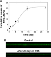
Results: The coprecipitation approach provided well retention of LN within biomimetic apatite up to 28 days, and supported the adhesion and proliferation of NSCs without cytotoxicity. For long-term cultivation, NSCs formed neurosphere-like aggregates on non-functionalized biomimetic apatite. A monolayer of proliferated NSCs on biomimetic apatite with coprecipitated LN was observed and even more stable than the positive control of LN coated tissue-culture treated polystyrene (TCP).
Conclusion: The simple and reproducible method of coprecipitation suggests that biomimetic apatite is an ideal carrier to functionalize materials with biological molecules for neural-related applications.
Keywords: adhesion; biomimetic apatite; coprecipitation; laminin; neural stem cell; proliferation.
Conflict of interest statement
Disclosure The authors report no conflicts of interest in this work.
Figures


Similar articles
-
Velvet antler polypeptide combined with calcium phosphate coating to protect peripheral nerve cells from oxidative stress.J Mol Histol. 2022 Dec;53(6):915-923. doi: 10.1007/s10735-022-10099-1. Epub 2022 Aug 29. J Mol Histol. 2022. PMID: 36036305 Free PMC article.
-
Laminin-apatite composite coating to enhance cell adhesion to ethylene-vinyl alcohol copolymer.J Biomed Mater Res A. 2005 Feb 1;72(2):168-74. doi: 10.1002/jbm.a.30205. J Biomed Mater Res A. 2005. PMID: 15578653
-
In vitro response of MC3T3-E1 pre-osteoblasts within three-dimensional apatite-coated PLGA scaffolds.J Biomed Mater Res B Appl Biomater. 2005 Oct;75(1):81-90. doi: 10.1002/jbm.b.30261. J Biomed Mater Res B Appl Biomater. 2005. PMID: 16001421
-
Biomimetic collagen/apatite coating formation on Ti6Al4V substrates.J Biomed Mater Res B Appl Biomater. 2012 Apr;100(3):871-81. doi: 10.1002/jbm.b.31970. Epub 2011 Nov 21. J Biomed Mater Res B Appl Biomater. 2012. PMID: 22102365 Review.
-
Adhesion of mesenchymal stem cells to biomimetic polymers: A review.Mater Sci Eng C Mater Biol Appl. 2017 Feb 1;71:1192-1200. doi: 10.1016/j.msec.2016.10.013. Epub 2016 Oct 14. Mater Sci Eng C Mater Biol Appl. 2017. PMID: 27987676 Review.
Cited by
-
siRNA-Loaded Hydroxyapatite Nanoparticles for KRAS Gene Silencing in Anti-Pancreatic Cancer Therapy.Pharmaceutics. 2021 Sep 8;13(9):1428. doi: 10.3390/pharmaceutics13091428. Pharmaceutics. 2021. PMID: 34575504 Free PMC article.
-
The Role of Lutheran/Basal Cell Adhesion Molecule in Hematological Diseases and Tumors.Int J Mol Sci. 2024 Jul 2;25(13):7268. doi: 10.3390/ijms25137268. Int J Mol Sci. 2024. PMID: 39000374 Free PMC article. Review.
References
-
- Grabel L. Developmental origin of neural stem cells: the glial cell that could. Stem Cell Rev. 2012;8(2):577–585. - PubMed
-
- Hsieh J, Schneider JW. Neuroscience. Neural stem cells, excited. Science. 2013;339(6127):1534–1535. - PubMed
-
- Péron S, Berninger B. Imported stem cells strike against stroke. Cell Stem Cell. 2015;17(5):501–502. - PubMed
MeSH terms
Substances
LinkOut - more resources
Full Text Sources
Molecular Biology Databases

