. 2018 Oct 8:9:2181.
doi: 10.3389/fmicb.2018.02181. eCollection 2018.

Common Cervicovaginal Microbial Supernatants Alter Cervical Epithelial Function: Mechanisms by Which Lactobacillus crispatus Contributes to Cervical Health

Affiliations

Common Cervicovaginal Microbial Supernatants Alter Cervical Epithelial Function: Mechanisms by Which Lactobacillus crispatus Contributes to Cervical Health

Lauren Anton et al. Front Microbiol. .

Abstract

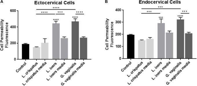
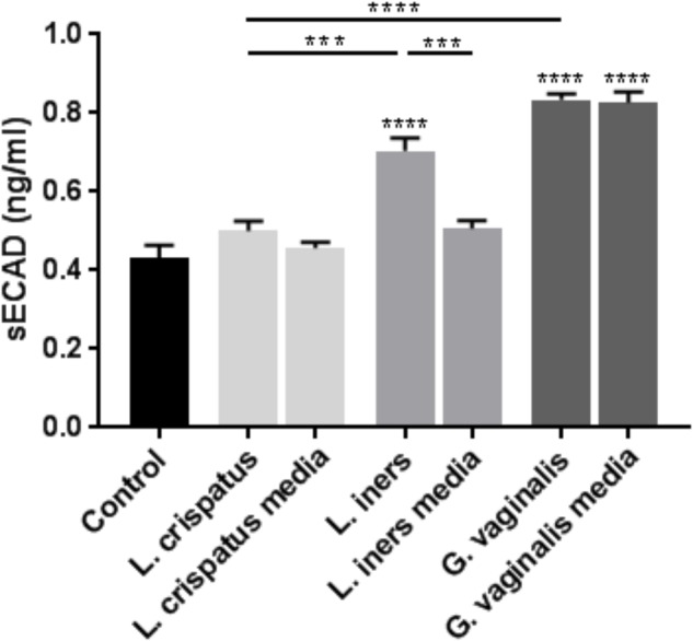
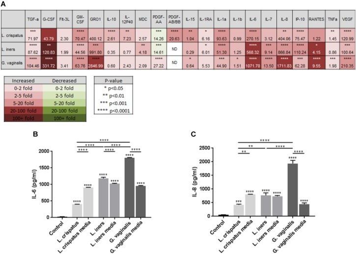
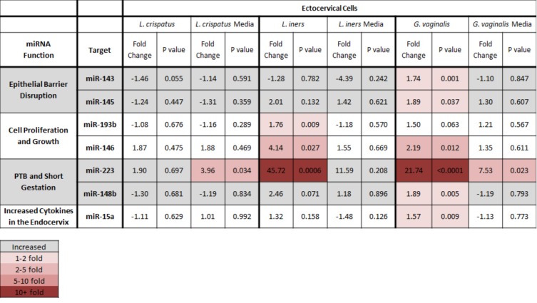
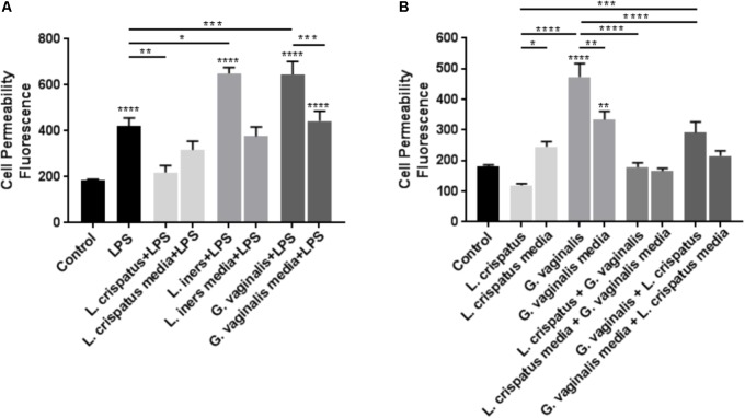
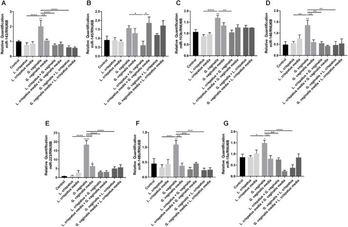
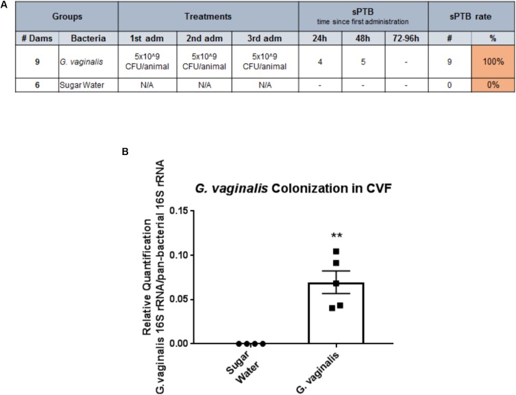
Cervicovaginal (CV) microbiota is associated with vaginal health and disease in non-pregnant women. Recent studies in pregnant women suggest that specific CV microbes are associated with preterm birth (PTB). While the associations between CV microbiota and adverse outcomes have been demonstrated, the mechanisms regulating the associations remain unclear. As the CV space contains an epithelial barrier, we postulate that CV microbiota can alter the epithelial barrier function. We investigated the biological, molecular, and epigenetic effects of Lactobacillus crispatus, Lactobacillus iners, and Gardnerella vaginalis on the cervical epithelial barrier function and determined whether L. crispatus mitigates the effects of lipopolysaccharide (LPS) and G. vaginalis on the cervical epithelial barrier as a possible mechanism by which CV microbiota mitigates disease risk. Ectocervical and endocervical cells treated with L. crispatus, L. iners, and G. vaginalis bacteria-free supernatants alone or combined were used to measure cell permeability, adherens junction proteins, inflammatory mediators, and miRNAs. Ectocervical and endocervical permeability increased after L. iners and G. vaginalis exposure. Soluble epithelial cadherin increased after exposure to L. iners but not G. vaginalis or L. crispatus. A Luminex cytokine/chemokine panel revealed increased proinflammatory mediators in all three bacteria-free supernatants with L. iners and G. vaginalis having more diverse inflammatory effects. L. iners and G. vaginalis altered the expression of cervical-, microbial-, and inflammatory-associated miRNAs. L. crispatus mitigated the LPS or G. vaginalis-induced disruption of the cervical epithelial barrier and reversed the G. vaginalis-mediated increase in miRNA expression. G. vaginalis colonization of the CV space of a pregnant C57/B6 mouse resulted in 100% PTB. These findings demonstrate that L. iners and G. vaginalis alter the cervical epithelial barrier by regulating adherens junction proteins, cervical immune responses, and miRNA expressions. These results provide evidence that L. crispatus confers protection to the cervical epithelial barrier by mitigating LPS- or G. vaginalis-induced miRNAs associated with cervical remodeling, inflammation, and PTB. This study provides further evidence that the CV microbiota plays a role in cervical function by altering the cervical epithelial barrier and initiating PTB. Thus, targeting the CV microbiota and/or its effects on the cervical epithelium may be a potential therapeutic strategy to prevent PTB.

Keywords: Gardnerella vaginalis; Lactobacillus crispatus; Lactobacillus iners; cervix; epithelial barrier; inflammation; miRNA; preterm birth.


Figures

FIGURE 1
FIGURE 1
Cell permeability is increased in ectocervical and endocervical cells after exposure to L. iners and G. vaginalis but not L. crispatus bacteria-free supernatants. Cell permeability was measured in ectocervical cells (A) and endocervical cells (B) after a 48 h exposure to bacteria-free supernatants (10% v/v) from L. crispatus, L. iners, and G. vaginalis compared to non-treated control cells. Bacterial growth media alone acted as a negative control for the three bacteria-free supernatants tested. Cell permeability is expressed as fluorescence OD measurements from a fluorescent plate reader and is indicative of the movement of FITC-dextran from the top to the bottom insert of a transwell chamber system. Values are mean ± SEM. Asterisks over the individual bars represent comparisons to control; asterisks over solid lines represent comparisons between treatment groups. p < 0.05, ∗∗p < 0.01, ∗∗∗p < 0.001, ∗∗∗∗p < 0.0001.
FIGURE 2
FIGURE 2
Lactobacillus iners and G. vaginalis but not L. crispatus bacteria-free supernatants increase epithelial-cadherin cleavage in ectocervical cells. Soluble epithelial-cadherin (sECAD) levels were measured after ectocervical cells were exposed to L. crispatus, L. iners, and G. vaginalis bacteria-free supernatants (10% v/v) for 48 h. Bacterial growth media alone acted as a negative control for the three bacteria-free supernatants tested. sECAD was measured in ectocervical cell culture supernatants using a commercially available sandwich ELISA. Values are mean ± SEM. Asterisks over the individual bars represent comparisons to control; asterisks over solid lines represent comparisons between treatment groups. p < 0.05, ∗∗p < 0.01, ∗∗∗p < 0.001, ∗∗∗∗p < 0.0001.
FIGURE 3
FIGURE 3
Lactobacillus crispatus, L. iners, and G. vaginalis bacteria-free supernatants differentially alter the host epithelial immune response in ectocervical cells. Immune cytokines/chemokines released from ectocervical cells after exposure to L. crispatus, L. iners, and G. vaginalis bacteria-free supernatants (10% v/v) for 48 h were measured by Luminex (A). Heat map depicts fold change by color, p-value by asterisks, and concentration (pg/ml) of each analyte by the number value present in each corresponding box. Validation of Luminex findings were performed for IL-6 (B) and IL-8 (C) in ectocervical cells exposed to L. crispatus, L. iners, and G. vaginalis bacteria-free supernatants or bacterial growth media alone. IL-6 and IL-8 were measured in ectocervical cell culture supernatants using a commercially available sandwich ELISA. Values are mean ± SEM. Asterisks over the individual bars represent comparisons to control; asterisks over solid lines represent comparisons between treatment groups. p < 0.05, ∗∗p < 0.01, ∗∗∗p < 0.001, ∗∗∗∗p < 0.0001.
FIGURE 4
FIGURE 4
Lactobacillus crispatus, L. iners, and G. vaginalis bacteria-free supernatants differentially alter the miRNA expression profile of ectocervical cells. miRNA expression was measured by QPCR in ectocervical cells after exposure to L. crispatus, L. iners, and G. vaginalis bacteria-free supernatants (10% v/v) for 48 h. Bacterial growth media alone acted as a negative control for the three bacteria-free supernatants tested. The ΔΔCT method was used for relative expression quantification using the endogenous reference gene RNU6B. Modified heat map shows fold change versus control (non-treated cells) and corresponding p-values. Color distinguishes level of fold change. Values are mean ± SEM.
FIGURE 5
FIGURE 5
Lactobacillus crispatus bacteria-free supernatants mitigate the LPS- or G. vaginalis-induced increases in cell permeability in ectocervical cells. (A) Cell permeability was measured in ectocervical cells after exposure to bacterial free supernatants (10% v/v) from L. crispatus, L. iners, and G. vaginalis for 24 h followed by LPS (25 μg/ml) exposure for additional 24 h. Bacterial growth media containing LPS acted as a negative control for the three bacteria-free supernatants tested. (B) Cell permeability was measured in ectocervical cells after exposure to bacterial free supernatants from L. crispatus and G. vaginalis alone or in combination. Ectocervical cells were exposed to L. crispatus supernatants (5% v/v) on day 1 followed by G. vaginalis supernatants (5% v/v) on day 2 or vice versa for 24–48 h. Bacterial growth media alone acted as a negative control for the three bacteria-free supernatants tested. Cell permeability is expressed as fluorescence OD measurements from a fluorescent plate reader and is indicative of the movement of FITC-dextran from the top to the bottom insert of a transwell chamber system. Values are mean ± SEM. Asterisks over the individual bars represent comparisons to control; asterisks over solid lines represent comparisons between treatment groups. p < 0.05, ∗∗p < 0.01, ∗∗∗p < 0.001, ∗∗∗∗p < 0.0001.
FIGURE 6
FIGURE 6
Lactobacillus crispatus bacteria-free supernatants mitigate the G. vaginalis-induced miRNA expression profile in ectocervical cells. miRNA expression (A–G) was measured by QPCR in ectocervical cells after exposure to L. crispatus supernatants (5% v/v) on day 1 followed by G. vaginalis supernatants (5% v/v) on day 2 or vice versa for 24–48 h. Bacterial growth media alone acted as a negative control for the three bacteria-free supernatants tested. The ΔΔCT method was used for relative expression quantification using the endogenous reference gene RNU6B. Values are mean ± SEM. Asterisks over the individual bars represent comparisons to control; asterisks over solid lines represent comparisons between treatment groups. p < 0.05, ∗∗p < 0.01, ∗∗∗p < 0.001, ∗∗∗∗p < 0.0001.
FIGURE 7
FIGURE 7
Intravaginal inoculation of G. vaginalis results in PTB and colonization of the cervicovaginal space. (A) G. vaginalis (5 × 10⁹ CFU/animal) was inoculated intravaginally into timed pregnant C57/B6 mice on embryonic day E13, E14, and E15. Sugar water inoculated animals acted as controls. Animals were observed for PTB daily until term. Table shows the dose of G. vaginalis inoculation at each administration, the number of dams delivering preterm on each day following the first inoculation, and the PTB percentage. (B). G. vaginalis (5 × 10⁹ CFU/animal) was inoculated intravaginally into timed pregnant C57/B6 mice on E13. Animals were sacrificed 24 h after inoculation. Cervicovaginal fluid (CVF) was collected for gDNA isolation and measurement of the 16S gene of G. vaginalis by QPCR. Sugar water inoculated animals acted as controls. Values are mean ± SEM. ∗∗p < 0.01.

References

    1. Abreu M. T. (2010). Toll-like receptor signalling in the intestinal epithelium: how bacterial recognition shapes intestinal function. Nat. Rev. Immunol. 10 131–144. 10.1038/nri2707. - DOI - PubMed
    1. Anton L., DeVine A., Sierra L. -J., Brown A. G., Elovitz M. A. (2017). miR-143 and miR-145 disrupt the cervical epithelial barrier through dysregulation of cell adhesion, apoptosis and proliferation. Sci. Rep. 7:3020. 10.1038/s41598-017-03217-7. - DOI - PMC - PubMed
    1. Atassi F., Brassart D., Grob P., Graf F., Servin A. L. (2006). Lactobacillus strains isolated from the vaginal microbiota of healthy women inhibit Prevotella bivia and Gardnerella vaginalis in coculture and cell culture. FEMS Immunol. Med. Microbiol. 48 424–432. 10.1111/j.1574-695X.2006.00162.x. - DOI - PubMed
    1. Barnum C. E., Fey J. L., Weiss S. N., Barila G., Brown A. G., Connizzo B. K., et al. (2017). Tensile mechanical properties and dynamic collagen fiber re-alignment of the murine cervix are dramatically altered throughout pregnancy. J. Biomech. Eng. 139:061008. 10.1115/1.4036473. - DOI - PMC - PubMed
    1. Behrman R., Butler A. (2007). Institute of Medicine (US) Committee on Understanding Premature Birth and Assuring Healthy Outcomes. Preterm Birth: Causes, Consequences, and Prevention. Washington, DC: National Academies Press (US). - PubMed