. 2018 Sep 7:15:179-188.
doi: 10.2142/biophysico.15.0_179. eCollection 2018.

Mutational analysis of the conserved carboxylates of anion channelrhodopsin-2 (ACR2) expressed in Escherichia coli and their roles in anion transport

Affiliations

Mutational analysis of the conserved carboxylates of anion channelrhodopsin-2 (ACR2) expressed in Escherichia coli and their roles in anion transport

Keiichi Kojima et al. Biophys Physicobiol. .

Abstract

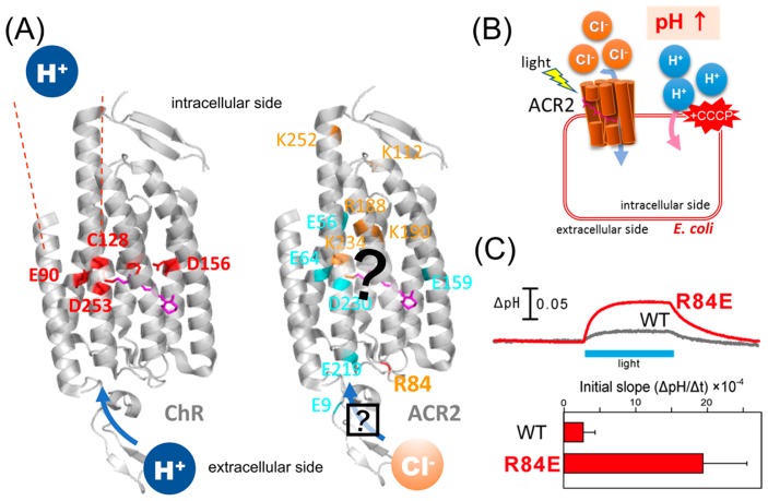
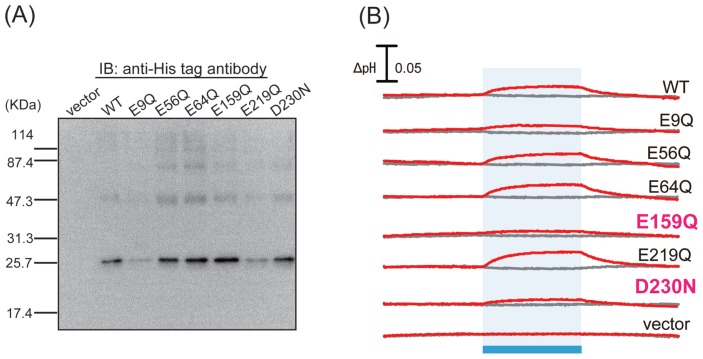
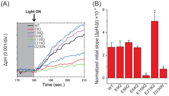
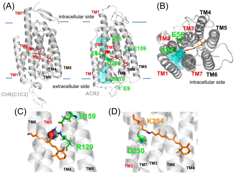
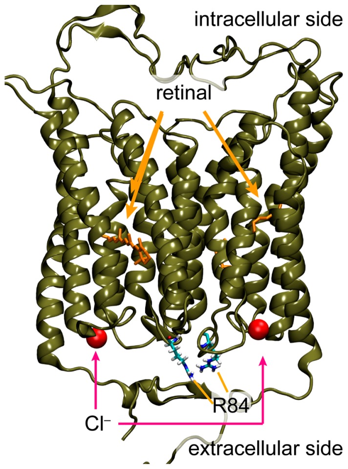
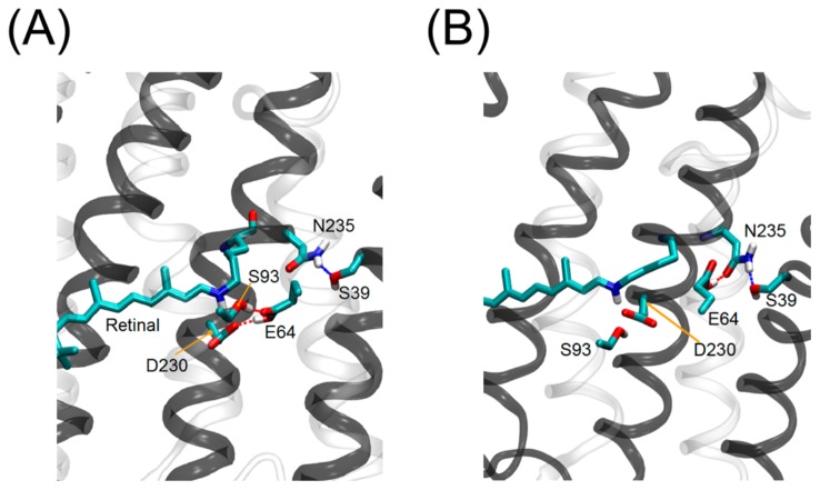
Anion channelrhodopsin-2 (ACR2), a light-gated channel recently identified from the cryptophyte alga Guillardia theta, exhibits anion channel activity with exclusive selectivity. In addition to its novel function, ACR2 has become a focus of interest as a powerful tool for optogenetics. Here we combined experimental and computational approaches to investigate the roles of conserved carboxylates on the anion transport activity of ACR2 in Escherichia coli membrane. First, we replaced six conserved carboxylates with a neutral residue (i.e. E9Q, E56Q, E64Q, E159Q, E219Q and D230N), and measured anion transport activity using E. coli expression system. E159Q and D230N exhibited significantly lower anion transport activity compared with wild-type ACR2 (1/12~1/3.4), which suggests that E159 and D230 play important roles in the anion transport. Second, to explain its molecular aspects, we constructed a homology model of ACR2 based on the crystal structure of a cation channelrhodopsin (ChR). The model structure showed a cavity formed by four transmembrane helices (TM1, TM2, TM3 and TM7) similar to ChRs, as a putative anion conducting pathway. Although E159 is not located in the putative pathway, the model structure showed hydrogen bonds between E159 and R129 with a water molecule. D230 is located in the pathway near the protonated Schiff base (PSB) of the chromophore retinal, which suggests that there is an interaction between D230 and the PSB. Thus, we demonstrated the functional importance and the hypothetical roles of two conserved carboxylates, E159 and D230, in the anion transport activity of ACR2 in E. coli membrane.

Keywords: anion channel; ion transport; microbial rhodopsin; retinal.


Conflict of interest statement

Conflict of Interest There are no conflicts of interest to declare. The authors declare no competing financial interests.

Figures

Figure 1
Figure 1
Characteristics of ChRs and ACRs. (A) (Left) Crystal structure of ChR (PDB;3UG9) showing that the cation conducting pathway is formed by TM1, TM2, TM3 and TM7. Key residues for the cation transport in ChRs are colored red. (Right) High resolution structure and the anion conducting pathway of ACR2 are still unclear. Conserved basic and acidic amino acid residues are colored orange and cyan, respectively. (B) Light-induced secondary proton movement across the cell membrane by the inward chloride channeling activity of ACR2 expressed in E. coli cells, which is facilitated by the addition of CCCP. The proton movement results in increases in extracellular pH. (C) (Upper) Light-induced pH changes of E. coli cells expressing wild-type ACR2 or the R84E mutant in a solution containing 300 mM NaCl in the presence of CCCP. The cell suspensions were illuminated with blue light (480±10 nm) for 3 min (blue stripe). (Lower) Comparison of the anion transport activity of wild-type ACR2 and the R84E mutant. These data are taken from our previous study [25].
Figure 2
Figure 2
Expression and measurement of anion transport of wild-type ACR2 and its mutants. (A) Western blotting analysis of wild-type ACR2 and its mutants. Cells harboring the pET22b vector alone were used as a negative control. (B) Light-induced pH changes of E. coli cells expressing wild-type ACR2 or its mutants in a solution containing 300 mM NaCl in the absence or presence of CCCP (gray and red lines, respectively). The cell suspensions were illuminated with blue light (480±10 nm) for 3 min (blue stripe).
Figure 3
Figure 3
Comparison of anion transport activity of wild-type ACR2 and its mutants. (A) Light-induced pH changes of wild-type ACR2 and its mutants in the presence of CCCP. The pH values are offset by the initial pH values. (B) Comparison of the anion transport activity of wild-type ACR2 and its mutants. The initial slope amplitudes of the light-induced pH changes, which were obtained from the data in Panel A, were normalized with the band intensities of the western blotting analysis (Fig. 2A) as an index of the protein expression level. All error bars represent the SEM of more than three independent measurements. Asterisks (*) indicate a significant difference from the wild-type (P<0.05; Dunnett’s test).
Figure 4
Figure 4
Homology model structure of ACR2. (A) Crystal structure of the chimeric ChR (PDB; 3UG9) and the homology model of ACR2. In both models, the cavity (cyan colored) seems to be formed by TM1, TM2, TM3 and TM7. (B) Intracellular side of the homology model of ACR2. The cavity is colored cyan. (C, D) Detailed structures around E159 (C) and D230 (D).
Figure 5
Figure 5
Hypothetical chloride binding site identified in the MD simulations. Overview of the hypothetical binding site in the dimeric structure of the homology model of ACR2.
Figure 6
Figure 6
Sidechain orientations of E64 in ACR2. Two sidechain orientations of E64 were observed in the MD simulations. E64 interacts with D230 (A) and N235 (B) in the model structure of ACR2.

Similar articles

Cited by

References

    1. Ernst OP, Lodowski DT, Elstner M, Hegemann P, Brown LS, Kandori H. Microbial and animal rhodopsins: structures, functions, and molecular mechanisms. Chem Rev. 2014;114:126–163. - PMC - PubMed
    1. Shichida Y, Matsuyama T. Evolution of opsins and photo-transduction. Philos Trans R Soc Lond, B, Biol Sci. 2009;364:2881–2895. - PMC - PubMed
    1. Yau KW, Hardie RC. Photo-transduction motifs and variations. Cell. 2009;139:246–264. - PMC - PubMed
    1. Kurihara M, Sudo Y. Microbial rhodopsins: wide distribution, rich diversity and great potential. Biophys Physicobiol. 2015;12:121–129. - PMC - PubMed
    1. Oesterhelt D, Stoeckenius W. Rhodopsin-like protein from the purple membrane of Halobacterium halobium. Nature New Biol. 1971;233:149–152. - PubMed