. 2018 Sep 15;5(10):ofy237.
doi: 10.1093/ofid/ofy237. eCollection 2018 Oct.

Human Thelaziasis: Emerging Ocular Pathogen in Nepal

Affiliations

Human Thelaziasis: Emerging Ocular Pathogen in Nepal

Ranjit Sah et al. Open Forum Infect Dis. .

Abstract

Thelaziasis is an ocular arthropod-borne, zoonotic disease of the eye infecting the conjunctival sac, lacrimal duct, and lacrimal gland caused by a nematode of the genus Thelazia. We report the first case of human ocular thelaziasis in Nepal in a 6-month-old child from a Rukum district, Nepal. The infant presented with conjunctivitis, and his visual acuity and dilated fundal examination were normal. A total of 6 worms were removed for identification. Collected nematodes were identified based on morphological keys as Thelazia callipaeda. The patient's symptoms improved after removal of the nematodes.

Keywords: Nepal; Phortica variegata; Thelazia callipaeda; Thelaziasis; zoonotic disease.


Figures

Figure 1.
Figure 1.
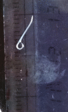
Adult worm of Thelazia callipaeda in the right eye of the child.
Figure 2.
Figure 2.
Thelazia callipaeda measuring approximately 10–12 mm.
Figure 3.
Figure 3.
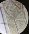
Anterior end of Thelazia callipaeda showing esophagointestinal junction.
Figure 4.
Figure 4.
Anterior end of female Thelazia callipaeda showing buccal capsule, esophagus, vulval opening near to mouth and anterior to the esophageal-intestinal junction with cuticular transverse striations.
Figure 5.
Figure 5.
Middle portion of the worm showing the reproductive system with embryonated eggs and alimentary canal.
Figure 6.
Figure 6.
Middle portion of the worm showing the reproductive system with embryonated eggs and alimentary canal.
Figure 7.
Figure 7.
Distal end of Thelazia callipaeda.
Figure 8.
Figure 8.
Caudal end of male Thelazia callipaeda showing characteristics curved tail end with shorter spicule and cloacal opening with pre- and postcloacal papillae.

Similar articles

Cited by

References

    1. Otranto D, Dutto M. Human thelaziasis, Europe. Emerg Infect Dis 2008; 14:647–9. - PMC - PubMed
    1. Viriyavejakul P, Krudsood S, Monkhonmu S, et al. . Thelazia callipaeda: a human case report. Southeast Asian J Trop Med Public Health 2012; 43:851–6. - PubMed
    1. Giangaspero A, Traversa D, Otranto D. Ecology of Thelazia spp. in cattle and their vectors in Italy. Parassitologia 2004; 46:257–9. - PubMed
    1. Mihalca AD, Amico GD, Scurtu L, et al. . Further spreading of canine oriental eye worm in Europe: first report of Thelazia callipaeda in Romania. Parasit Vectors 2015; 8:48. - PMC - PubMed
    1. Otranto D, Dantas-Torres F. Transmission of the eyeworm Thelazia callipaeda: between fantasy and reality. Parasit Vectors 2015; 8:273. - PMC - PubMed