

Identification and characterization of novel mutations implicated in congenital fibrinogen disorders
- PMID: 30349899
- PMCID: PMC6178649
- DOI: 10.1002/rth2.12127
Identification and characterization of novel mutations implicated in congenital fibrinogen disorders
Abstract
Introduction: Fibrinogen is a complex molecule comprised of two sets of Aα, Bβ, and γ chains. Fibrinogen deficiencies can lead to the development of bleeding or thromboembolic events. The objective of this study was to perform DNA sequence analysis of patients with clinical fibrinogen abnormalities, and to perform genotype-phenotype correlations.
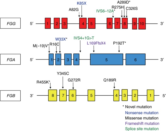
Materials and methods: DNA from 31 patients was sequenced to evaluate disease-causing mutations in the three fibrinogen genes: FGA,FGB, and FGG. Clinical data were extracted from medical records or from consultation with referring hematologists. Fibrinogen antigen and functional (Clauss method) assays, as well as reptilase time (RT) and thrombin time (TT) were obtained for each patient. Molecular modeling was used to simulate the functional impact of specific missense variants on the overall protein structure.
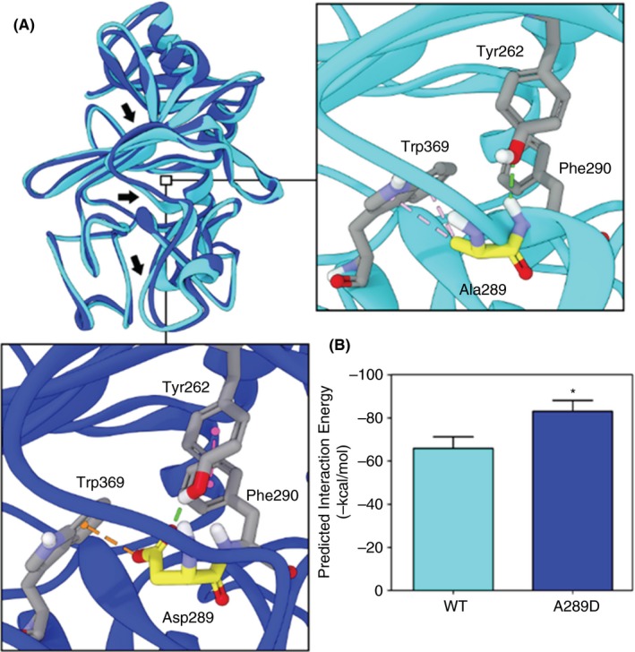
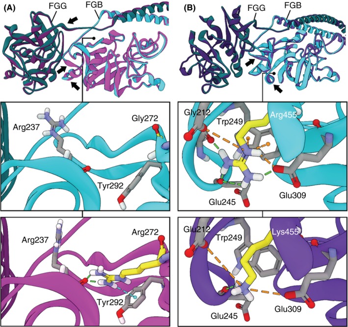
Results: Seventeen mutations, including six novel mutations, were identified in the three fibrinogen genes. There was little correlation between genotype and phenotype. Molecular modeling predicted a substantial conformational change for a novel variant, FGG p.Ala289Asp, leading to a more rigid molecule in a region critical for polymerization and alignment of the fibrin monomers. This mutation is associated with both bleeding and clotting in the two affected individuals.
Conclusions: Robust genotype-phenotype correlations are difficult to establish for fibrinogen disorders. Molecular modeling might represent a valuable tool for understanding the function of certain missense fibrinogen mutations but those should be followed by functional studies. It is likely that genetic and environmental modifiers account for the incomplete penetrance and variable expressivity that characterize fibrinogen disorders.
Keywords: afibrinogenemia; dysfibrinogenemia; fibrinogen disorders; fibrinogen mutations; molecular modeling.
Figures
References
-
- Mosesson MW, Siebenlist KR, Hainfeld JF, Wall JS. The covalent structure of factor XIIIa crosslinked fibrinogen fibrils. J Struct Biol. 1995;115:88–101. - PubMed
-
- Meh DA, Siebenlist KR, Mosesson MW. Identification and characterization of the thrombin binding sites on fibrin. J Biol Chem. 1996;271:23121–5. - PubMed
Grants and funding
LinkOut - more resources
Full Text Sources
Miscellaneous
