Case Reports
. 2019 Apr;18(2):245-254.
doi: 10.1007/s12311-018-0987-5.

TRIM9 and TRIM67 Are New Targets in Paraneoplastic Cerebellar Degeneration

Affiliations
Case Reports

TRIM9 and TRIM67 Are New Targets in Paraneoplastic Cerebellar Degeneration

Le Duy Do et al. Cerebellum. 2019 Apr.

Abstract

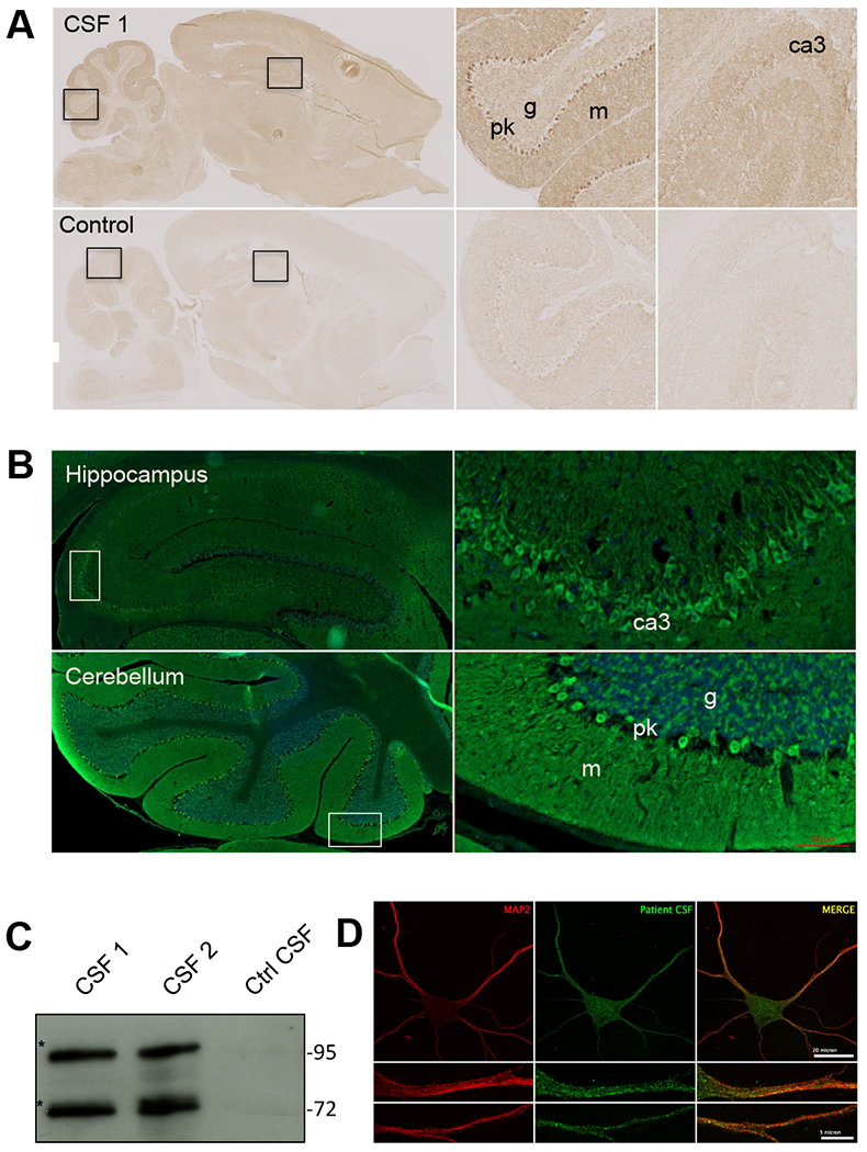
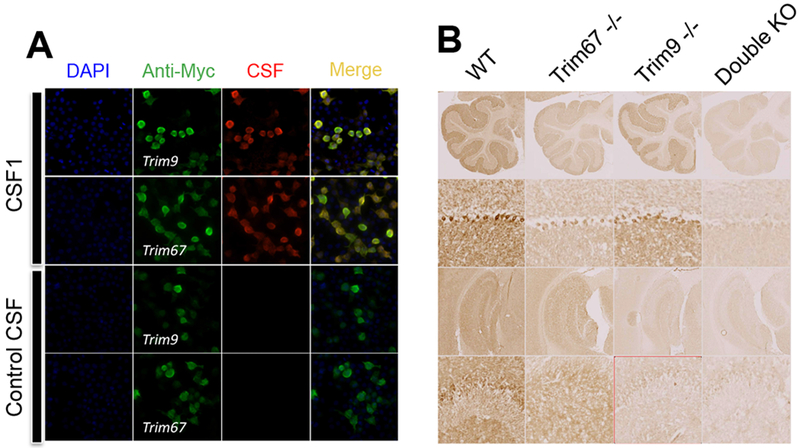
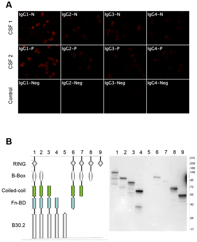
To describe autoantibodies (Abs) against tripartite motif-containing (TRIM) protein 9 and 67 in two patients with paraneoplastic cerebellar degeneration (PCD) associated with lung adenocarcinoma. Abs were characterized using immunohistochemistry, Western blotting, cultures of murine cortical, and hippocampal neurons, immunoprecipitation, mass spectrometry, knockout mice for Trim9 and 67, and cell-based assay. Control samples included sera from 63 patients with small cell lung cancer without any paraneoplastic neurological syndrome, 36 patients with lung adenocarcinoma and PNS, CSF from 100 patients with autoimmune encephalitis, and CSF from 165 patients with neurodegenerative diseases. We found Abs targeting TRIM9 and TRIM67 at high concentration in the serum and the cerebrospinal fluid (CSF) of a 78-year-old woman and a 65-year-old man. Both developed subacute severe cerebellar ataxia. Brain magnetic resonance imaging found no abnormality and no cerebellar atrophy. Both had CSF inflammation with mild pleiocytosis and a few oligoclonal bands. We identified a pulmonary adenocarcinoma, confirming the paraneoplastic neurological syndrome in both patients. They received immunomodulatory and cancer treatments without improvement of cerebellar ataxia, even though both were in remission of their cancer (for more than 10 years in one patient). Anti-TRIM9 and anti-TRIM67 Abs were specific to these two patients. All control serum and CSF samples tested were negative for anti-TRIM9 and 67. Anti-TRIM9 and anti-TRIM67 Abs appeared to be specific biomarkers of PCD and should be added to the panel of antigens tested when this is suspected.

Keywords: Autoantibodies; Lung cancer; Paraneoplastic cerebellar disorders; TRIM67; TRIM9.


Conflict of interest statement

Conflict of Interest: The authors declare that they have no conflict of interest.

Figures

Figure 1:
Figure 1:
(A) Immunohistochemistry of adult rat brain with cerebrospinal fluid (CSF) from patient 1 (upper panels) or control CSF (lower panels). Note a strong staining of CA3 pyramidal cells in the hippocampus and the granular layer and Purkinje cell layer in the cerebellum. (B) Fluorescent staining of rat brain with CSF from patient 1. Note a strong reactivity with the cell bodies and proximal dendrite of the pyramidal cells in the hippocampus (upper panels) and those of Purkinje cell in the cerebellum (lower panel). (C) Immunolabeling of embryonic hippocampal neurons (at div15) with CSF from patient 1. Abs in the CSF recognized cytosolic proteins organized in small speckles in the cell bodies and dendrites. (D) Western blotting showing reactivity of patient CSF. Targeted proteins were enriched by immunoprecipitation using CSF from patient 1. Bands at 72 and 95 kDa were excised and analyzed by mass spectrometry. m: molecular layer, g: granular layer, pk: Purkinje cell layer.
Figure 2:
Figure 2:
(A) Cell-based assay test to confirm the identity of Abs targets. HEK cells were transfected with plasmids coding for Myc-tagged TRIM9 or Myc-tagged TRIM67 for transient overexpression. Transfected cells were fixed, permeabilized, and immunolabeled with patient CSF (red) or commercial anti-Myc antibody (green). Colocalization of the two stains confirmed that Abs recognized TRIM9 and TRIM67. (B) Immunostaining of single or double knock-out (KO) mouse brain with CSF from patient 1. TRIM9 was expressed widely in the adult brain while TRIM67 is more restricted to the cerebellum.
Figure 3:
Figure 3:
(A) Analysis of the IgG subclass of anti-TRIM9 Abs in patient CSF. HEK cells expressing TRIM9 were incubated with patient CSF and revealed with subclass-specific anti-human IgG antibodies. Positive signal was observed for all IgG subclasses; the strongest signal was found for IgG1. (B) Epitope mapping of anti-TRIM9 Abs. HEK cells were transfected with plasmids coding for different domains of TRIM9 and then lysed and immuno-blotted with CSF from patient 1 that recognized all TRIM9 domains except the B30.2 domain. The Abs are polyclonal.
Figure 4:
Figure 4:
Effects of Patient CSF on neuronal viability and development. E15.5 cortical neurons from wildtype (+/+) and Trim9−/−:Trim67−/− (double KO) mice were treated with patient CSF (1:100) for 3 days or 1 day prior to fixation. Neurons were stained for βIII tubulin (green), filamentous actin (red), and DNA (blue). Several parameters were measured per neuron, including density of axon branches, primary axon length, total axonal material, dendritic material, primary dendrite number, and dendrite branch points. In the box and whisker plots: the cyan boxes for each genotype are untreated. The second two were incubated from the 24th to the 96th hour (3 days) with 1:100 CSF from patient 1 (magenta boxes) and patient 2 (yellow boxes), respectively, the next two were incubated from the 72nd to the 96th hour (1 day), patient 1 and patient 2, respectively. Boxplots depict median +/− the interquartile range (IQR), whiskers reach minimum and maximum values. P-values were determined by ANOVA, comparisons made to untreated control, with Bonnferroni post hoc corrections of the number of comparisons made. P- values are considered significant when p<0.05. No consistent significant effects of patients’ CSF on neurons were observed.

References

    1. Honnorat J, Cartalat-Carel S. Advances in paraneoplastic neurological syndromes. Curr Opin Oncol. 2004;16(6):614–620. - PubMed
    1. Jarius S, Wildemann B. ‘Medusa head ataxia’: the expanding spectrum of Purkinje cell antibodies in autoimmune cerebellar ataxia. J Neuroinflammation. 2015, 12:166–168 - PMC - PubMed
    1. Greenlee JE, Brashear HR. Antibodies to cerebellar Purkinje cells in patients with paraneoplastic cerebellar degeneration and ovarian carcinoma. Ann Neurol 1983; 14(6): 609–13. - PubMed
    1. Ducray F, Demarquay G, Graus F, Decullier E, Antoine JC, Giometto B, Psimaras D, Delattre JY, Carpentier AF, Honnorat J. Seronegative paraneoplastic cerebellar degeneration: the PNS Euronetwork experience. Eur J Neurol. 2014. May;21(5):731–5. - PubMed
    1. Winkle CC, McClain LM, Valtschanoff JG, Park CS, Maglione C, Gupton SL. A novel Netrin-1–sensitive mechanism promotes local SNARE-mediated exocytosis during axon branching. J Cell Biol. 2014, 205(2):217–32. - PMC - PubMed

Publication types

MeSH terms

Supplementary concepts