Review
. 2020 Jan;38(1):9-23.
doi: 10.5534/wjmh.180066. Epub 2018 Oct 22.

Microtubular Dysfunction and Male Infertility

Affiliations
Review

Microtubular Dysfunction and Male Infertility

Sezgin Gunes et al. World J Mens Health. 2020 Jan.

Abstract

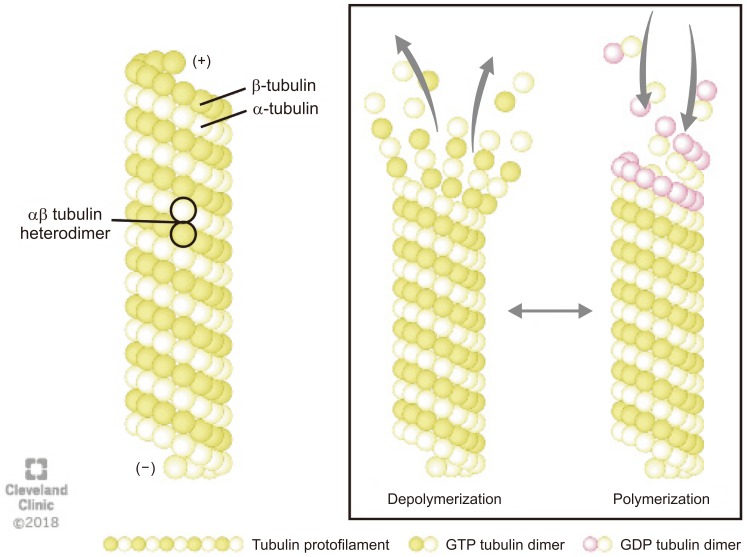
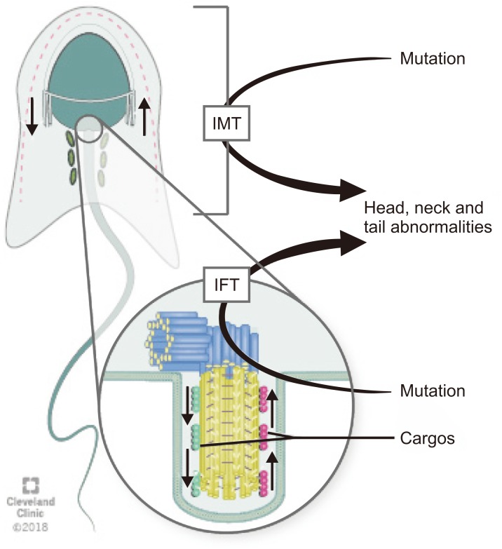
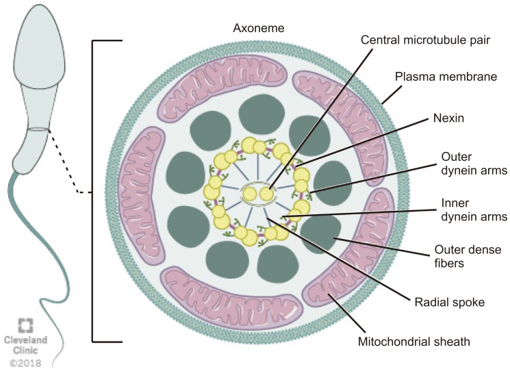
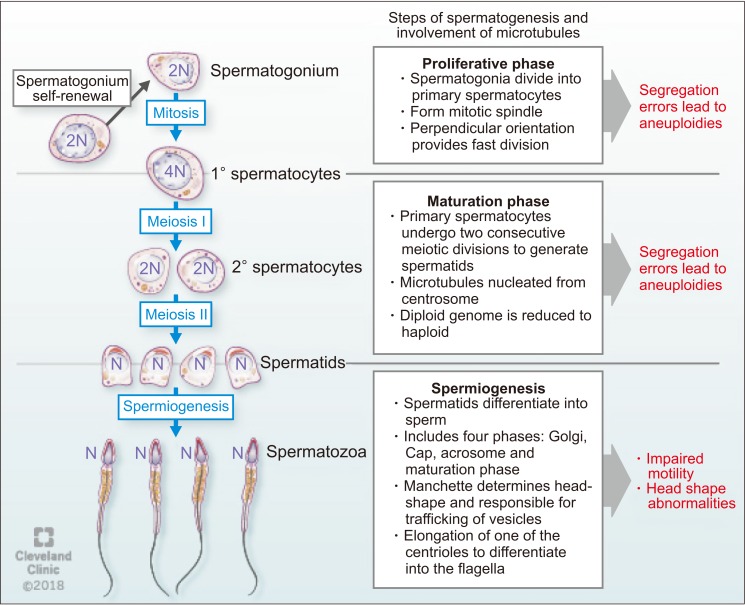
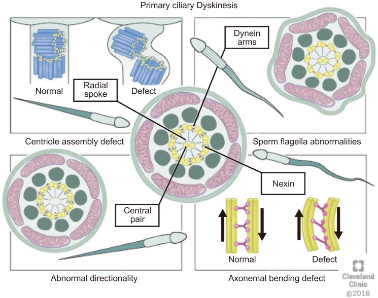
Microtubules are the prime component of the cytoskeleton along with microfilaments. Being vital for organelle transport and cellular divisions during spermatogenesis and sperm motility process, microtubules ascertain functional capacity of sperm. Also, microtubule based structures such as axoneme and manchette are crucial for sperm head and tail formation. This review (a) presents a concise, yet detailed structural overview of the microtubules, (b) analyses the role of microtubule structures in various male reproductive functions, and (c) presents the association of microtubular dysfunctions with male infertility. Considering the immense importance of microtubule structures in the formation and maintenance of physiological functions of sperm cells, this review serves as a scientific trigger in stimulating further male infertility research in this direction.

Keywords: Axoneme; Infertility, male; Kartagener syndrome; Microtubule-associated proteins.


Conflict of interest statement

The authors have no potential conflicts of interest to disclose.

Figures

Fig. 1
Fig. 1. The structure and dynamics of microtubule: Microtubules are composed of α- and β-tubulin heterodimers that polymerizes using guanosine-5′-triphosphate (GTP) to form a single proto-filament. The tubulins are arranged in a polarized manner whereby the α-tubulins are exposed at the negative (−) and the β-tubulins at the positive (+) end. The (−) end is more stable, whereas the (+) end can polymerize (grow) and depolymerize (shrink) rapidly, thus rendering microtubules highly dynamic structures. GDP: guanosine 5′-diphosphate.
Fig. 2
Fig. 2. Schematic diagram of the transport pathways of manchette. Intra-manchette transport (IMT) transfers structural and functional proteins via the microtubule tracks and motor proteins to the basal body region where they are stored. During intra-flagellar transport (IFT), proteins are transported from the sperm cell body to the tip of the flagellum and then back to the sperm head along the axonemal microtubules. Outward or anterograde movement (from the sperm head to the tail) is directly associated with microtubule motor kinesin-2 while inward or retrograde movement is related with dynein 1b. Abnormalities of IMT or IFT microtubule structure could lead to sperm head, neck and tail aberrations because of disruption of the protein delivery to the correct assembly site during spermiogenesis.
Fig. 3
Fig. 3. Schematic representation of flagellum structure. Schematic representation of flagellum structure. A cross-section of flagellum mid-piece shows plasma membrane and mitochondrial sheath surrounding the outer dense fibers. The axoneme displays the characteristic 9+2 arrangement of the microtubules with nine microtubule doublets around the periphery and two singlet microtubules in the center. Adjacent microtubule doublet are connected by Nexin.
Fig. 4
Fig. 4. The function of microtubules during spermatogenesis.
Fig. 5
Fig. 5. Abnormalities of the cilia (1) dynein arms abnormalities involve either partial or complete absence of outer dynein arm/inner dynein arm, (2) radial spokes abnormalities including the absence of spokes or deviated central microtubules, (3) inappropriate directionality of cilia, caused by partial or complete absence of the central microtubules, and (4) abnormal number of peripheral microtubules. Atypical arrangements for example 8+1, 8+2, 8+3, or 7+2 have been observed under the microscope on cross-section.

References

    1. Tuttelmann F, Gromoll J, Kliesch S. Genetics of male infertility. Urologe A. 2008;47:1561–1562. 1564–1567. - PubMed
    1. Beyaz CC, Gunes S, Onem K, Kulac T, Asci R. Partial deletions of Y-chromosome in infertile men with non-obstructive azoospermia and oligoasthenoteratozoospermia in a Turkish population. In Vivo. 2017;31:365–371. - PMC - PubMed
    1. Cooper TG, Noonan E, von Eckardstein S, Auger J, Baker HW, Behre HM, et al. World Health Organization reference values for human semen characteristics. Hum Reprod Update. 2010;16:231–245. - PubMed
    1. Kaverina I, Straube A. Regulation of cell migration by dynamic microtubules. Semin Cell Dev Biol. 2011;22:968–974. - PMC - PubMed
    1. Watanabe T, Noritake J, Kaibuchi K. Regulation of microtubules in cell migration. Trends Cell Biol. 2005;15:76–83. - PubMed