

4-Sodium phenyl butyric acid has both efficacy and counter-indicative effects in the treatment of Col4a1 disease
- PMID: 30351356
- PMCID: PMC6360271
- DOI: 10.1093/hmg/ddy369
4-Sodium phenyl butyric acid has both efficacy and counter-indicative effects in the treatment of Col4a1 disease
Abstract
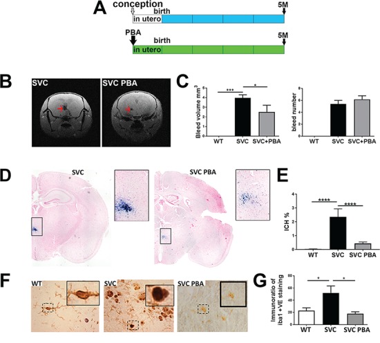
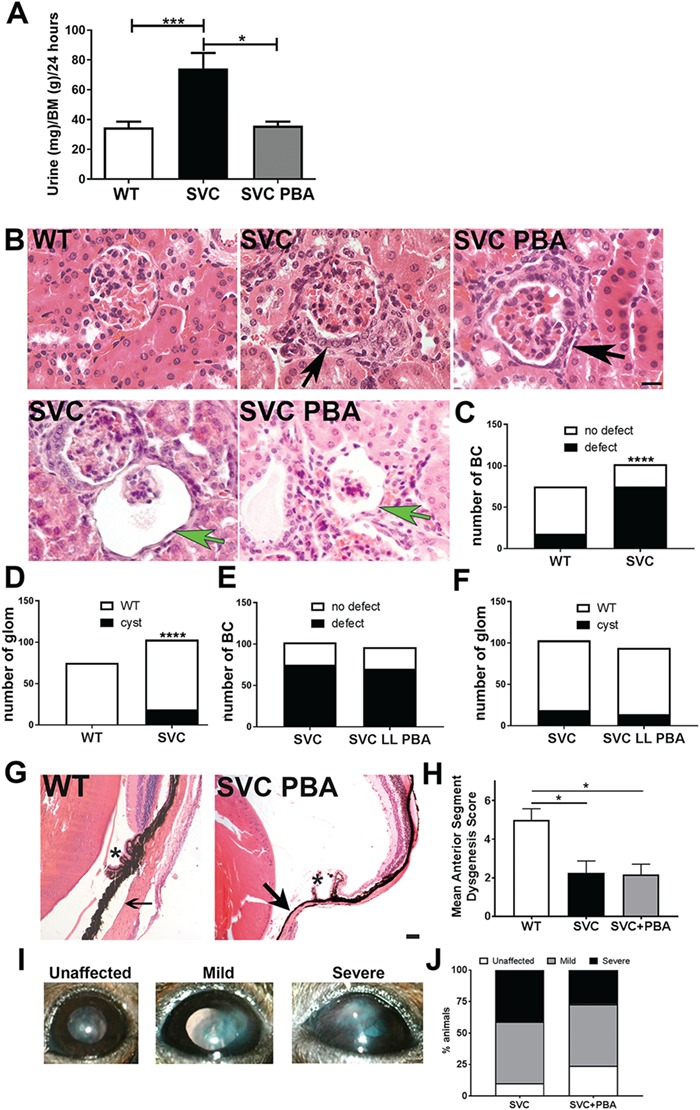
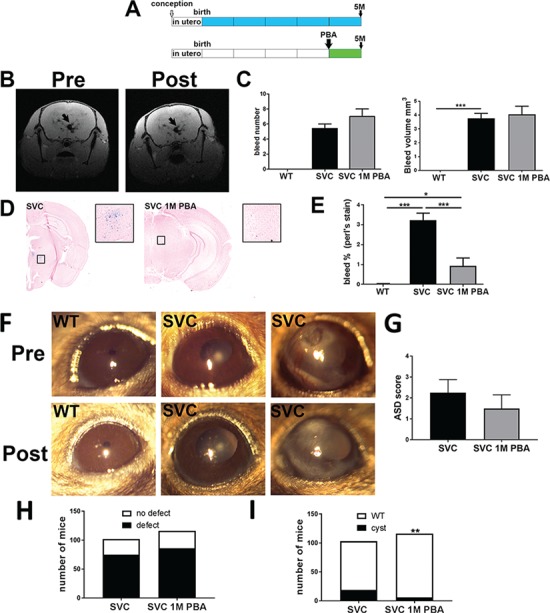
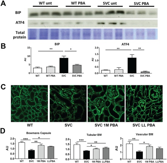
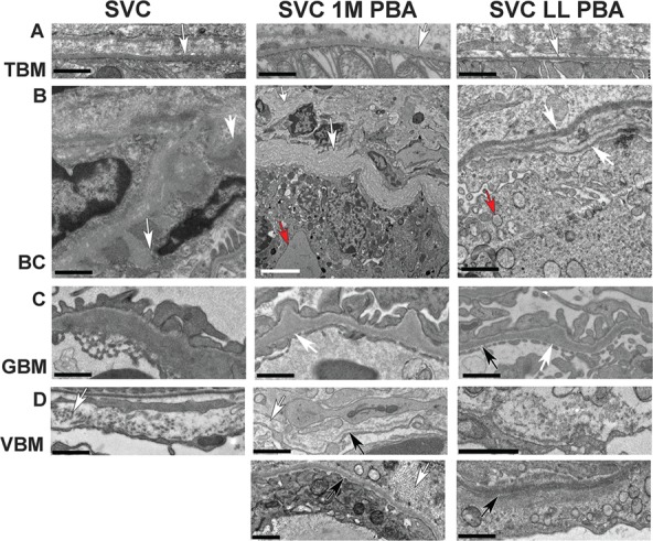
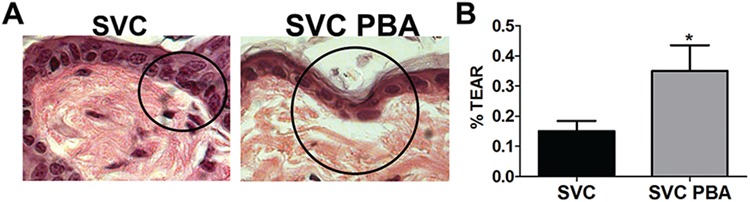
Mutations in the collagen genes COL4A1 and COL4A2 cause Mendelian eye, kidney and cerebrovascular disease including intracerebral haemorrhage (ICH), and common collagen IV variants are a risk factor for sporadic ICH. COL4A1 and COL4A2 mutations cause endoplasmic reticulum (ER) stress and basement membrane (BM) defects, and recent data suggest an association of ER stress with ICH due to a COL4A2 mutation. However, the potential of ER stress as a therapeutic target for the multi-systemic COL4A1 pathologies remains unclear. We performed a preventative oral treatment of Col4a1 mutant mice with the chemical chaperone phenyl butyric acid (PBA), which reduced adult ICH. Importantly, treatment of adult mice with the established disease also reduced ICH. However, PBA treatment did not alter eye and kidney defects, establishing tissue-specific outcomes of targeting Col4a1-derived ER stress, and therefore this treatment may not be applicable for patients with eye and renal disease. While PBA treatment reduced ER stress and increased collagen IV incorporation into BMs, the persistence of defects in BM structure and reduced ability of the BM to withstand mechanical stress indicate that PBA may be counter-indicative for pathologies caused by matrix defects. These data establish that treatment for COL4A1 disease requires a multipronged treatment approach that restores both ER homeostasis and matrix defects. Alleviating ER stress is a valid therapeutic target for preventing and treating established adult ICH, but collagen IV patients will require stratification based on their clinical presentation and mechanism of their mutations.
Figures
Similar articles
-
Chemical chaperone treatment reduces intracellular accumulation of mutant collagen IV and ameliorates the cellular phenotype of a COL4A2 mutation that causes haemorrhagic stroke.Hum Mol Genet. 2014 Jan 15;23(2):283-92. doi: 10.1093/hmg/ddt418. Epub 2013 Sep 2. Hum Mol Genet. 2014. PMID: 24001601 Free PMC article.
-
Use of sodium 4-phenylbutyrate to define therapeutic parameters for reducing intracerebral hemorrhage and myopathy in Col4a1 mutant mice.Dis Model Mech. 2018 Jul 4;11(7):dmm034157. doi: 10.1242/dmm.034157. Dis Model Mech. 2018. PMID: 29895609 Free PMC article.
-
COL4A2 mutations impair COL4A1 and COL4A2 secretion and cause hemorrhagic stroke.Am J Hum Genet. 2012 Jan 13;90(1):91-101. doi: 10.1016/j.ajhg.2011.11.022. Epub 2011 Dec 29. Am J Hum Genet. 2012. PMID: 22209247 Free PMC article.
-
The expanding phenotype of COL4A1 and COL4A2 mutations: clinical data on 13 newly identified families and a review of the literature.Genet Med. 2015 Nov;17(11):843-53. doi: 10.1038/gim.2014.210. Epub 2015 Feb 26. Genet Med. 2015. PMID: 25719457 Review.
-
Role of COL4A1 in basement-membrane integrity and cerebral small-vessel disease. The COL4A1 stroke syndrome.Curr Med Chem. 2010;17(13):1317-24. doi: 10.2174/092986710790936293. Curr Med Chem. 2010. PMID: 20166936 Review.
Cited by
-
Genetic Causes of Cerebral Small Vessel Diseases: A Practical Guide for Neurologists.Neurology. 2023 Apr 18;100(16):766-783. doi: 10.1212/WNL.0000000000201720. Epub 2022 Dec 19. Neurology. 2023. PMID: 36535782 Free PMC article. Review.
-
Material-driven fibronectin assembly rescues matrix defects due to mutations in collagen IV in fibroblasts.Biomaterials. 2020 Sep;252:120090. doi: 10.1016/j.biomaterials.2020.120090. Epub 2020 May 3. Biomaterials. 2020. PMID: 32413593 Free PMC article.
-
A novel human iPSC model of COL4A1/A2 small vessel disease unveils a key pathogenic role of matrix metalloproteinases.Stem Cell Reports. 2023 Dec 12;18(12):2386-2399. doi: 10.1016/j.stemcr.2023.10.014. Epub 2023 Nov 16. Stem Cell Reports. 2023. PMID: 37977146 Free PMC article.
-
Identification of an Altered Matrix Signature in Kidney Aging and Disease.J Am Soc Nephrol. 2021 Jul;32(7):1713-1732. doi: 10.1681/ASN.2020101442. Epub 2021 May 28. J Am Soc Nephrol. 2021. PMID: 34049963 Free PMC article.
-
Reducing Hypermuscularization of the Transitional Segment Between Arterioles and Capillaries Protects Against Spontaneous Intracerebral Hemorrhage.Circulation. 2020 Jun 23;141(25):2078-2094. doi: 10.1161/CIRCULATIONAHA.119.040963. Epub 2020 Mar 18. Circulation. 2020. PMID: 32183562 Free PMC article.
References
-
- Bateman J.F., Boot-Handford R.P. and Lamandé S.R. (2009) Genetic diseases of connective tissues: cellular and extracellular effects of ECM mutations. Nat. Rev. Genet., 10, 173–183. - PubMed
-
- Kalluri R. (2003) Basement membranes: structure, assembly and role in tumour angiogenesis. Nat. Rev. Cancer, 3, 422–433. - PubMed
-
- Gould D.B., Phalan F.C., Breedveld G.J., Mil S.E., Smith R.S., Schimenti J.C., Aguglia U., Knaap M.S., Heutink P. and John S.W. (2005) Mutations in Col4a1 cause perinatal cerebral hemorrhage and porencephaly. Science, 308, 1167–1171. - PubMed
-
- Sibon I., Coupry I., Menegon P., Bouchet J.P., Gorry P., Burgelin I., Calvas P., Orignac I., Dousset V., Lacombe D. et al. (2007) COL4A1 mutation in Axenfeld–Rieger anomaly with leukoencephalopathy and stroke. Ann. Neurol., 62, 177–184. - PubMed
Publication types
MeSH terms
Substances
Grants and funding
LinkOut - more resources
Full Text Sources
Molecular Biology Databases
