. 2019 Mar 15;115(4):752-764.
doi: 10.1093/cvr/cvy257.

Regional diastolic dysfunction in post-infarction heart failure: role of local mechanical load and SERCA expression

Affiliations

Regional diastolic dysfunction in post-infarction heart failure: role of local mechanical load and SERCA expression

Åsmund T Røe et al. Cardiovasc Res. .

Abstract

Aims: Regional heterogeneities in contraction contribute to heart failure with reduced ejection fraction (HFrEF). We aimed to determine whether regional changes in myocardial relaxation similarly contribute to diastolic dysfunction in post-infarction HFrEF, and to elucidate the underlying mechanisms.

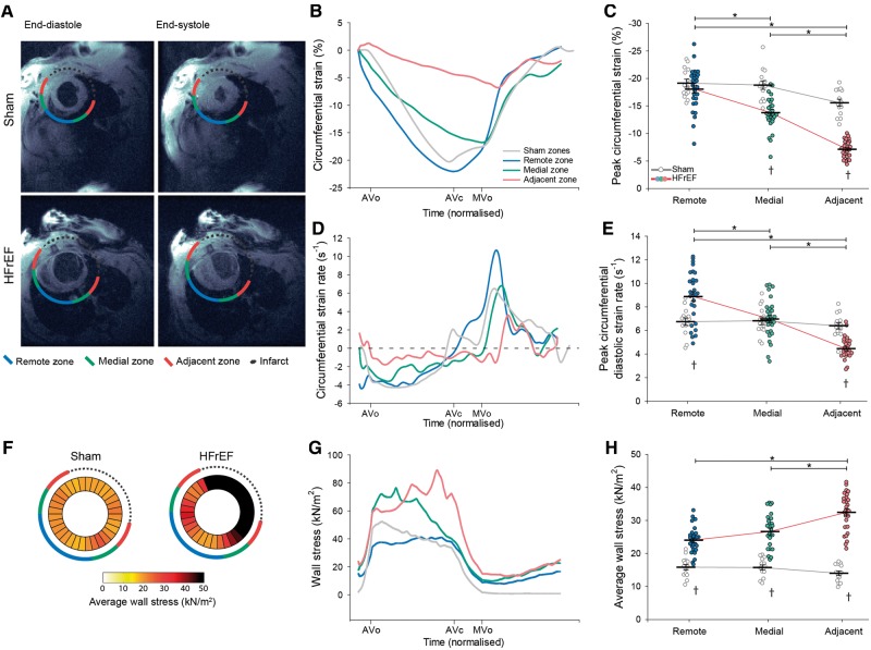
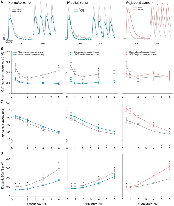
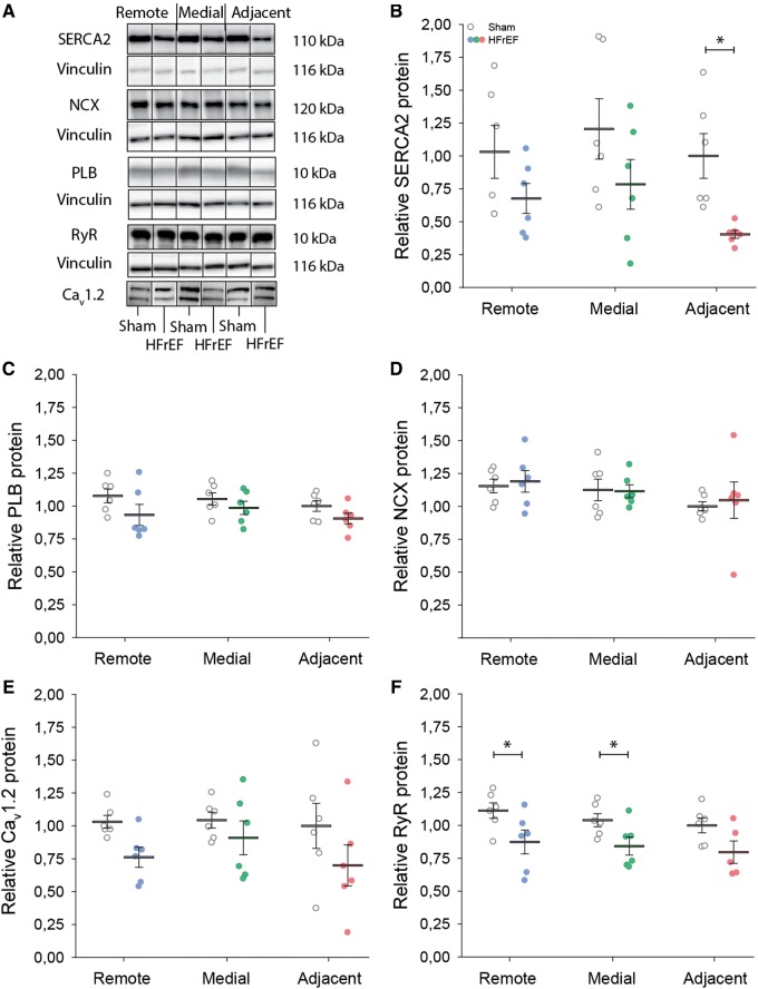
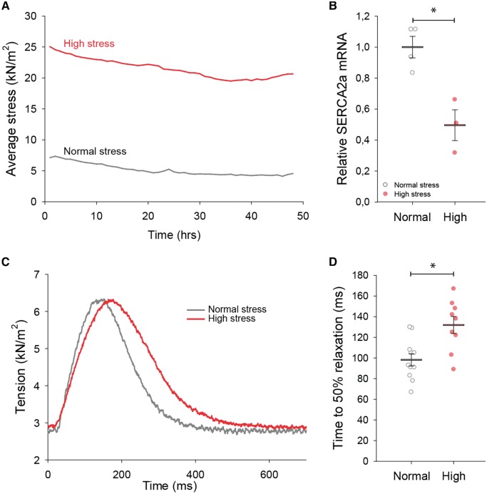
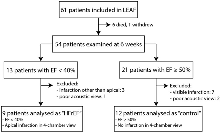
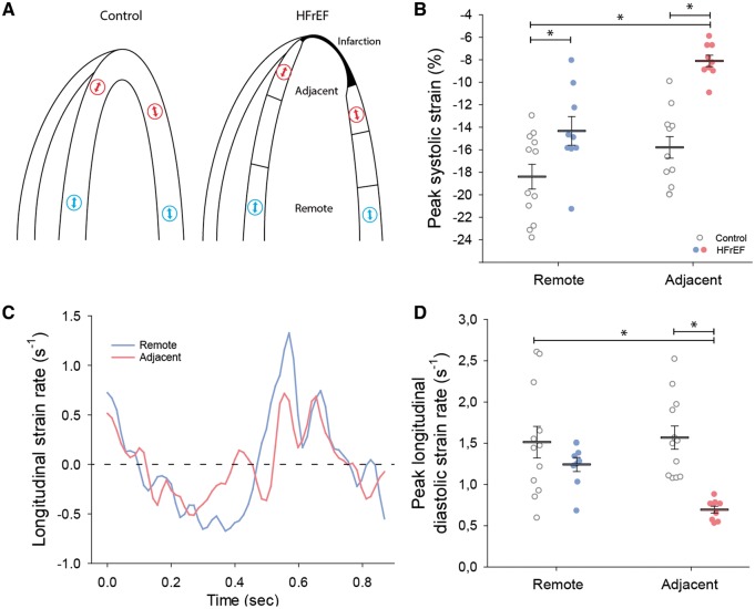
Methods and results: Using the magnetic resonance imaging phase-contrast technique, we examined local diastolic function in a rat model of post-infarction HFrEF. In comparison with sham-operated animals, post-infarction HFrEF rats exhibited reduced diastolic strain rate adjacent to the scar, but not in remote regions of the myocardium. Removal of Ca2+ within cardiomyocytes governs relaxation, and we indeed found that Ca2+ transients declined more slowly in cells isolated from the adjacent region. Resting Ca2+ levels in adjacent zone myocytes were also markedly elevated at high pacing rates. Impaired Ca2+ removal was attributed to a reduced rate of Ca2+ sequestration into the sarcoplasmic reticulum (SR), due to decreased local expression of the SR Ca2+ ATPase (SERCA). Wall stress was elevated in the adjacent region. Using ex vivo experiments with loaded papillary muscles, we demonstrated that high mechanical stress is directly linked to SERCA down-regulation and slowing of relaxation. Finally, we confirmed that regional diastolic dysfunction is also present in human HFrEF patients. Using echocardiographic speckle-tracking of patients enrolled in the LEAF trial, we found that in comparison with controls, post-infarction HFrEF subjects exhibited reduced diastolic train rate adjacent to the scar, but not in remote regions of the myocardium.

Conclusion: Our data indicate that relaxation varies across the heart in post-infarction HFrEF. Regional diastolic dysfunction in this condition is linked to elevated wall stress adjacent to the infarction, resulting in down-regulation of SERCA, disrupted diastolic Ca2+ handling, and local slowing of relaxation.

Keywords: Cardiomyocyte calcium cycling; Diastolic dysfunction; Heart failure; Post-infarction remodelling; Wall stress.


Figures

Figure 1
Figure 1
Slowing of relaxation and elevated wall stress in the adjacent region of HFrEF rats. MRI images illustrate the division of HFrEF rat hearts into three regions (adjacent, medial, and remote) according to their proximity to the infarction (A). Comparison was made to equivalent regions in sham-operated controls. Representative recordings (B) and mean data (C) revealed lower peak circumferential systolic strain adjacent to the infarct in HFrEF. The rate of relaxation was dyssynchronous across the failing heart, as indicated by measurements of circumferential strain rate (D). Specifically, peak diastolic strain rate was only reduced in the adjacent region of HFrEF hearts, and was increased in the remote region (E). Wall stress was calculated across sham and HFrEF hearts based on intraventricular pressure measurements and local ventricular geometry (see Methods section). Measurements are presented as bullseye plots (F) and tracings of wall stress values during the cardiac cycle (G). Mean data demonstrate that while integrated wall stress was high in all regions of HFrEF hearts, values were particularly elevated in the adjacent region (H). (nhearts = 14, 29 in sham, HFrEF). *P < 0.05 calculated by two-way ANOVA with a post hoc Bonferroni t-test. AVc, aortic valve closure; AVo, aortic valve opening; MVo, mitral valve opening.
Figure 2
Figure 2
Impaired diastolic Ca2+ handling in cardiomyocytes from the adjacent region. Representative recordings of whole-cell, wide-field Ca2+ transients (A, 1 Hz and 6 Hz stimulation, fluo-4), and mean data (B) revealed that, relative to sham, Ca2+ transient magnitude was maintained in cardiomyocytes isolated from the adjacent zone of HFrEF hearts, but reduced in the medial and remote zones. However, HFrEF cells from the adjacent region exhibited significantly slower Ca2+ transient decay (C), and at high-pacing frequencies, a significant elevation of diastolic Ca2+ levels (D). (nhearts = 4, 5 in sham, HFrEF). Comparisons between HFrEF regions are presented in Supplementary material online, Figure S1. *P < 0.05 vs. sham calculated with nested ANOVA.
Figure 3
Figure 3
Reduced SERCA activity in the adjacent region. Representative recordings of Ca2+ transients are illustrated for cardiomyocytes during 1 Hz pacing, followed by rapid application of 10 mmol/L caffeine (A). The magnitude of caffeine-elicited Ca2+ release, an indicator of SR Ca2+ content, tended to be lower in the medial and distal regions (B). Fits of the declining phases of 1 Hz and caffeine transients revealed slowed SR Ca2+ reuptake in HFrEF cardiomyocytes from the adjacent region (C), while the rate of NCX Ca2+ extrusion tended to be higher in the remote zone (D) (nhearts = 4, 5 in sham, HFrEF). (E) Caffeine transients recorded in the presence of 5 mmol/L Ni2+ revealed no alterations in Ca2+ fluxes via slow extrusion pathways (nhearts = 2, 3 in sham, HFrEF). (F) Simulations of Ca2+ transients at physiological frequency (6 Hz) demonstrate the consequences of enhancing NCX activity (blue), as observed in myocytes from the distal zone, or reducing SERCA activity (red), as observed in the adjacent zone. *P < 0.05, calculated by nested ANOVA.
Figure 4
Figure 4
Lower SERCA2 expression in the adjacent region. Consistent with impaired SERCA function in the adjacent region, representative immunoblots (A, vinculin as loading control), and mean data (B) show that SERCA2 expression was significantly reduced in the adjacent region. By contrast, expression of phospholamban (PLB, C), NCX (D), the L-type Ca2+ channel (Cav1.2, E), and the ryanodine receptor (RyR, F) were not markedly altered across HFrEF and sham hearts. Complete blots are presented in Supplementary material online, Figure S4 (nhearts = 6, 6 in sham, HFrEF). *P < 0.05, calculated by two-way ANOVA with a post hoc Bonferroni t-test.
Figure 5
Figure 5
High mechanical stress down-regulates SERCA2 and slows relaxation in ex vivo myocardial tissue. Excised rat papillary muscles were stretched to reproduce high wall stress values comparable to those in the adjacent region of HFrEF hearts, and maintained during 48 h of culture. Comparison was made with muscles subjected to low wall stress conditions approximating those present in the normal heart. Average tension during the protocol is illustrated in A for representative muscles. High stress triggered a significant down-regulation of SERCA2 mRNA (B) (nmuscles = 4 in low group, 3 in high). Returning high stress muscles to normal levels at the completion of the protocol revealed that SERCA2 down-regulation was associated with marked slowing of force decline (representative recordings in C; mean data in D) (nmuscles = 11, 9 in low, high). *P < 0.05 vs. low stress calculated with Student’s t-test.
Figure 6
Figure 6
Selection of LEAF trial participants. HFrEF patients were compared with non-failing ‘control’ individuals (EF ≥50%) who were without a detectable myocardial infarction at 6 weeks of follow-up.
Figure 7
Figure 7
HFrEF patients exhibited regional diastolic dysfunction adjacent to the infarction. Four-chamber, apical echocardiographic images of HFrEF patients were examined to divide the viable myocardium into regions adjacent and remote to the myocardial infarction (schematically illustrated in A, right panel). Using speckle-tracking to examine local strain at indicated regions (arrows), comparison was made with regionally matched values in control hearts (A, left panel). Peak strain was reduced in HFrEF hearts, particularly in the adjacent region (B). Representative recordings of strain rate (C) and mean measurements (D) reveal that diastolic strain rate was also reduced adjacent to the myocardial infarction in HFrEF, but similar to control values in the remote region (nhearts = 12, 9 in control, HFrEF). *P < 0.05 with two-way ANOVA with a post hoc Bonferroni t-test.

Comment in

Similar articles

Cited by

References

    1. Ponikowski P, Voors AA, Anker SD, Bueno H, Cleland JG, Coats AJ, Falk V, Gonzalez-Juanatey JR, Harjola VP, Jankowska EA, Jessup M, Linde C, Nihoyannopoulos P, Parissis JT, Pieske B, Riley JP, Rosano GM, Ruilope LM, Ruschitzka F, Rutten FH, van der Meer P; Authors/Task Force Members. 2016 ESC guidelines for the diagnosis and treatment of acute and chronic heart failure. Eur Heart J 2016;37:2129–2200. - PubMed
    1. Kramer CM, Lima JA, Reichek N, Ferrari VA, Llaneras MR, Palmon LC, Yeh IT, Tallant B, Axel L.. Regional differences in function within noninfarcted myocardium during left ventricular remodeling. Circulation 1993;88:1279–1288. - PubMed
    1. Frisk M, Ruud M, Espe EK, Aronsen JM, Roe AT, Zhang L, Norseng PA, Sejersted OM, Christensen GA, Sjaastad I, Louch WE.. Elevated ventricular wall stress disrupts cardiomyocyte t-tubule structure and calcium homeostasis. Cardiovasc Res 2016;112:443–451. - PMC - PubMed
    1. Bursi F, Weston SA, Redfield MM, Jacobsen SJ, Pakhomov S, Nkomo VT, Meverden RA, Roger VL.. Systolic and diastolic heart failure in the community. JAMA 2006;296:2209–2216. - PubMed
    1. Brucks S, Little WC, Chao T, Kitzman DW, Wesley-Farrington D, Gandhi S, Shihabi ZK.. Contribution of left ventricular diastolic dysfunction to heart failure regardless of ejection fraction. Am J Cardiol 2005;95:603–606. - PubMed

Publication types

MeSH terms

Substances