. 2018 Dec 3;15(12):5585-5590.
doi: 10.1021/acs.molpharmaceut.8b00765. Epub 2018 Oct 30.

Endocytosis Pathways of Endothelial Cell Derived Exosomes

Endocytosis Pathways of Endothelial Cell Derived Exosomes

Anna B Banizs et al. Mol Pharm. .

Abstract

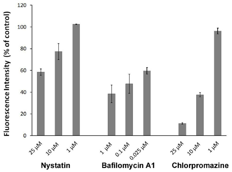
Nanosized extracellular vesicles (EVs) possess the natural machinery needed to enter selectively and transmit complex molecular messages efficiently into targeted cells. The intracellular fate of the vesicular cargos depends on the route of internalization. Therefore, understanding the mechanism of attachment and subsequent intake of these vesicles (before and after exerting any modification) is imperative. Here the extent of communication, the uptake kinetics, and the pathways of endothelial EVs into endothelial cells in the presence of specific pharmacological inhibitors were assessed by imaging flow cytometry. The results showed that the uptake of endothelial EVs into endothelial cells was largely an energy-dependent process using predominantly a receptor-mediated, clathrin-dependent pathway.

Keywords: endocytosis; endothelial cells; exosome; extracellular vesicles; nanomedicine.


Figures

Figure 1.
Figure 1.
Potential interactions and uptake mechanisms of extracellular vesicles (EVs) into cells. At least five communication pathways are distinguished between EVs and cells: direct fusion of the vesicles with plasma membrane, micropinocytosis, receptor-mediated clathrin-dependent internalization, caveolin-dependent pathway and lipid raft endocytosis.
Figure 2.
Figure 2.
Characterization of EVs. Transmission electron micrographs show endothelial EVs before (A) and after (B) labeling with DiO membrane dye. Average of five recorded curves represents the size distribution of endothelial EVs before (C) and after (D) labeling with DiO membrane dye determined by NanoSight platform. Scale bar represents 100 nanometers.
Figure 3.
Figure 3.
Internalization kinetics of DiO-labeled endothelial EVs analyzed by imaging flow cytometry. Representative images (upper panel, A) and scatter plots (lower panel, A) demonstrate endothelial cells treated with DiO-labeled EVs at 37°C and 4°C temperatures recorded by Amnis ImageStreamX Flow Cytometer. Graph depicts uptake kinetics of DiO-labeled EVs in the function of concentration of EVs at 37°C and 4°C temperatures (B).
Figure 4.
Figure 4.
Fluorescence microscopy images showing interaction of DiO-labeled EVs with endothelial cells in the absence or presence of Chlorpromazine, Bafilomycin A1 and Nystatin, respectively. Green fluorescence represents DiO-labeled endothelial EVs, blue is DAPI nuclear staining. Images were recorded with 100 × oil objective.
Figure 5.
Figure 5.
Effect of pharmacologic pathway specific inhibitors on the internalization of DiO-labeled endothelial EVs into endothelial cells. Graph depicts dose-dependent inhibitory effect of the Chlorpromazine, Bafilomycin A1 and Nystatin, respectively on the relative uptake of EVs obtained from imaging flow cytometry.

References

    1. Théry C, Boussac M, Véron P, Ricciardi-Castagnoli P, Raposo G, Garin J and Amigorena S. Proteomic Analysis of Dendritic Cell-Derived Exosomes: A Secreted Subcellular Compartment Distinct from Apoptotic Vesicles. J. Immunol 2001, 166(12), 7309–7318. - PubMed
    1. Barros FM, Carneiro F, Machado JC, Melo SA. Exosomes and Immune Response in Cancer: Friends or Foes? Front. Immunol 2018, 9, 730. doi: 10.3389/fimmu.2018.00730. - DOI - PMC - PubMed
    1. Karasu E, Eisenhardt SU, Harant J, Huber-Lang M. Extracellular Vesicles: Packages Sent With Complement. Front. Immunol 2018, 9, 721. doi: 10.3389/fimmu.2018.00721. - DOI - PMC - PubMed
    1. Alvarez-Erviti L, Seow YQ, Yin HF, Betts C, Lakhai S, Wood MJA Delivery of siRNA to the mouse brain by systemic injection of targeted exosomes, Nature Biotechnol. 2011, 29, 341. - PubMed
    1. Marcus ME, Leonard JN, FedExosomes: engineering therapeutic biological nanoparticles that truly deliver. Pharmaceuticals. 2013, 6, 659–680. - PMC - PubMed

Publication types

MeSH terms