. 2018 Oct 23;13(1):266.
doi: 10.1186/s13018-018-0977-9.

Evaluation of bone marrow-derived mesenchymal stem cell quality from patients with congenital pseudoarthrosis of the tibia

Affiliations

Evaluation of bone marrow-derived mesenchymal stem cell quality from patients with congenital pseudoarthrosis of the tibia

Ismail Hadisoebroto Dilogo et al. J Orthop Surg Res. .

Abstract

Background: The treatment of congenital pseudoarthrosis of the tibia (CPT) remains challenging in pediatric orthopedics due to the difficulties in bone union, continuous angulation, joint stiffness, and severe limb length discrepancy. Mesenchymal stem cells (MSCs) therapy offers a complementary approach to improve the conventional surgical treatments. Although the autologous MSC treatment shows a promising strategy to promote bone healing in CPT patients, the quality of MSCs from CPT patients has not been well studied. The purpose of this study is to investigate the quality of MSCs isolated from patients with CPT.

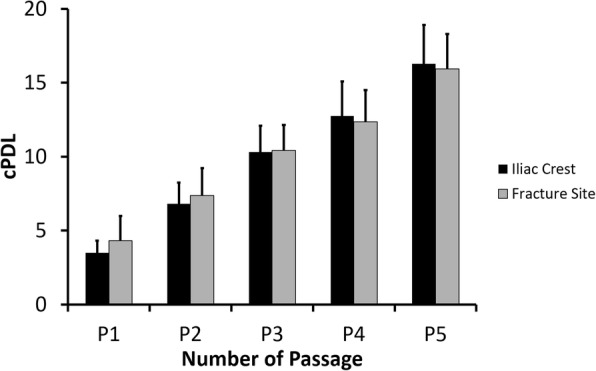
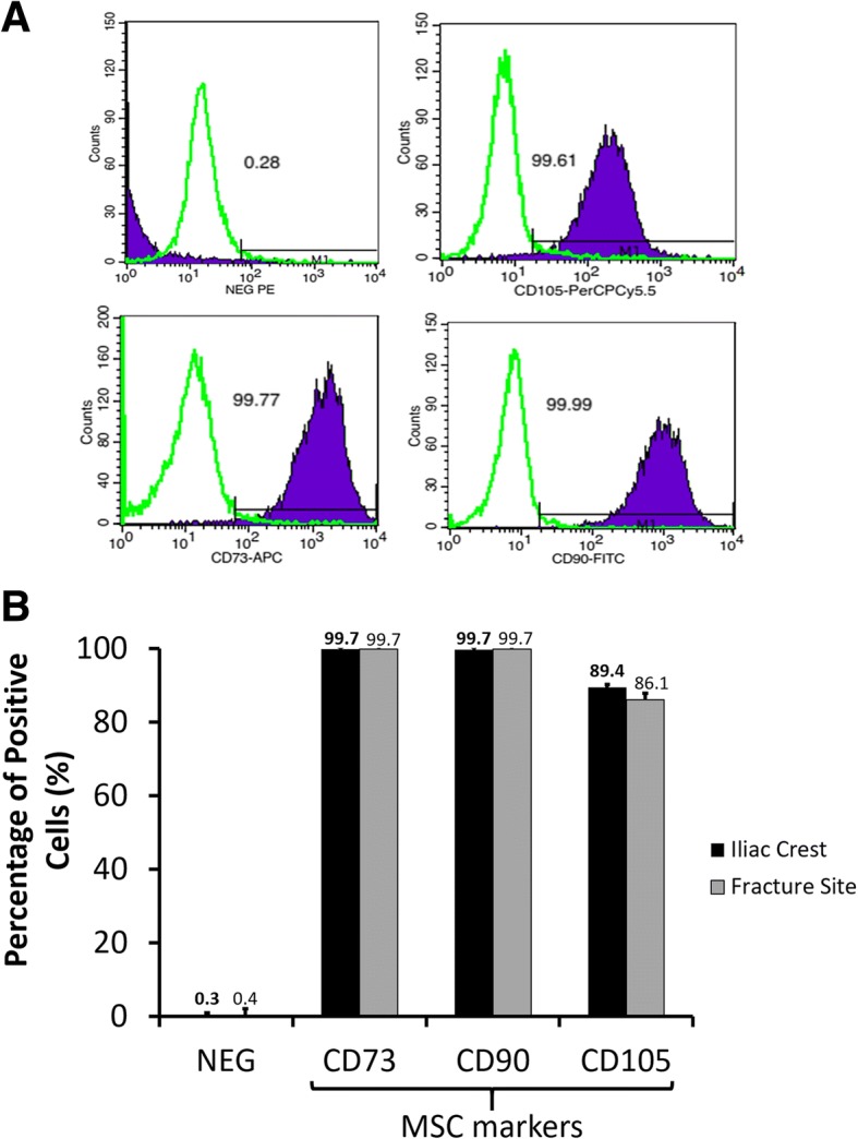
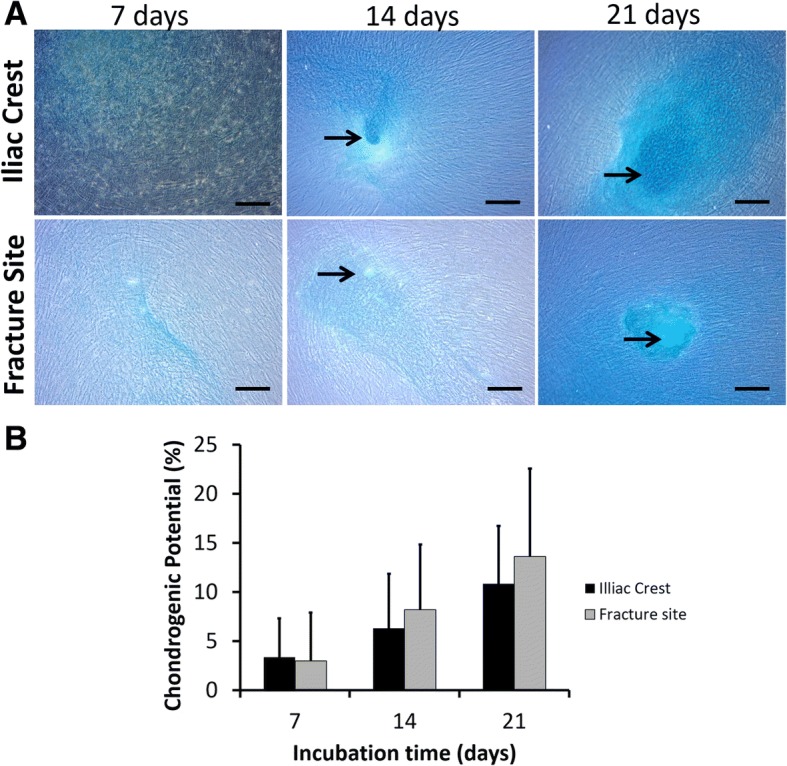
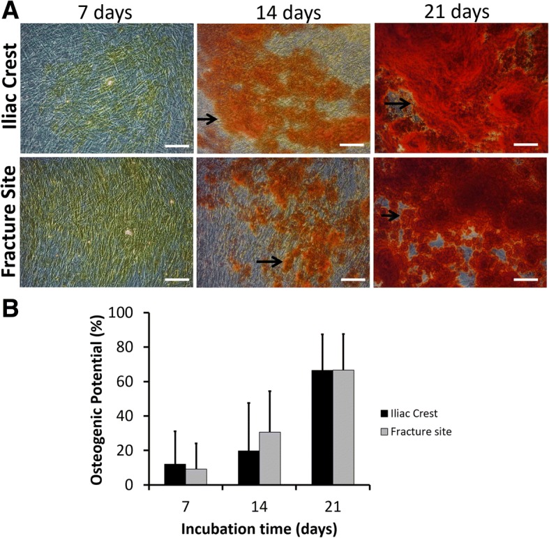
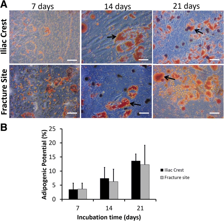
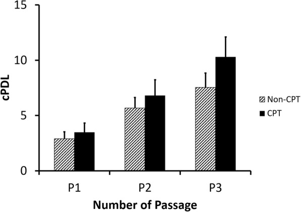
Methods: The bone marrow-derived MSCs from the fracture site and iliac crest of six CPT patients were isolated and compared. The cumulative population doubling level (cPDL), phenotype characteristics, and trilineage differentiation potency were observed to assess the quality of both MSCs.

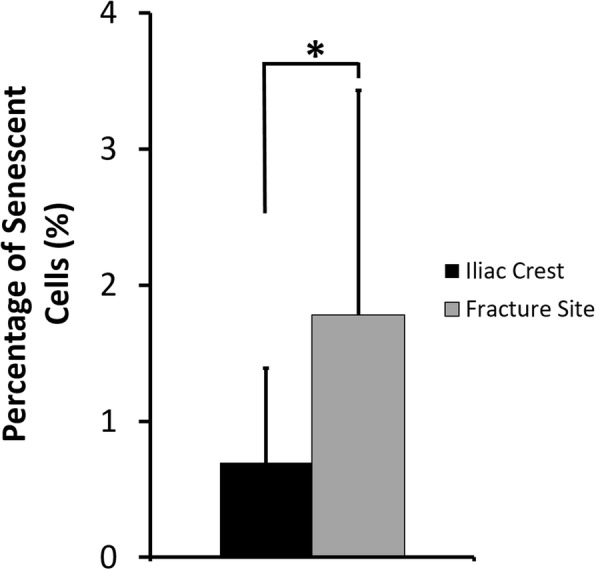
Results: There were no significant differences of the MSCs derived from the fracture site and the MSCs from the iliac crest of the subjects, in terms of cPDL, phenotype characteristics, and trilineage differentiation potency (all p > 0.05). However, MSCs from the fracture site had a higher senescence tendency than those from the iliac crest.

Conclusion: MSC quality is not the main reason for delayed bone regeneration in those with CPT. Thus, autologous MSC is a promising source for treating CPT patients.

Keywords: Cell differentiation; Mesenchymal stem cells; Osteocytes; Pseudoarthrosis.


Conflict of interest statement

Ethics approval and consent to participate

All participants were given an informed consent prior to the bone marrow aspiration. The protocols of this study were approved by the Ethics Committee of the Faculty of Medicine, Universitas Indonesia–Dr. Cipto Mangunkusumo General Hospital, and the studies were conducted in compliance with the Helsinki Declaration.

Consent for publication

Not applicable.

Competing interests

The authors declare that they have no competing interests.

Publisher’s Note

Springer Nature remains neutral with regard to jurisdictional claims in published maps and institutional affiliations.

Figures

Fig. 1
Fig. 1
The cPDL of bone marrow-derived MSCs from CPT patients. Data were shown as means, and the vertical bars indicated standard deviations (n = 6)
Fig. 2
Fig. 2
MSC purity of the fifth passaged culture cells isolated from the iliac crest and fracture site of CPT patients. a Typical flow cytometer histograms of positive markers (CD105, CD73, CD90) and cocktail of negative markers (NEG) for MSC characterization. b Percentage of MSC and non-MSC positive cells. Data were shown as means, and the vertical bars indicated standard deviations (n = 6)
Fig. 3
Fig. 3
Chondrogenic differentiation of MSCs isolated from the iliac crest and fracture site of the tibia from CPT patients. a Representative microscopic images of chondrogenic assays. Bars and black arrows indicate 100 μm and chondrocytes population, respectively. b Percentage of chondrogenic differentiation potential. Data were shown as means, and the vertical bars indicated standard deviations (n = 6)
Fig. 4
Fig. 4
Osteogenic differentiation of MSCs isolated from the iliac crest and fracture site of the tibia from CPT patients. a Representative microscopic images of osteogenic assays. Bars and black arrows indicate 100 μm and osteocytes population, respectively. b Percentage of osteogenic differentiation potential. Data were shown as means, and the vertical bars indicated standard deviations (n = 6)
Fig. 5
Fig. 5
Adipogenic differentiation of MSCs isolated from the iliac crest and fracture site of the tibia from CPT patients. a Representative microscopic images of adipogenic assays. Bars and black arrows indicate 100 μm and adipocytes population, respectively. b Percentage of adipogenic differentiation potential. Data were shown as means and the vertical bars indicated standard deviations (n = 6)
Fig. 6
Fig. 6
Percentage of senescent cells from the fifth passaged MSCs isolated from CPT patients. Data were shown as means, and the vertical bars indicated standard deviations (n = 6). The value of p < 0.05 (*) was indicated based on a paired t test
Fig. 7
Fig. 7
The cPDL of iliac crest-derived MSCs from healthy (non-CPT) and CPT patients. Data were shown as means and the vertical bars indicated standard deviations (n = 6)

Similar articles

Cited by

References

    1. Hardinge K. Congenital anterior bowing of the tibia. The significance of the different types in relation to pseudarthrosis. Ann R Coll Surg Engl. 1972;51(1):17–30. - PMC - PubMed
    1. Hefti F, Bollini G, Dungl P, Fixsen J, Grill F, Ippolito E, Romanus B, Tudisco C, Wientroub S. Congenital pseudarthrosis of the tibia: history, etiology, classification, and epidemiologic data. J Pediatr Orthop B. 2000;9:11–15. doi: 10.1097/01202412-200001000-00003. - DOI - PubMed
    1. Andersen KS. Radiological classification of congenital pseudarthrosis of the tibia. Acta Orthop Scand. 1973;44(6):719–727. doi: 10.3109/17453677308989112. - DOI - PubMed
    1. DeClue JE, Cohen BD, Lowy DR. Identification and characterization of the neurofibromatosis type 1 protein product. P Natl Acad Sci USA. 1991;88:9914–9918. doi: 10.1073/pnas.88.22.9914. - DOI - PMC - PubMed
    1. Abramowicz A, Gos M. Neurofibromin in neurofibromatosis type 1 mutations in NF1 gene as a cause of disease. Dev Period Med. 2014;18(3):297–306. - PubMed

Publication types

Supplementary concepts

LinkOut - more resources