. 2018 Nov 6;115(45):E10615-E10624.
doi: 10.1073/pnas.1811624115. Epub 2018 Oct 23.

Evolutionarily conserved Tbx5- Wnt2/2b pathway orchestrates cardiopulmonary development

Affiliations

Evolutionarily conserved Tbx5- Wnt2/2b pathway orchestrates cardiopulmonary development

Jeffrey D Steimle et al. Proc Natl Acad Sci U S A. .

Abstract

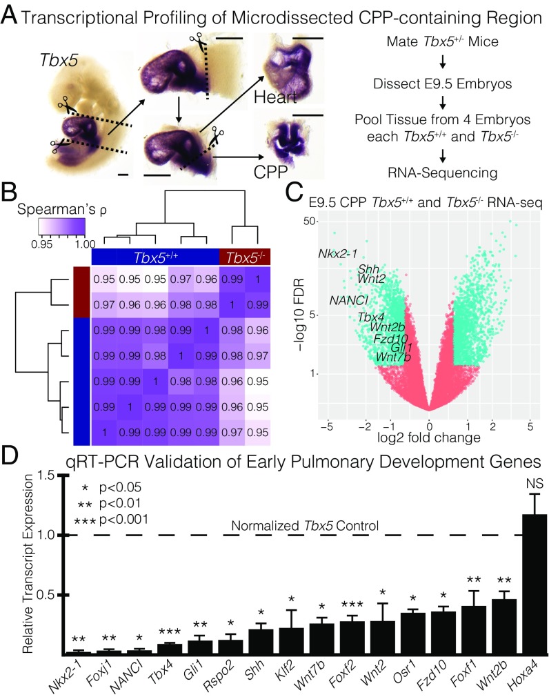
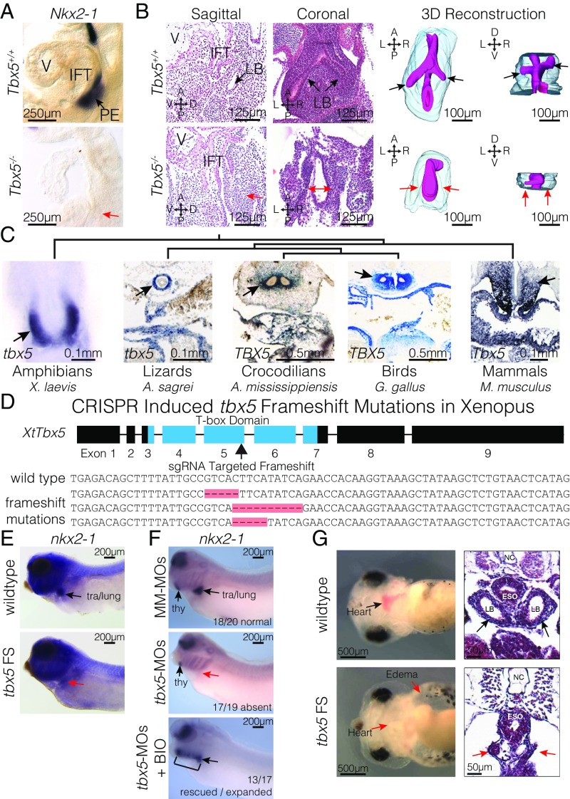
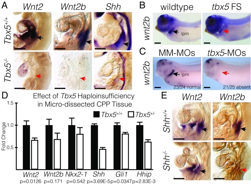
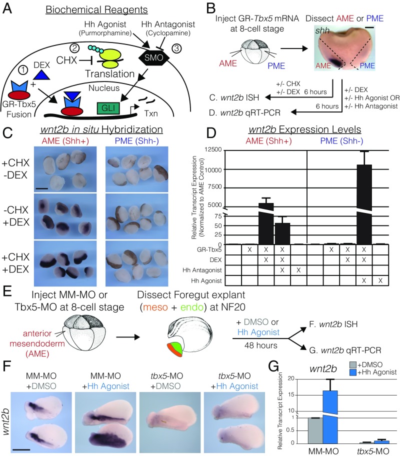
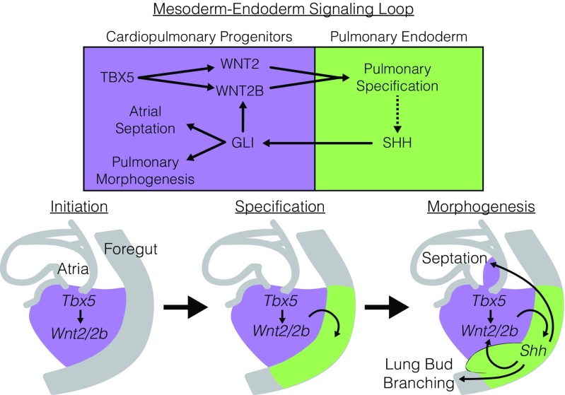
Codevelopment of the lungs and heart underlies key evolutionary innovations in the transition to terrestrial life. Cardiac specializations that support pulmonary circulation, including the atrial septum, are generated by second heart field (SHF) cardiopulmonary progenitors (CPPs). It has been presumed that transcription factors required in the SHF for cardiac septation, e.g., Tbx5, directly drive a cardiac morphogenesis gene-regulatory network. Here, we report instead that TBX5 directly drives Wnt ligands to initiate a bidirectional signaling loop between cardiopulmonary mesoderm and the foregut endoderm for endodermal pulmonary specification and, subsequently, atrial septation. We show that Tbx5 is required for pulmonary specification in mice and amphibians but not for swim bladder development in zebrafish. TBX5 is non-cell-autonomously required for pulmonary endoderm specification by directly driving Wnt2 and Wnt2b expression in cardiopulmonary mesoderm. TBX5 ChIP-sequencing identified cis-regulatory elements at Wnt2 sufficient for endogenous Wnt2 expression domains in vivo and required for Wnt2 expression in precardiac mesoderm in vitro. Tbx5 cooperated with Shh signaling to drive Wnt2b expression for lung morphogenesis. Tbx5 haploinsufficiency in mice, a model of Holt-Oram syndrome, caused a quantitative decrement of mesodermal-to-endodermal Wnt signaling and subsequent endodermal-to-mesodermal Shh signaling required for cardiac morphogenesis. Thus, Tbx5 initiates a mesoderm-endoderm-mesoderm signaling loop in lunged vertebrates that provides a molecular basis for the coevolution of pulmonary and cardiac structures required for terrestrial life.

Keywords: Hedgehog signaling; TBX5; Wnt signaling; heart development; lung development.


Conflict of interest statement

The authors declare no conflict of interest.

Figures

Fig. 1.
Fig. 1.
Transcriptional profiling of microdissected CPPs identifies a critical role for Tbx5 in pulmonary specification and lung development. (A, Left) Demonstration of microdissection methodology used for embryonic mouse experiments on an E9.5 embryo probed for Tbx5 RNA by ISH. (Scale bars: 0.25 mm.) (Right) Transcriptional profiling strategy used to measure the Tbx5-dependent transcriptome in the CPP-containing tissue by RNA-seq. (B) Spearman’s correlation of RNA-seq replicates. (C) Volcano plot of transcriptional profiling results with significantly dysregulated genes (teal) from the comparison of Tbx5+/+ and Tbx5−/− CPPs. Early markers of the PE and canonical Wnt and Shh signaling are identified. (D) qRT-PCR validation of 16 early lung-development genes that were significantly dysregulated in the RNA-seq.
Fig. 2.
Fig. 2.
Tbx5 is required for lung development in mice and frogs. (A) RNA ISH for Nkx2-1 in E9.5 Tbx5+/+ and Tbx5−/− embryos. The PE, inflow tract (IFT), and cardiac ventricle (V) are labeled. (B) Histology from both sagittal and coronal perspectives (Left) and 3D reconstruction (Right) of E10.5 lungs from Tbx5+/+ and Tbx5−/− embryos. Black and red arrows point to the lung bud (LB) branches or lack of branches off the foregut. (C) ISH stains of Tbx5 across vertebrate species. Arrows indicate expression in the mesodermal derivatives surrounding the PE. (D, Upper) Strategy for generating biallelic frameshift mutations using sgRNA targeted to the fifth exon of X. tropicalis to disrupt the T-box domain. (Lower) Examples of sequences recovered from tbx5 FS mutants. (E and F) RNA ISH of NF35 tadpoles for nkx2-1 in wild type and tbx5 FS mutants (E) and tadpoles injected with mismatched morpholinos (MM-MOs), tbx5-MOs, or tbx5-MOs cotreated with BIO (F). (G) Live images (Left) and H&E-stained transverse sections (Right) of NF42 tadpoles, depicting anatomical defects induced by CRISPR-mediated mutation of tbx5.
Fig. 3.
Fig. 3.
Tbx5 is required for Wnt2/2b and Shh expression. (A) RNA ISH for Wnt2, Wnt2b, and Shh in E9.5 Tbx5+/+ and Tbx5−/− mouse embryos. Black and red arrows point to positive and negative staining, respectively, in the lung-forming region. (B and C) RNA ISH for wnt2b performed in wild-type or tbx5 FS NF35 tadpoles (B) and in NF35 tadpoles injected with mismatched morpholinos (MM-MOs) or tbx5-MOs (C). The stained region corresponds with the lateral plate mesoderm (lpm). (D) qRT-PCR of microdissected CPP tissue from E9.5 Tbx5+/+ or Tbx5+/− mouse embryos. (E) RNA ISH for Wnt2 and Wnt2b in E9.5 Shh+/+ and Shh−/− mouse embryos. (Scale bars: 250 µm.)
Fig. 4.
Fig. 4.
tbx5 directly regulates wnt2b expression for lung development in the presence of Hh signaling. (A) To study the interaction of Tbx5 and Hh signaling, we utilized (1) a DEX-inducible GR-Tbx5 fusion protein; (2) CHX to inhibit protein synthesis; and (3) pharmacological agonists (purmorphamine) and antagonists (cyclopamine) of Smoothened (SMO) to activate or repress Hh signaling. (B) Strategy used in C and D for examining the regulation of wnt2b by Tbx5 in the presence or absence of Shh signaling in Xenopus. The AME (red) corresponds to shh-expressing tissue, and the PME (blue) corresponds to shh-negative tissue. (C) RNA ISH for wnt2b in AME or PME explants treated with CHX, DEX, or both. Note that the ISH signal is black; the brown color is pigment. (D) qRT-PCR of wnt2b in AME or PME explants, with or without GR-Tbx5, and treated with combinations of DEX, Hh agonist, and Hh antagonist. (E) Strategy used in F and G to examine the regulation of wnt2b by Hh signaling in the presence or absence of tbx5. AME explants were treated with DMSO (gray) or the Hh agonist purmorphamine (blue) for 48 h. (F) RNA ISH for wnt2b on DMSO- or Hh agonist-treated explants from embryos injected with mismatched morpholinos (MM-MO) or tbx5-MO. (G) qRT-PCR of wnt2b normalized to DMSO-treated MM-MO–injected explants. (Scale bars: B, 200 µm; C and E, 400 µm.)
Fig. 5.
Fig. 5.
Identification of TBX5-bound cis-regulatory elements for Wnt2. (A, Upper) We identified 3,883 peaks by TBX5 ChIP-seq in the E9.5 heart that correspond to 3,060 distal and 823 proximal sites. (Lower) Heatmaps of fold enrichment plotted for TBX5, H3K4me1, and H3K4me3 by ChIP-seq and ATAC-seq from the heart and CPP microdissections at each of the 3,883 summits ± 2,000 bp. (B) Overlap of the 1,318 down-regulated genes in the CPP of Tbx5−/− embryos by RNA-seq and the 3,883 genes nearest to TBX5 ChIP-seq peaks. The 162 genes in the intersection include Wnt2 and Wnt2b. (C) Genome browser view of Wnt2 and St7 (mm10 chr6:17,920,000–18,048,500) with TBX5 ChIP-seq (both from A and published in refs. and 60), H3K4me1 ChIP-seq, and ATAC-seq in the heart and pSHF. Tracks depict fold-enriched signal, and bars below represent significant peak calls. Cloned enhancers W2E1, -2, and -3 are shaded in gray. (D) ChIP-PCR for TBX5 at W2E1, -2, and -3 in the human IMR90 lung fibroblast cell line. Significance is calculated relative to IgG control. (E) Luciferase assay examining activation of W2E1, -2, and -3 in HEK293T cells provided a vector containing Tbx5 relative to a control vector. (F) Transgenic embryos were generated using an Hsp68-LacZ reporter construct upstream of W2E1, -2, or -3 and were stained at E9.5.
Fig. 6.
Fig. 6.
Cis-regulatory elements are required for Wnt2 expression. (A) Dose-dependent gene-expression changes in mESC-derived cardiogenic progenitors harboring a DOX-dependent Tbx5 construct (Tbx5OE-mESC) measured by qRT-PCR. Cells were treated with DOX for 24 h before analysis. (B, Upper) sgRNAs were designed to induce a 4.5-kb deletion within the last intron of St7, removing a majority of W2E1 and W2E2 in the Tbx5OE-mESC via CRISPR-Cas9. This design maintains the last exon and the predicted splice branch for St7 while removing the TBX5-bound sites in W2E1 and W2E2. (Lower) Amplification and sequencing across the target site demonstrate successful deletion. (C) Expression levels of Tbx5, Wnt2, and St7 were compared in W2E mutants and isogenic controls following differentiation to cardiogenic progenitors by qRT-PCR. (D) Changes in Tbx5, Wnt2, and St7 expression following 24 h of 100 ng/mL DOX relative 0 ng/mL DOX in the W2E mutant and isogenic controls differentiated to cardiogenic precursors.
Fig. 7.
Fig. 7.
Tbx5 is required for a mesoderm–endoderm bidirectional signaling loop for cardiopulmonary development. Model of genetic interaction between Tbx5, canonical Wnt signaling, and Hh signaling for PE specification, pulmonary morphogenesis, and cardiac septation. TBX5, expressed in the CPPs (purple) initiates the bidirectional signaling loop through direct activation of Wnt2 and Wnt2b expression. Canonical Wnt signaling drives pulmonary specification in the foregut endoderm and Nkx2-1 expression. SHH, derived from the PE (green) signals back to the CPPs where it cooperatively activates Wnt2b but not Wnt2. Shh signaling drives both atrial septation and lung bud morphogenesis through previously described downstream targets.

Similar articles

Cited by

References

    1. Hsia CC, Schmitz A, Lambertz M, Perry SF, Maina JN. Evolution of air breathing: Oxygen homeostasis and the transitions from water to land and sky. Compr Physiol. 2013;3:849–915. - PMC - PubMed
    1. Farmer CG. Evolution of the vertebrate cardio-pulmonary system. Annu Rev Physiol. 1999;61:573–592. - PubMed
    1. Jensen B, Spicer DE, Sheppard MN, Anderson RH. Development of the atrial septum in relation to postnatal anatomy and interatrial communications. Heart. 2017;103:456–462. - PubMed
    1. Jensen B, Wang T, Christoffels VM, Moorman AF. Evolution and development of the building plan of the vertebrate heart. Biochim Biophys Acta. 2013;1833:783–794. - PubMed
    1. Peng T, et al. Coordination of heart and lung co-development by a multipotent cardiopulmonary progenitor. Nature. 2013;500:589–592. - PMC - PubMed

Publication types