. 2019 Feb 1;25(3):967-976.
doi: 10.1158/1078-0432.CCR-18-2039. Epub 2018 Oct 23.

Clonal Relatedness and Mutational Differences between Upper Tract and Bladder Urothelial Carcinoma

Affiliations

Clonal Relatedness and Mutational Differences between Upper Tract and Bladder Urothelial Carcinoma

François Audenet et al. Clin Cancer Res. .

Abstract

Purpose: To investigate genomic differences between urothelial carcinomas of the upper tract (UTUC) and bladder (UCB), with a focus on defining the clonal relatedness of temporally distinct tumors.

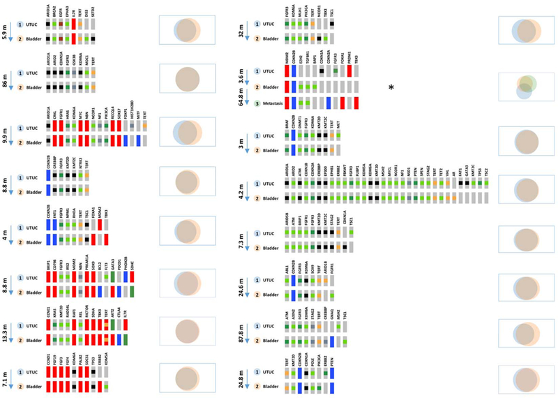
Experimental design: We prospectively sequenced tumors and matched germline DNA using targeted next-generation sequencing methods. The cohort included 195 UTUC patients and 454 UCB patients. For a subgroup of 29 patients with UTUC and a history of a subsequent UCB, both tumors were analyzed to assess their clonal relatedness.

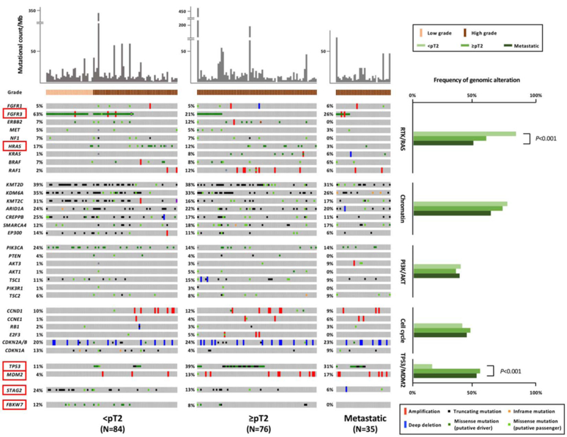
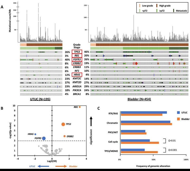
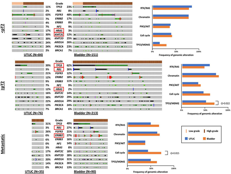
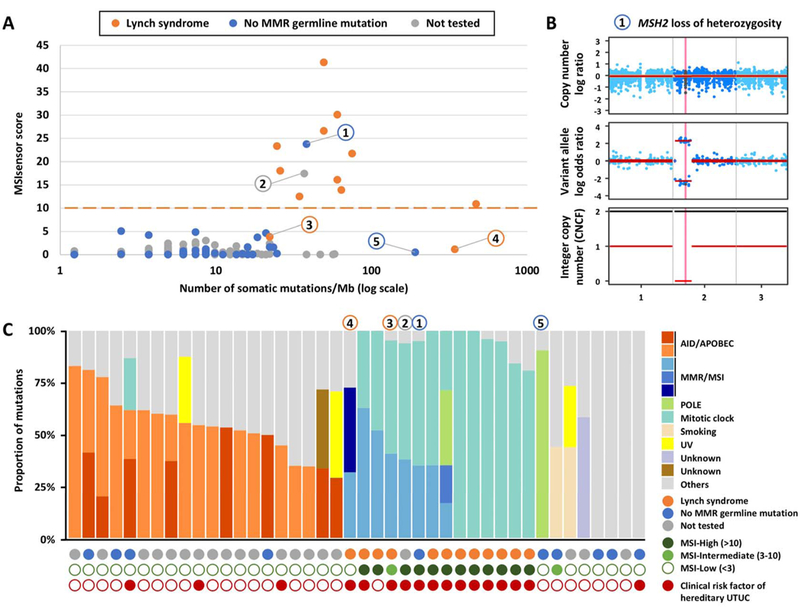
Results: With the progression to higher UTUC clinical state, there were fewer alterations in the RTK/RAS pathway but more alterations in TP53/MDM2. Compared with UCB, TP53, RB1, and ERBB2 were less frequently altered in UTUC (26% vs. 46%, 3% vs. 20%, 8% vs. 19%, respectively; Q < 0.001), whereas FGFR3 and HRAS were more frequently altered (40% vs. 26%, 12% vs. 4%, respectively; Q < 0.001). On the basis of an integrated analysis of tumor mutational burden, MSIsensor score and mutational signature, 7.2% of UTUC tumors were classified as MSI-high/MMR-deficient (MSI-H/dMMR). The risk of bladder recurrence after UTUC was significantly associated with mutations in FGFR3, KDM6A, CCND1, and TP53. Comparison of UCB with corresponding UTUC tumors from the same patient supports their clonal relatedness.

Conclusions: UTUC and UCB exhibit significant differences in the prevalence of common genomic alterations. In individual patients with a history of both tumors, UCB and UTUC were always clonally related. Genomic characterization of UTUC provides information regarding the risk of bladder recurrence and can identify tumors associated with Lynch syndrome.


Conflict of interest statement

Conflicts of interest: will be declared electronically by each author.

Figures

Figure 1:
Figure 1:. Overview of the genomic landscape of UTUC, stratified by clinical state and molecular pathways.
The genes with significant differences in genomic alteration frequencies across clinical states are highlighted by red boxes.
Figure 2:
Figure 2:. Genomic differences between UTUC and UCB.
A. Comparison of the differences in the prevalence of genomic alterations between UTUC and UCB, for the genes present at a frequency of ≥10% in any clinical state. The genes with significant differences after multiple comparison testing using the Bonferroni method are highlighted by red boxes. B.Volcano-plot of the frequency of gene alterations (log2(OR)) by significance (-log10(P-value)) in urothelial carcinomas of the upper tract (UTUC) or bladder. The horizontal doted line indicates an adjusted P-value <0.001. C.Comparison of genomic alteration frequencies for molecular pathways commonly mutated in urothelial carcinomas.
Figure 3:
Figure 3:. Stage by stage comparison of the differences in the prevalence of genomic alterations between UTUC and UCB, for the genes identified at a frequency of ≥10% at any clinical state.
The genes with significant differences after multiple comparison using the Bonferroni method are highlighted by red boxes.
Figure 4:
Figure 4:. Use of NGS to identify microsatellite instability high (MSI-H)/mismatch repair deficient (dMMR) UTUC tumors.
A. MSIsensor scores of UTUC samples were calculated using MSIsensor (version 0.2) and are displayed as a function of the number of somatic mutations/Mb, and its association with Lynch syndrome as determined by analysis of germline DNA. B. FACETS analysis of case 1 showing MSH2 loss of heterozygosity. This patient had 29 somatic mutations/Mb, an MSIsensor score of 23.7, a predominant MMR/MSI signature by mutational decomposition analysis and loss of MSH2 and MSH6 expression by immunohistochemistry in the absence of a germline mutation in these two genes, all consistent with a somatic Lynch-like MMR-deficient tumor. C. Mutational decomposition analysis of the UTUC samples with more than 10 somatic mutations. Case 2 had no consent for germline analysis. However, the patient’s personal history of colon cancer, the tumor mutational burden (TMB) of 21 somatic mutations/Mb, the elevated MSIsensor score of 17.4 and the predominant MMR/MSI signature by mutational decomposition indicated that this patient had an MMR-deficient tumor and thus germline testing would have been indicated. Notably, one patient with no clinical risk factors for hereditary UTUC but with a MSI-High tumor was found to have Lynch syndrome. Two of the 12 patients with pathogenic/likely pathogenic germline mutations in Lynch Syndrome-associated genes had low or intermediate MSIsensor scores but elevated tumor mutational burden in the setting of a predominant MMR/MSI signature by mutational decomposition. Case 3 was 45 years old at diagnosis, had a TMB of 22 somatic mutations/Mb, an MSIsensor score of 3.8, a germline mutation in MSH6, absence of both MSH6 and MSH2 expression by immunohistochemistry (IHC) and a predominant MMR/MSI signature. Case 4 had a familial history of Lynch-related cancers, a UTUC with 343 somatic mutations/Mb, an MSIsensor score of 1.1, a germline mutation in MSH2, absence of MSH2 and MSH6 expression by IHC and a predominant MMR/MSI signature.
Figure 5:
Figure 5:. Oncoprints and Venn diagrams for the matched-pair comparison between UTUC and UCB tumors in patients with no prior history of a bladder tumor before resection of the UTUC.
The arrows show the time between sample collection in months. Patient * underwent radical nephroureterectomy for pT2N0 UTUC and had a bladder recurrence 4 months later (pTa High grade). He was followed regularly and 65 months after the initial RNU and ultimately progressed to metastatic disease. The molecular profile of the metastasis was closer to the bladder tumor than to the radical nephroureterectomy specimen. However, all three tumor samples shared a clonal origin.

Comment in

Similar articles

Cited by

References

    1. Siegel RL, Miller KD, Jemal A. Cancer statistics, 2018. CA Cancer J Clin. 2018;68:7–30. - PubMed
    1. Babjuk M, Böhle A, Burger M, Capoun O, Cohen D, Compérat EM, et al. EAU Guidelines on Non-Muscle-invasive Urothelial Carcinoma of the Bladder: Update 2016. Eur Urol. 2017;71:447–61. - PubMed
    1. Munoz JJ, Ellison LM. Upper tract urothelial neoplasms: incidence and survival during the last 2 decades. J Urol. 2000;164:1523–5. - PubMed
    1. Green DA, Rink M, Xylinas E, Matin SF, Stenzl A, Rouprêt M, et al. Urothelial carcinoma of the bladder and the upper tract: disparate twins. J Urol. 2013;189:1214–21. - PubMed
    1. Chiou HY, Chiou ST, Hsu YH, Chou YL, Tseng CH, Wei ML, et al. Incidence of transitional cell carcinoma and arsenic in drinking water: a follow-up study of 8,102 residents in an arseniasis-endemic area in northeastern Taiwan. American journal of epidemiology. 2001;153:411–8. - PubMed

Publication types

MeSH terms

Substances