. 2018 Oct 23;9(5):e01905-18.
doi: 10.1128/mBio.01905-18.

Identification of Novel Protein Lysine Acetyltransferases in Escherichia coli

Affiliations

Identification of Novel Protein Lysine Acetyltransferases in Escherichia coli

David G Christensen et al. mBio. .

Erratum in

Abstract

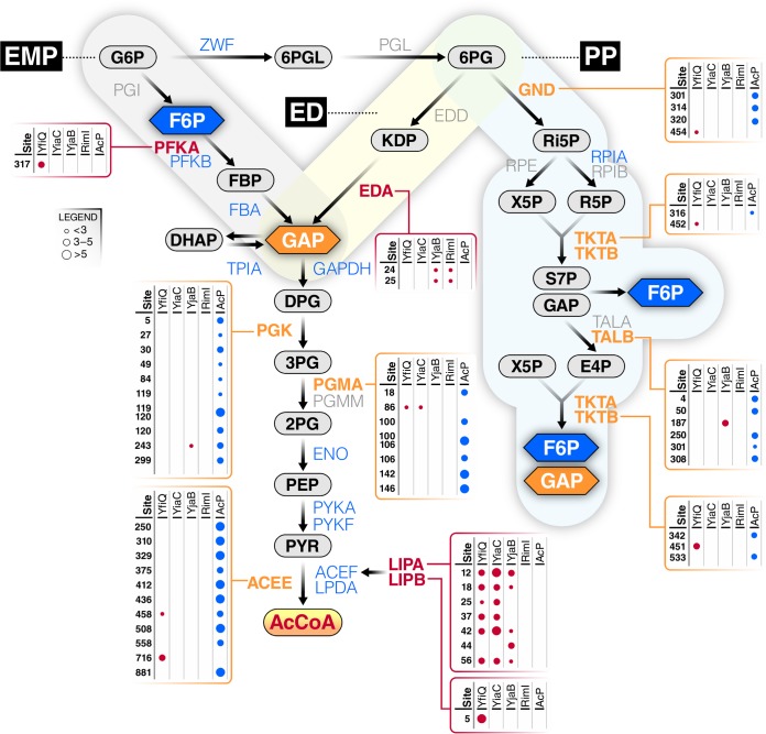
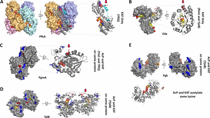
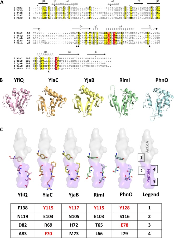
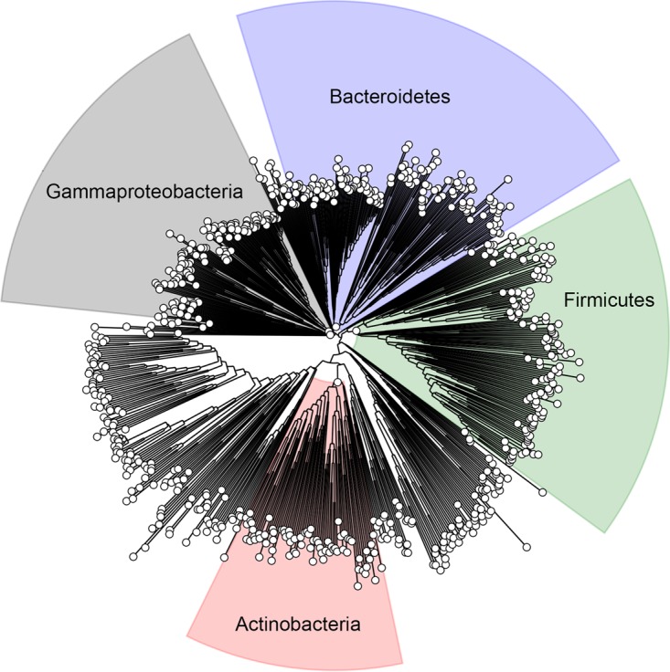
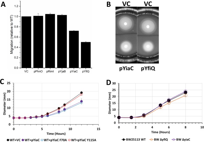
Posttranslational modifications, such as Nε-lysine acetylation, regulate protein function. Nε-lysine acetylation can occur either nonenzymatically or enzymatically. The nonenzymatic mechanism uses acetyl phosphate (AcP) or acetyl coenzyme A (AcCoA) as acetyl donor to modify an Nε-lysine residue of a protein. The enzymatic mechanism uses Nε-lysine acetyltransferases (KATs) to specifically transfer an acetyl group from AcCoA to Nε-lysine residues on proteins. To date, only one KAT (YfiQ, also known as Pka and PatZ) has been identified in Escherichia coli Here, we demonstrate the existence of 4 additional E. coli KATs: RimI, YiaC, YjaB, and PhnO. In a genetic background devoid of all known acetylation mechanisms (most notably AcP and YfiQ) and one deacetylase (CobB), overexpression of these putative KATs elicited unique patterns of protein acetylation. We mutated key active site residues and found that most of them eliminated enzymatic acetylation activity. We used mass spectrometry to identify and quantify the specificity of YfiQ and the four novel KATs. Surprisingly, our analysis revealed a high degree of substrate specificity. The overlap between KAT-dependent and AcP-dependent acetylation was extremely limited, supporting the hypothesis that these two acetylation mechanisms play distinct roles in the posttranslational modification of bacterial proteins. We further showed that these novel KATs are conserved across broad swaths of bacterial phylogeny. Finally, we determined that one of the novel KATs (YiaC) and the known KAT (YfiQ) can negatively regulate bacterial migration. Together, these results emphasize distinct and specific nonenzymatic and enzymatic protein acetylation mechanisms present in bacteria.IMPORTANCENε-Lysine acetylation is one of the most abundant and important posttranslational modifications across all domains of life. One of the best-studied effects of acetylation occurs in eukaryotes, where acetylation of histone tails activates gene transcription. Although bacteria do not have true histones, Nε-lysine acetylation is prevalent; however, the role of these modifications is mostly unknown. We constructed an E. coli strain that lacked both known acetylation mechanisms to identify four new Nε-lysine acetyltransferases (RimI, YiaC, YjaB, and PhnO). We used mass spectrometry to determine the substrate specificity of these acetyltransferases. Structural analysis of selected substrate proteins revealed site-specific preferences for enzymatic acetylation that had little overlap with the preferences of the previously reported acetyl-phosphate nonenzymatic acetylation mechanism. Finally, YiaC and YfiQ appear to regulate flagellum-based motility, a phenotype critical for pathogenesis of many organisms. These acetyltransferases are highly conserved and reveal deeper and more complex roles for bacterial posttranslational modification.

Keywords: GNAT; acetylation; acetyltransferase; bacteria; lysine acetyltransferase; mass spectrometry; protein acetyltransferase; proteomics.


Figures

FIG 1
FIG 1
Inactivation of the two known acetylation mechanisms in E. coli eliminates the majority of acetylation. Wild-type (WT) E. coli (strain BW25113) and an isogenic Δpta yfiQ acs cobB mutant (Gutted) were aerated in TB7 supplemented with 0.4% glucose for 10 h. Whole-cell lysates were analyzed (A) by Coomassie blue-stained SDS-polyacrylamide gel to ensure equivalent loading and (B) by anti-acetyllysine Western blotting.
FIG 2
FIG 2
Overexpression of five GNAT family members results in altered lysine acetylation patterns by anti-acetyllysine Western blotting. The gutted strain (BW25113 Δpta yfiQ acs cobB) was transformed with the pCA24n vector control (negative [−] control) or pCA24n containing the indicated genes under an IPTG-inducible promoter (69). As a positive (+) control, an isogenic strain that retained the WT allele of acspta yfiQ cobB) was transformed with pCA24n containing YfiQ. The resulting strains were aerated in TB7 supplemented with 0.4% glucose, 50 μM IPTG, and 25 μg/ml chloramphenicol for 10 h. Whole-cell lysates were analyzed (right panels) by Coomassie blue-stained SDS-polyacrylamide gel electrophoresis to ensure equivalent loading and (left panels) by anti-acetyllysine Western blotting. Note that the band in RimJ was not reproducible. The positive control contains one additional YfiQ-dependent band around 72 kDa, which corresponds to Acs (82). YncA and AstA each produce an acetylated band that can be observed in the Coomassie blue-stained gel at the expected molecular weight of these proteins.
FIG 3
FIG 3
Mutation of conserved catalytic amino acids prevents RimI-, PhnO-, YjaB-, and YiaC-dependent acetylation. The gutted strain (BW25113 Δpta yfiQ acs cobB) was transformed with the pCA24n vector control, pCA24n carrying the wild-type allele for each putative KAT, or mutant alleles for each putative KAT with alanine substitutions of the indicated residues. The resulting strains were grown in TB7 supplemented with 0.4% glucose, 100 μM IPTG, and 25 μg/ml chloramphenicol for 8 h. Crude lysates harvested after 4 h were analyzed for expression of the KAT proteins. Whole-cell lysates harvested after 8 h were analyzed for acetylation. Coomassie blue-stained SDS-PAGE gels (A and C) served as loading controls for anti-His (B) and anti-acetyllysine (D) Western blot analysis.
FIG 4
FIG 4
Identification of site-specific regulation of acetylation sites by KATs. (A) Cartoon showing workflow used to identify KAT target sites. (B) Overview of the significant sites and proteins regulated by at least one putative KAT. Significance defined as FDR of <0.01 within-set and log2(FC) of >2. (C) Venn diagram showing the complementary nature of trypsin and GluC digestion in terms of significant acetylation sites. (D) Heat map of all significant changes; acetylation sites are grouped by unsupervised hierarchical clustering.
FIG 5
FIG 5
Most of central metabolism is differentially acetylated by acetyl-P and/or KATs. The three glycolytic pathways, Embden-Parnas-Meyerhof (EMP), Entner-Dourdoroff (ED), and pentose phosphate (PP), are shown with metabolites and enzymes indicated. Some enzymes are not acetylated (gray), while others are acetylated by acetyl-P alone (blue), KATs alone (red), or both (orange). Enzymes with boxes were modified by at least one KAT (as indicated); some were also acetylated by acetyl-P (AcP). The size of the dot indicates the fold upregulation for each lysine by either a KAT or AcP.
FIG 6
FIG 6
Structural analysis of selected proteins modified by KATs and/or AcP. (A) PfkA (PDB ID 1pfk) structure. Each monomer of the tetramer is tinted in pink, orange, cyan, and violet and shown as a surface representation. The ligand ADP is bound to the allosteric site and the ligand fructose 1,6-bisphosphate is bound to the active site; both are shown as spheres. One monomer is also shown as a ribbon representation. A red arrow indicates the location of K317. The C terminus that is disordered in the 2pfk structure is shown in cyan. (B) Eda (PDB ID 1eua) structure. A surface representation of the trimer is shown in gray. One monomer of the trimer is also shown as a ribbon representation, and a red arrow indicates the two adjacent sites of acetylation. Pyruvate is shown as spheres. The active site residues are colored in yellow, and K24 and K25 are colored in red. (C) PgmA (PDB ID 1e58) structure. The dimer is shown as a surface representation, and one monomer of the dimer is shown as a ribbon representation. Sulfate is shown as spheres in the active site. K86, which is acetylated by YfiQ and YiaC, is shown in red and indicated by a red arrow. K18, 100, 106, 142, and 146 are acetylated by AcP and shown in blue. (D) TalB (PDB ID 4s2c) structure. The dimer is shown in both surface and ribbon representations. K187 is acetylated by YjaB and shown in red with a red arrow. K4, 50, 250, 301, and 308 are acetylated by AcP and shown in blue. Fructose 6-phosphate is shown as spheres in the active site, and surrounding residues are shown as yellow sticks. (E) Pgk (PDB ID 1zmr) structure. The monomer is shown as a surface and ribbon representation where K243, which is acetylated by both YjaB and AcP, is shown in orange and an orange arrow points to its location. K5, 27, 30, 49, 84, 119, 120, and 299 are acetylated by AcP and shown in blue. Phosphoaminophosphonic acid-adenylate ester and 3-phosphoglycerate are shown as spheres in the active site and modeled from the 1vpe structure.
FIG 7
FIG 7
Sequence and structural comparison of KAT proteins and their key catalytic residues. (A) Sequence alignment of all five E. coli KATs. Only the GNAT portion of the sequence for YfiQ is shown. The structural elements above the sequences are based on the 5isv RimI structure. Red highlighting represents 100% identity, whereas yellow highlighting shows a global score of 70% identity based on ESPript 3.0 parameters. Black arrowheads beneath the sequences indicate the residues selected for structural comparison in panel C. (B) Comparison of overall structures and homology models of E. coli YfiQ (pink), YiaC (orange), YjaB (yellow), RimI (green; full C terminus not shown in the figure), and PhnO (blue) proteins in a ribbon representation. 3D structures of YjaB and RimI were determined previously (PDB IDs 2kcw and 5isv, respectively). We built homology models of the remaining KATs using the following structures as the templates: 4nxy for YfiQ, 2kcw for YiaC, and 1z4e for PhnO. Only the GNAT portion of the YfiQ protein sequence was used for the homology model. Further details regarding parameters for building and selecting representative homology models for these proteins are described in Materials and Methods. (C) Comparison of select active site residues potentially important for substrate recognition and catalysis in GNATs. The crystal structure of RimI (5isv) has the C terminus of one monomer bound in the active site of the second monomer. A surface representation of this portion of the protein that encompasses the AcCoA donor (gray) and peptide acceptor (purple) site is shown. Each of the KAT homology models and structures was aligned using TopMatch and PyMOL. Four active site residues are shown. A table beneath the structures shows the specific residue numbers for each KAT. Residues that were mutated are shown in red.
FIG 8
FIG 8
YjaB is highly conserved across bacteria. A representative phylogenetic tree showing bacterial species that contain a gene homologous to yjaB from E. coli. The trees for rimI and yiaC are comparable.
FIG 9
FIG 9
YiaC and YfiQ inhibit migration. Cultures were grown overnight in TB medium supplemented with chloramphenicol and 50 μM IPTG. Five microliters of each normalized culture was spotted on low-percentage TB plates supplemented with chloramphenicol and 50 μM IPTG. The diameter of the cell spot was measured hourly. (A) Final diameter relative to vector control (VC) after 12 h is shown for wild-type E. coli BW25113 strains carrying the indicated plasmids. (B) Representative motility plates of BW25113 carrying VC, pYiaC, or pYfiQ. (C) Hourly migration of wild-type E. coli BW25113 strains carrying pCA24n encoding YiaC, YiaC mutants, or vector control (VC). (D) Hourly migration of wild-type E. coli strain BW25113 or isogenic deletion mutants on low-percentage TB plates without supplement.

References

    1. Drazic A, Myklebust LM, Ree R, Arnesen T. 2016. The world of protein acetylation. Biochim Biophys Acta 1864:1372–1401. doi: 10.1016/j.bbapap.2016.06.007. - DOI - PubMed
    1. Soppa J. 2010. Protein acetylation in archaea, bacteria, and eukaryotes. Archaea 2010:1. doi: 10.1155/2010/820681. - DOI - PMC - PubMed
    1. Wolfe AJ. 2016. Bacterial protein acetylation: new discoveries unanswered questions. Curr Genet 62:335–341. doi: 10.1007/s00294-015-0552-4. - DOI - PMC - PubMed
    1. Hentchel KL, Escalante-Semerena JC. 2015. Acylation of biomolecules in prokaryotes: a widespread strategy for the control of biological function and metabolic stress. Microbiol Mol Biol Rev 79:321–346. doi: 10.1128/MMBR.00020-15. - DOI - PMC - PubMed
    1. Kuhn ML, Zemaitaitis B, Hu LI, Sahu A, Sorensen D, Minasov G, Lima BP, Scholle M, Mrksich M, Anderson WF, Gibson BW, Schilling B, Wolfe AJ. 2014. Structural, kinetic and proteomic characterization of acetyl phosphate-dependent bacterial protein acetylation. PLoS One 9:e94816. doi: 10.1371/journal.pone.0094816. - DOI - PMC - PubMed

Publication types

MeSH terms