

Theranostics Aspects of Various Nanoparticles in Veterinary Medicine
- PMID: 30352960
- PMCID: PMC6274759
- DOI: 10.3390/ijms19113299
Theranostics Aspects of Various Nanoparticles in Veterinary Medicine
Abstract
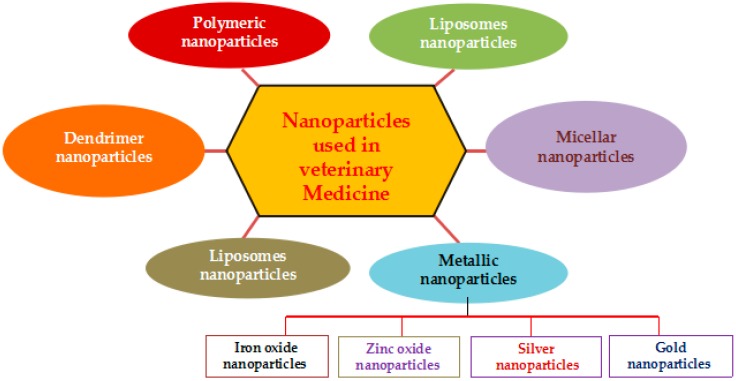
Nanoscience and nanotechnology shows immense interest in various areas of research and applications, including biotechnology, biomedical sciences, nanomedicine, and veterinary medicine. Studies and application of nanotechnology was explored very extensively in the human medical field and also studies undertaken in rodents extensively, still either studies or applications in veterinary medicine is not up to the level when compared to applications to human beings. The application in veterinary medicine and animal production is still relatively innovative. Recently, in the era of health care technologies, Veterinary Medicine also entered into a new phase and incredible transformations. Nanotechnology has tremendous and potential influence not only the way we live, but also on the way that we practice veterinary medicine and increase the safety of domestic animals, production, and income to the farmers through use of nanomaterials. The current status and advancements of nanotechnology is being used to enhance the animal growth promotion, and production. To achieve these, nanoparticles are used as alternative antimicrobial agents to overcome the usage alarming rate of antibiotics, detection of pathogenic bacteria, and also nanoparticles being used as drug delivery agents as new drug and vaccine candidates with improved characteristics and performance, diagnostic, therapeutic, feed additive, nutrient delivery, biocidal agents, reproductive aids, and finally to increase the quality of food using various kinds of functionalized nanoparticles, such as liposomes, polymeric nanoparticles, dendrimers, micellar nanoparticles, and metal nanoparticles. It seems that nanotechnology is ideal for veterinary applications in terms of cost and the availability of resources. The main focus of this review is describes some of the important current and future principal aspects of involvement of nanotechnology in Veterinary Medicine. However, we are not intended to cover the entire scenario of Veterinary Medicine, despite this review is to provide a glimpse at potential important targets of nanotechnology in the field of Veterinary Medicine. Considering the strong potential of the interaction between the nanotechnology and Veterinary Medicine, the aim of this review is to provide a concise description of the advances of nanotechnology in Veterinary Medicine, in terms of their potential application of various kinds of nanoparticles, secondly we discussed role of nanomaterials in animal health and production, and finally we discussed conclusion and future perspectives of nanotechnology in veterinary medicine.
Keywords: animal production; antimicrobial; diagnostic; drug delivery; livestock; nanoparticles.
Conflict of interest statement
The authors report no conflicts of interest in this work.
Figures

Similar articles
-
Advanced applications of nanotechnology in veterinary medicine.Environ Sci Pollut Res Int. 2020 Jun;27(16):19073-19086. doi: 10.1007/s11356-018-3913-y. Epub 2018 Dec 13. Environ Sci Pollut Res Int. 2020. PMID: 30547342 Review.
-
Current and future prospects for nanotechnology in animal production.J Anim Sci Biotechnol. 2017 Mar 14;8:26. doi: 10.1186/s40104-017-0157-5. eCollection 2017. J Anim Sci Biotechnol. 2017. PMID: 28316783 Free PMC article. Review.
-
2D Nanomaterials for Cancer Theranostic Applications.Adv Mater. 2020 Apr;32(13):e1902333. doi: 10.1002/adma.201902333. Epub 2019 Jul 28. Adv Mater. 2020. PMID: 31353752 Review.
-
Application of modelling and nanotechnology-based approaches: The emergence of breakthroughs in theranostics of central nervous system disorders.Life Sci. 2017 Aug 1;182:93-103. doi: 10.1016/j.lfs.2017.06.001. Epub 2017 Jun 3. Life Sci. 2017. PMID: 28583367 Review.
-
Veterinary nanomedicine: Pros and cons.Vet Med Sci. 2023 Jan;9(1):494-506. doi: 10.1002/vms3.1050. Epub 2022 Dec 29. Vet Med Sci. 2023. PMID: 36580403 Free PMC article. Review.
Cited by
-
ESR as a monitoring method of the interactions between TEMPO-functionalized magnetic nanoparticles and yeast cells.Sci Rep. 2019 Dec 10;9(1):18733. doi: 10.1038/s41598-019-55335-z. Sci Rep. 2019. PMID: 31822759 Free PMC article.
-
Fate of Biodegradable Engineered Nanoparticles Used in Veterinary Medicine as Delivery Systems from a One Health Perspective.Molecules. 2021 Jan 20;26(3):523. doi: 10.3390/molecules26030523. Molecules. 2021. PMID: 33498295 Free PMC article. Review.
-
Nanosilver-Biopolymer-Silica Composites: Preparation, and Structural and Adsorption Analysis with Evaluation of Antimicrobial Properties.Int J Mol Sci. 2024 Dec 18;25(24):13548. doi: 10.3390/ijms252413548. Int J Mol Sci. 2024. PMID: 39769310 Free PMC article.
-
Beneficial and toxicological aspects of zinc oxide nanoparticles in animals.Vet Med Sci. 2022 Jul;8(4):1769-1779. doi: 10.1002/vms3.814. Epub 2022 May 19. Vet Med Sci. 2022. PMID: 35588498 Free PMC article. Review.
-
Potential of Nanomaterial Applications in Dietary Supplements and Foods for Special Medical Purposes.Nanomaterials (Basel). 2019 Feb 19;9(2):296. doi: 10.3390/nano9020296. Nanomaterials (Basel). 2019. PMID: 30791492 Free PMC article. Review.
References
Publication types
MeSH terms
LinkOut - more resources
Full Text Sources
Other Literature Sources
