. 2018 Oct 23;9(1):4404.
doi: 10.1038/s41467-018-06750-9.

Dynamic coordination of two-metal-ions orchestrates λ-exonuclease catalysis

Affiliations

Dynamic coordination of two-metal-ions orchestrates λ-exonuclease catalysis

Wonseok Hwang et al. Nat Commun. .

Abstract

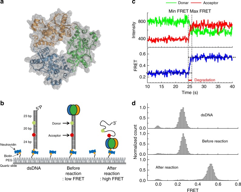
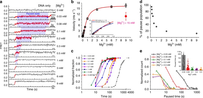
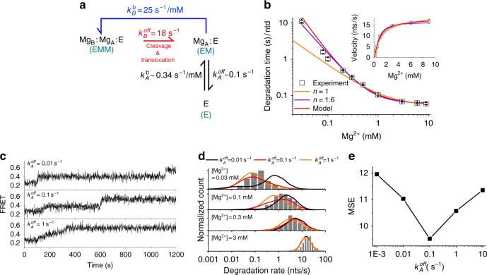
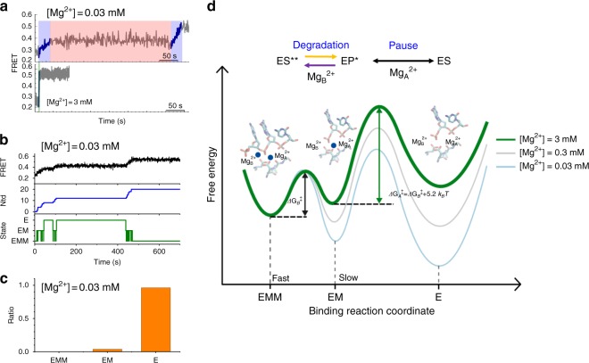
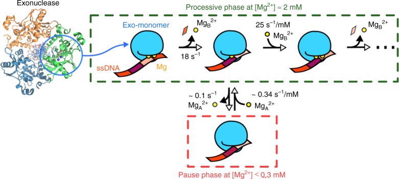
Metal ions at the active site of an enzyme act as cofactors, and their dynamic fluctuations can potentially influence enzyme activity. Here, we use λ-exonuclease as a model enzyme with two Mg2+ binding sites and probe activity at various concentrations of magnesium by single-molecule-FRET. We find that while MgA2+ and MgB2+ have similar binding constants, the dissociation rate of MgA2+ is two order of magnitude lower than that of MgB2+ due to a kinetic-barrier-difference. At physiological Mg2+ concentration, the MgB2+ ion near the 5'-terminal side of the scissile phosphate dissociates each-round of degradation, facilitating a series of DNA cleavages via fast product-release concomitant with enzyme-translocation. At a low magnesium concentration, occasional dissociation and slow re-coordination of MgA2+ result in pauses during processive degradation. Our study highlights the importance of metal-ion-coordination dynamics in correlation with the enzymatic reaction-steps, and offers insights into the origin of dynamic heterogeneity in enzymatic catalysis.


Conflict of interest statement

The authors declare no competing interests

Figures

Fig. 1
Fig. 1
Real-time measurements of processive degradation by λ-exonuclease. a The crystal structure of λ-exonuclease (PDB entry 1AVQ). b Experimental layout, depicting the DNA, protein binding to DNA, and processive degradation. c Schematic showing how the degradation time is measured using FRET signal. d Single-molecule FRET histogram obtained as in b (top: dsDNA only; middle: before the degradation in the absence of Mg2+; and bottom: after the degradation in the presence of Mg2+)
Fig. 2
Fig. 2
Mg2+-dependent degradation of DNA by λ-exonuclease. a Representative traces showing that degradation reaction slows down at lower Mg2+ concentrations (0 mM, 0.03 mM, 0.08 mM, 0.1 mM, 0.2 mM, 0.3 mM, 0.5 mM, 1 mM, 3 mM, 6 mM, and 9 mM Mg2+: top to bottom) at the fixed 16 nM λ-exonuclease (trimer concentration unless otherwise stated). b The degradation rate (velocity) versus Mg2+ concentrations following a Hill fit (red line) with a maximum velocity of 17.2 nt s−1, a Km value of 0.885 mM of Mg2+, and n = 1.6. Inset, blowing up at lower Mg2+ concentrations showing fitting to a sigmoidal kinetics (red) versus a Michaelis Menten kinetics (blue). Error bars denote the standard error of the mean (SEM). The velocity at [Mg2 + ] = 15 mM is highlighted in red. c Time-dependent fractional growth at various Mg2+ concentrations. d The proportion of the pause population as a function of the Mg2+ concentration. e Distribution of pause times with varying concentrations of Mg2+
Fig. 3
Fig. 3
Inhibition of λ-exonuclease activity by Ca2+ ions. a Single-molecule FRET histograms with various concentrations of Ca2+ at the fixed concentration of 3 mM Mg2+. b Ca2+-substitution-induced pause behavior fitted with an automated step-finding algorithm. c Pause histograms obtained from various Ca2+ concentrations at the fixed concentration of 3 mM Mg2+. d Three degradation patterns with pauses and backtracking when inhibited by Ca2+. e The proportion of pause and backtracking as a function of Ca2+ concentration. f Increase in the degradation time with increasing concentration of Ca2+
Fig. 4
Fig. 4
Quantitative analysis of two-metal dynamics. a A kinetic model for the two-metal-ion dynamics consisting of three states: EMM (exonuclease:MgB2+:MgA2+ complex), EM (exonuclease:MgA2+ complex), and E (exonuclease only). In the model, MgB2+ dissociates upon DNA cleavage and translocation (kBoff, red arrow, EMM to EM) whereas MgA2+ dissociates stochastically (kAoff, EM to E). The single cycle is completed upon rebinding of MgB2+ (kBb, EM to EMM). b The degradation time per nucleotide (τ1) versus Mg2+ concentrations. The data are fitted to three models: (1) Michaelis Menten equation (orange line), (2) Hill equation (purple line, n = 1.6), and (3) Eq. 1 for the model shown in a (red line). Inset shows velocity ( = τ1-1) versus Mg2+ concentrations data and their fits (solid lines). c Representative FRET time traces from simulations performed at three different choices of kAoff: 0.01 s−1 (top panel), 0.1 s−1 (middle panel), and 1 s−1 (bottom panel). [Mg2+] is set to 0.03 mM. d Degradation rate histograms at four different [Mg2+] conditions: 0.03 mM, 0.1 mM, 0.3 mM, and 3 mM. Gray bars represent experimental data, whereas solid lines are theoretical prediction calculated under three difference choices of kAoff: 0.01 s−1 (black), 0.1 s−1 (red), and 1 s−1 (orange). e Mean squared error (MSE) versus kAoff. The errors are calculated by summing the squares of the difference between the experimental and theoretically predicted results
Fig. 5
Fig. 5
FRET time trajectories and the free energy landscape during metal-ion dynamics. a Two experimental FRET time trajectories, showing MgB2+-dynamics-dependent slope change (purple) and a pause caused by the dissociation of two metal ions, MgA2+ and MgB2+ (pink). b A representative simulation trajectory, displaying three panels: a time-FRET trace (black line in the top panel), degradation position of exonuclease along DNA (the line in the middle panel), and its metal-ion state (green line in the bottom panel). The trajectory revealed that iteration between EM and EMM decreases the degradation slope in a Mg2+-dependent manner, and pauses occur due to the E state by dissociation of both metal ions. c Total fraction of single-molecule traces showing the pause state at three different Mg2+ states. d Free energy landscape along the reaction coordination of Mg2+ binding and dissociation. Processive degradation occurs during a repetitive cycle between the 1st and 2nd metal-ion binding states, whereas a pause occurs due to the dissociation of two metal ions. The energy barrier between the 2nd and 1st states is lower than the one between the 1st and 2nd states due to the different numbers of ligands surviving after the translocation (e.g., 1 versus 3 ligands with protein residues for MgB2+ and MgA2+, respectively). Transitions between ES** and EP* are indicated by two one-directional arrows to emphasize the transitions are non-reversible and not in equilibrium
Fig. 6
Fig. 6
The mechanism of two-metal-ion dynamics. The active site of one subunit of the homotrimer is shown in green whereas two Mg2+ ions and nucleotides are represented by yellow circles and rectangles in various colors, respectively. Metal-ion coordination to the catalytic active site is highly dynamic so that two Mg2+ ions (MgA2+ and MgB2+) can coordinate with and dissociate from the surrounding ligands. MgA2+ remains stably bound to the active site, but MgB2+, which is close to the 5′ terminal side of the scissile phosphate, dissociates during every round of catalytic cleavage. As a result, the dissociation of MgB2+ facilitates product release and exonuclease translocation, promoting the overall processivity of exonuclease activity. More specifically, at high [Mg2+] (≈2 mM), λ-exonuclease degrades processively and its cleavage activity is mainly controlled by MgB2+ dynamics (green dashed square). The fast unbinding/rebinding dynamics of MgB2+ and strict requirement of two Mg2+ ions for the catalytic step yield a Mg2+-concentration-dependent exonuclease activity. Conversely at low [Mg2+] (≤0.3 mM), occasional unbinding of MgA2+ from the catalytic site and slow rebinding of MgA2+ stalls the exonuclease activity, giving rise to a long pause (red dashed square), thus elucidating the molecular origin of dynamic heterogeneity in exonuclease activity. The dynamic variation in the coordination states of two metal ions orchestrates the multistep process of exonuclease activity

Similar articles

Cited by

References

    1. Cowan J. A. The Biological Chemistry of Magnesium (VCH, New York, 1995).
    1. Mordasini T, Curioni A, Andreoni W. Why do divalent metal ions either promote or inhibit enzymatic reactions? The case of BamHI restriction endonuclease from combined quantum-classical simulations. J. Biol. Chem. 2003;278:4381–4384. doi: 10.1074/jbc.C200664200. - DOI - PubMed
    1. Sigel H. Metal Ions in Biological Systems (Marcel Dekker, New York, 1974).
    1. Babu CS, Lim C. Protein/solvent medium effects on Mg2+ -carboxylate interactions in metalloenzymes. J. Am. Chem. Soc. 2010;132:6290–6291. doi: 10.1021/ja101494m. - DOI - PubMed
    1. Babu CS, Dudev T, Lim C. Differential role of the protein matrix on the binding of a catalytic aspartate to Mg2+vs Ca2+: application to ribonuclease H. J. Am. Chem. Soc. 2013;135:6541–6548. doi: 10.1021/ja4006095. - DOI - PubMed

Publication types