. 2018 Oct 23;8(1):15647.
doi: 10.1038/s41598-018-34092-5.

The Sema3A receptor Plexin-A1 suppresses supernumerary axons through Rap1 GTPases

Affiliations

The Sema3A receptor Plexin-A1 suppresses supernumerary axons through Rap1 GTPases

Nannan Wang et al. Sci Rep. .

Abstract

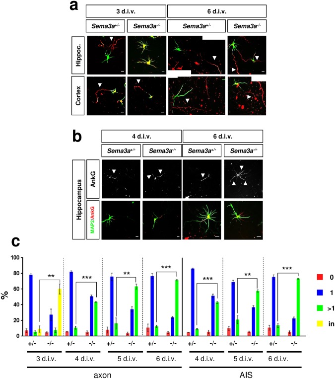
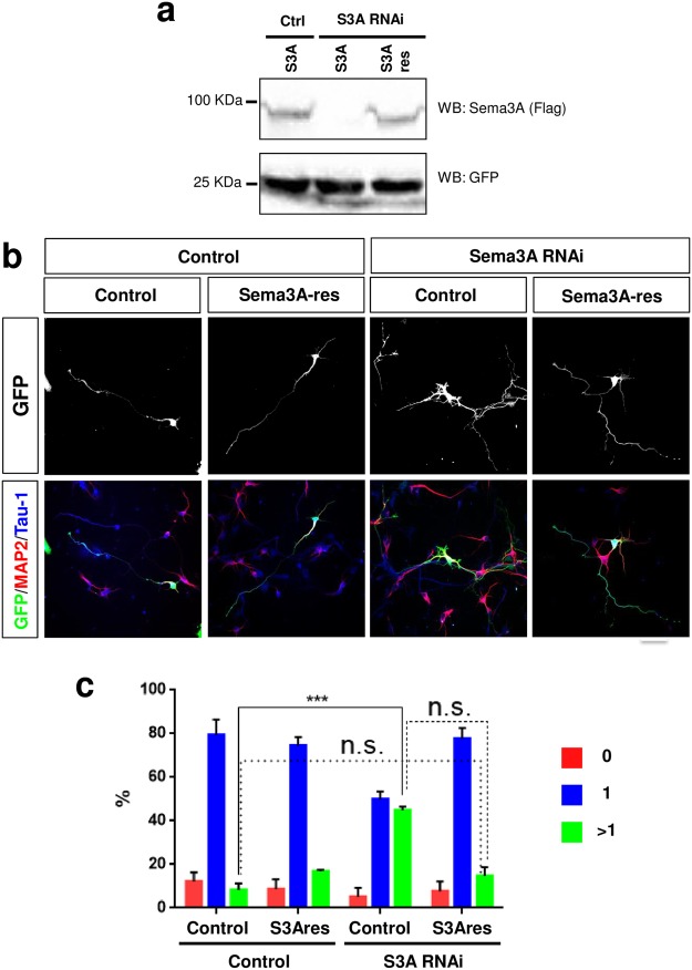
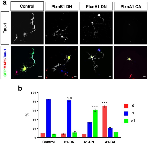
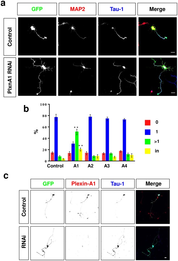
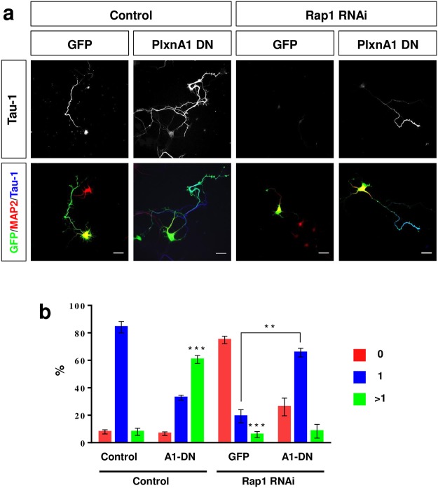
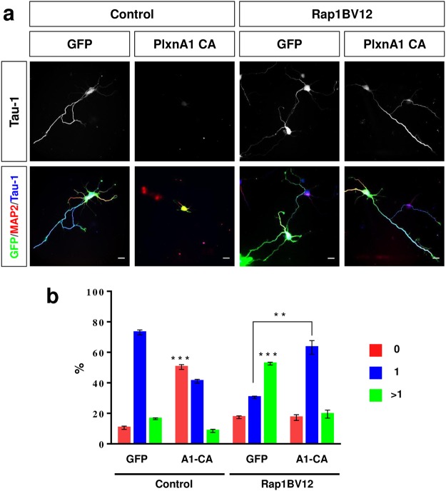
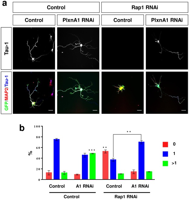
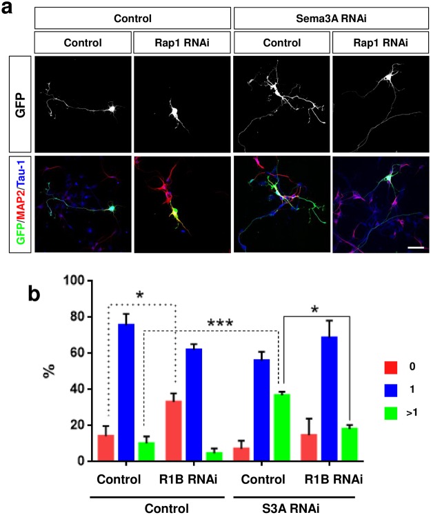
The highly conserved Rap1 GTPases perform essential functions during neuronal development. They are required for the polarity of neuronal progenitors and neurons as well as for neuronal migration in the embryonic brain. Neuronal polarization and axon formation depend on the precise temporal and spatial regulation of Rap1 activity by guanine nucleotide exchange factors (GEFs) and GTPases-activating proteins (GAPs). Several Rap1 GEFs have been identified that direct the formation of axons during cortical and hippocampal development in vivo and in cultured neurons. However little is known about the GAPs that limit the activity of Rap1 GTPases during neuronal development. Here we investigate the function of Sema3A and Plexin-A1 as a regulator of Rap1 GTPases during the polarization of hippocampal neurons. Sema3A was shown to suppress axon formation when neurons are cultured on a patterned substrate. Plexin-A1 functions as the signal-transducing subunit of receptors for Sema3A and displays GAP activity for Rap1 GTPases. We show that Sema3A and Plexin-A1 suppress the formation of supernumerary axons in cultured neurons, which depends on Rap1 GTPases.


Conflict of interest statement

The authors declare no competing interests.

Figures

Figure 1
Figure 1
Hippocampal neurons from Sema3a−/− embryos extend supernumerary axons in culture. (ac) Cultures of hippocampal or cortical neurons from E17 Sema3a+/− or Sema3a−/− embryos were analyzed at 3, 4, 5 and 6 d.i.v. by staining with an anti-MAP2 (green, dendrites) and the Tau-1 (a, red, axons) or an anti-AnkG antibody (b, red, AIS marked by arrow heads). Representative images of hippocampal neurons at the indicated times in culture are shown. The scale bar is 25 μm. (c) The percentage of unpolarized hippocampal neurons without an axon or AIS (0, red), polarized neurons with a single axon or AIS (1, blue), neurons with multiple axons or AIS (>1, green) and neurons with neurites that are positive for both axonal and dendritic markers (in, yellow) is shown (Student’s t-test and two-way ANOVA; n = 3 independent experiments with >150 neurons per genotype; values are means ± s.e.m., **p < 0.01, ***p < 0.001 compared to control as indicated).
Figure 2
Figure 2
Knockdown of Sema3A induces the formation of supernumerary axons. (a) HEK 293 T cells were transfected with vectors for FLAG-Sema3A (S3A) or RNAi-resistant FLAG-Sema3A-res (S3Ares) and an shRNA directed against Sema3A (S3A RNAi) or pcDNA6.2-GW/EmGFP-miR (Ctrl). The expression of Sema3A and GFP was analyzed by Western blot (WB) using an anti-FLAG antibody. The molecular weight is indicated in kDa. (b) Hippocampal neurons from E18 rat embryos were transfected with vectors for GFP (green), an shRNA against Sema3A (Sema3A RNAi) or pcDNA6.2-GW/EmGFP-miR (control), and a vector for RNAi-resistant FLAG-Sema3A-res (Sema3A-res) or pBK-CMV (control) as indicated. Neurons were analyzed at 3 d.i.v. by staining with an anti-MAP2 (red, dendrites) and the Tau-1 antibody (blue, axon). Representative images of transfected neurons are shown. The scale bar is 25 μm. (c) The percentage of unpolarized neurons without an axon (0, red), polarized neurons with a single axon (1, blue) and neurons with multiple axons (>1, green) is shown (Student’s t-test and two-way ANOVA; n = 3, independent experiments with >150 neurons per experiment; values are means ± s.e.m, ***p < 0.001 compared to control as indicated; n.s., not significant).
Figure 3
Figure 3
A-type plexins suppress the formation of axons. (a,b) Hippocampal neurons from E18 rat embryos were transfected with vectors for GFP (green, control), dominant-negative Plexin-B1Δcyt (PlxnB1-DN), Plexin-A1Δcyt (PlxnA1-DN) or constitutively active PlexinA1Δsema (PlxnA1-CA) and analyzed at 3 d.i.v. by staining with an anti-MAP2 (red, dendrites) and the Tau-1 antibody (blue, axon). Representative images of transfected neurons are shown. The scale bar is 25 μm. (b) The percentage of unpolarized neurons without an axon (0, red), polarized neurons with a single axon (1, blue) and neurons with multiple axons (>1, green) is shown (Student’s t-test and two-way ANOVA; n = 3, independent experiments with >150 neurons per experiment for; values are means ± s.e.m, ***p < 0.001 compared to control; n.s. not significant).
Figure 4
Figure 4
Knockdown of Plexin-A1 induces the formation of supernumerary axons. (a,b) Hippocampal neurons from E18 rat embryos were transfected with vectors for GFP (green) and shRNAs against Plexin-A1, -A2. -A3, or -A4 or pSUPER (control) and analyzed at 3 d.i.v. by staining with an anti-MAP2 (red, dendrites) and the Tau-1 antibody (blue, axon). Representative images of transfected neurons are shown. The scale bar is 25 μm. (b) The percentage of unpolarized neurons without an axon (0, red), polarized neurons with a single axon (1, blue), neurons with multiple axons (>1, green) and neurons with neurites that are positive for both axonal and dendritic markers (in, yellow) is shown (Student’s t-test and two-way ANOVA; n = 3, independent experiments with >150 neurons per experiment; values are means ± s.e.m, **p < 0.01 compared to control). (c) Hippocampal neurons from E18 rat embryos were transfected with vectors for GFP (green) and an shRNA against Plexin-A1 (RNAi) or pSUPER (control) and analyzed at 3 d.i.v. by staining with an anti-Plexin-A1 (red) and the Tau-1 antibody (blue, axon). Representative images of transfected neurons are shown. The scale bar is 25 μm.
Figure 5
Figure 5
Knockdown of Rap1B counteracts the induction of supernumerary axons by dominant-negative Plexin-A1. (a) Hippocampal neurons from E18 rat embryos were transfected with vectors for GFP (green, control), an shRNA against Rap1B or pSHAG-1 (control) and Plexin-A1Δcyt (PlxnA1-DN) or pBK-CMV (control). The establishment of neuronal polarity was analyzed at 3 d.i.v. by staining with an anti-MAP2 (red, dendrites) and the Tau-1 antibody (blue, axons). Representative images of transfected neurons are shown. The scale bar is 25 μm. (b) The percentage of unpolarized neurons without an axon (0, red), polarized neurons with a single axon (1, blue) and neurons with multiple axons (>1, green) is shown (Student’s t-test and two-way ANOVA; n = 3, independent experiments with >150 neurons per experiment for; values are means ± s.e.m, **p < 0.01; ***p < 0.001 compared to control and as indicated.
Figure 6
Figure 6
Active Rap1B recues the suppression of axon formation by constitutively active Plexin-A1. (a) Hippocampal neurons from E18 rat embryos were transfected with vectors for GFP (green) and Plexin-A1Δsema (PlxnA1-CA), Rap1BV12 or a combination of both and analyzed at 3 d.i.v. by staining with an anti-MAP2 (red, dendrites) and the Tau-1 antibody (blue, axon). pBK-CMV and pSHAG-1 were transfected as control. Representative images of transfected neurons are shown. The scale bar is 25 μm. (b) The percentage of unpolarized neurons without an axon (0, red), polarized neurons with a single axon (1, blue) and neurons with multiple axons (>1, green) is shown (Student’s t-test and two-way ANOVA; n = 3, independent experiments with >150 neurons per experiment; values are means ± s.e.m, **p < 0.01; ***p < 0.001 compared to control and as indicated).
Figure 7
Figure 7
Rap1B is required for the induction of supernumerary axons by the knockdown of Plexin-A1 in hippocampal neurons. (a) Hippocampal neurons from E18 rat embryos were transfected with vectors for GFP (green) and Plexin-A1Δsema (PlxnA1-CA), Rap1BV12, pSHAG-1 (control) or a combination of both and analyzed at 3 d.i.v. by staining with an anti-MAP2 (red, dendrites) and the Tau-1 antibody (blue, axon). Representative images of transfected neurons are shown. The scale bar is 25 μm. (b) The percentage of unpolarized neurons without an axon (0, red), polarized neurons with a single axon (1, blue) and neurons with multiple axons (>1, green) is shown (Student’s t-test and two-way ANOVA; n = 3, independent experiments with >150 neurons per experiment for; values are means ± s.e.m, **p < 0.01; ***p < 0.001 compared to control and as indicated).
Figure 8
Figure 8
Rap1B acts downstream of Sema3A in hippocampal neurons. (a) Hippocampal neurons from E18 rat embryos were transfected with vectors for GFP (green), an shRNA against Sema3A (S3A RNAi) or pcDNA6.2-GW/EmGFP-miR (control) and an shRNA against Rap1B (R1B RNAi) or pSHAG-1 (control) as indicated. Neurons were analyzed at 3 d.i.v. by staining with an anti-MAP2 (red, dendrites) and the Tau-1 antibody (blue, axon). Representative images of transfected neurons are shown. The scale bar is 25 μm. (b) The percentage of unpolarized neurons without an axon (0, red), polarized neurons with a single axon (1, blue) and neurons with multiple axons (>1, green) is shown (Student’s t-test and two-way ANOVA; n = 3, independent experiments with >150 neurons per experiment for; values are means ± s.e.m, ***p < 0.001 compared to control as indicated).

References

    1. Shah B, et al. Rap1 GTPases Are Master Regulators of Neural Cell Polarity in the Developing Neocortex. Cereb Cortex. 2017;27:1253–1269. - PubMed
    1. Franco SJ, Martinez-Garay I, Gil-Sanz C, Harkins-Perry SR, Muller U. Reelin regulates cadherin function via Dab1/Rap1 to control neuronal migration and lamination in the neocortex. Neuron. 2011;69:482–497. doi: 10.1016/j.neuron.2011.01.003. - DOI - PMC - PubMed
    1. Jossin Y, Cooper JA. Reelin, Rap1 and N-cadherin orient the migration of multipolar neurons in the developing neocortex. Nat Neurosci. 2011;14:697–703. doi: 10.1038/nn.2816. - DOI - PMC - PubMed
    1. Ye T, Ip JP, Fu AK, Ip NY. Cdk5-mediated phosphorylation of RapGEF2 controls neuronal migration in the developing cerebral cortex. Nat Commun. 2014;5:4826. doi: 10.1038/ncomms5826. - DOI - PMC - PubMed
    1. Shah B, Püschel AW. In vivo functions of small GTPases in neocortical development. Biol Chem. 2014;395:465–476. doi: 10.1515/hsz-2013-0277. - DOI - PubMed

Publication types

LinkOut - more resources