. 2018 Oct 23;8(1):15645.
doi: 10.1038/s41598-018-34026-1.

Inhibiting ex-vivo Th17 responses in Ankylosing Spondylitis by targeting Janus kinases

Affiliations

Inhibiting ex-vivo Th17 responses in Ankylosing Spondylitis by targeting Janus kinases

Ariane Hammitzsch et al. Sci Rep. .

Abstract

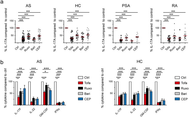
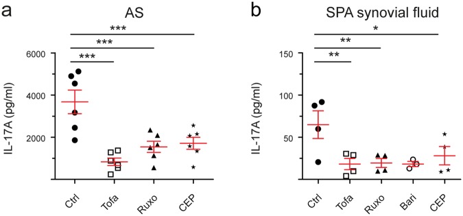
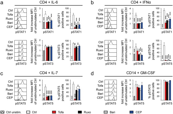
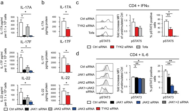
Treatment options for Ankylosing Spondylitis (AS) are still limited. The T helper cell 17 (Th17) pathway has emerged as a major driver of disease pathogenesis and a good treatment target. Janus kinases (JAK) are key transducers of cytokine signals in Th17 cells and therefore promising targets for the treatment of AS. Here we investigate the therapeutic potential of four different JAK inhibitors on cells derived from AS patients and healthy controls, cultured in-vitro under Th17-promoting conditions. Levels of IL-17A, IL-17F, IL-22, GM-CSF and IFNγ were assessed by ELISA and inhibitory effects were investigated with Phosphoflow. JAK1/2/3 and TYK2 were silenced in CD4+ T cells with siRNA and effects analyzed by ELISA (IL-17A, IL-17F and IL-22), Western Blot, qPCR and Phosphoflow. In-vitro inhibition of CD4+ T lymphocyte production of multiple Th17 cytokines (IL-17A, IL-17F and IL-22) was achieved with JAK inhibitors of differing specificity, as well as by silencing of JAK1-3 and Tyk2, without impacting on cell viability or proliferation. Our preclinical data suggest JAK inhibitors as promising candidates for therapeutic trials in AS, since they can inhibit multiple Th17 cytokines simultaneously. Improved targeting of TYK2 or other JAK isoforms may confer tailored effects on Th17 responses in AS.


Conflict of interest statement

The authors declare no competing interests.

Figures

Figure 1
Figure 1
JAK inhibitors inhibit CD4+ T cell “type 17” cytokine production in-vitro in Spondyloarthritis, Rheumatoid Arthritis and healthy controls. (a) IL-17A secretion from CD4+ T cells cultured under Th17-promoting conditions in-vitro in the presence of JAK inhibitors (Tofa, JAK3 > JAK1/2; Ruxo, JAK2 > JAK1; Bari, JAK1/2 > TYK2; CEP, JAK2) from day 0 to 3. Measured by supernatant ELISA and normalized to DMSO control (=100%) on day 3 (no.s AS = 43/Bari = 10, HC = 26/Bari = 14, PSA = 16/Bari = 3 and RA = 18/Bari = 9). (b) Inhibitory effects of JAK inhibitors on IL-17F, IL-22, GM-CSF and IFNγ secretion from AS (n = 10 – 8 – 9 – 6 respectively) and HC (n = 10 – 7 – 10 – 10) CD4+ T cells, measured by ELISA as in (a) and normalized to DMSO control (=100%). Statistical analysis: mean ± SEM, repeated measures 1-way ANOVA followed by Dunnett’s method for multiple comparisons (a) and 2-way ANOVA (b) followed by Bonferroni’s method for multiple comparisons.
Figure 2
Figure 2
JAK inhibitors work on “type 17” cytokine production in-vitro in Spondyloarthritis on established peripheral Th17 cells and on synovial fluid CD4+ T cells. (a) Reduction of IL-17A secretion by JAK inhibitors (Tofa, JAK3 > JAK1/2; Ruxo, JAK2 > JAK1; Bari, JAK1/2 > TYK2; CEP, JAK2) in AS CD4+ T cells (n = 6), primed under Th17-promoting conditions for 6 days, upon restimulation with anti-CD2/3/28 beads for 24 hours measured by ELISA. (b) Effects of JAK inhibitors on IL-17A secretion (ELISA) from synovial CD4+ T cells of SPA patients cultured for 3 days (n = 4, Bari n = 3). Statistical analysis: mean ± SEM, repeated measures 1-way ANOVA followed by Dunnett’s method for multiple comparisons.
Figure 3
Figure 3
JAK inhibitors inhibit multiple cytokine-driven STAT phosphorylation events. Inhibition of STAT phosphorylation by JAK inhibitors (Tofa, JAK3 > JAK1/2; Ruxo, JAK2 > JAK1; Bari, JAK1/2 > TYK2; CEP, JAK2) in freshly isolated AS PBMC (n = 5–6) upon cytokine stimulation assessed by intracellular Flow Cytometry. Stimulation with (a) IL-6, (b) IFNα and (c) IL-7 gated on CD4+ T cells and with (d) GM-CSF gated on CD14+ Monocytes. Panel on the left shows exemplary flow cytometry plot (light grey filled curves in each panel show unstimulated control staining), middle panel shows fold increase of mean fluorescence intensity (MFI) compared to unstimulated control and right panel shows frequency of phosphorylated STAT of parental population. Statistical analysis: mean ± SEM, 2-way ANOVA followed by Bonferroni’s method for multiple comparisons.
Figure 4
Figure 4
siRNA-mediated silencing of JAK1 and 3 and TYK2 inhibits “type 17” cytokine responses. (a) Reduction of IL-17A, IL-17F and IL-22 secretion (ELISA) upon siRNA-mediated silencing of JAK1/2/3 in HC CD4+ T cells cultured under Th17-promoting conditions for 3 days in-vitro. Data shown are normalized on cell number at the end of experiment = day 3 (n = 3–7). (b) Reduction of IL-17A, IL-17F and IL-22 secretion (ELISA) upon siRNA-mediated silencing of TYK2 in HC CD4+ T cells cultured as in (a). Data shown are normalized on mg protein (n = 5/4/3). (c) Inhibition of STAT5 phosphorylation by siRNA-mediated TYK2 silencing in HC CD4+ T cells upon IFNα stimulation 3 days post transfection compared to Tofa treatment (n = 2–5) and (d) of STAT3 phosphorylation by siRNA-mediated JAK1 and JAK2 silencing upon IL-6 stimulation (n = 1, triplicates). Panel on the left shows exemplary flow cytometry plot (light grey filled curves in each panel show unstimulated control staining), middle panel shows fold increase of MFI compared to unstimulated control and right panel shows frequency of phosphorylated STAT of parental population. Statistical analysis: mean ± SEM, paired t test.

References

    1. Stolwijk Carmen, van Onna Marloes, Boonen Annelies, van Tubergen Astrid. Global Prevalence of Spondyloarthritis: A Systematic Review and Meta-Regression Analysis. Arthritis Care & Research. 2016;68(9):1320–1331. doi: 10.1002/acr.22831. - DOI - PubMed
    1. Dougados M, Baeten D. Spondyloarthritis. Lancet. 2011;377:2127–2137. doi: 10.1016/S0140-6736(11)60071-8. - DOI - PubMed
    1. van der Heijde D, et al. Efficacy and safety of adalimumab in patients with ankylosing spondylitis: results of a multicenter, randomized, double-blind, placebo-controlled trial. Arthritis Rheum. 2006;54:2136–2146. doi: 10.1002/art.21913. - DOI - PubMed
    1. van der Heijde D, et al. Efficacy and safety of infliximab in patients with ankylosing spondylitis: results of a randomized, placebo-controlled trial (ASSERT) Arthritis Rheum. 2005;52:582–591. doi: 10.1002/art.20852. - DOI - PubMed
    1. Landewe R, et al. Efficacy of certolizumab pegol on signs and symptoms of axial spondyloarthritis including ankylosing spondylitis: 24-week results of a double-blind randomised placebo-controlled Phase 3 study. Ann Rheum Dis. 2014;73:39–47. doi: 10.1136/annrheumdis-2013-204231. - DOI - PMC - PubMed

Publication types

MeSH terms