. 2018 Oct 23;8(1):15642.
doi: 10.1038/s41598-018-33958-y.

Ionic and cellular mechanisms underlying TBX5/PITX2 insufficiency-induced atrial fibrillation: Insights from mathematical models of human atrial cells

Affiliations

Ionic and cellular mechanisms underlying TBX5/PITX2 insufficiency-induced atrial fibrillation: Insights from mathematical models of human atrial cells

Jieyun Bai et al. Sci Rep. .

Abstract

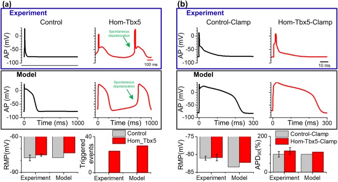
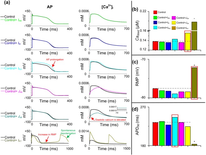
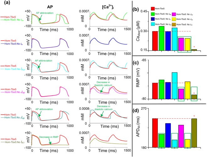
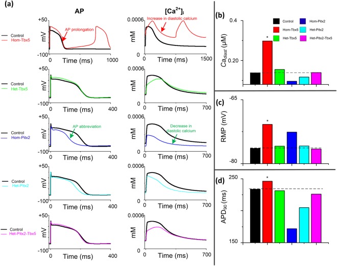
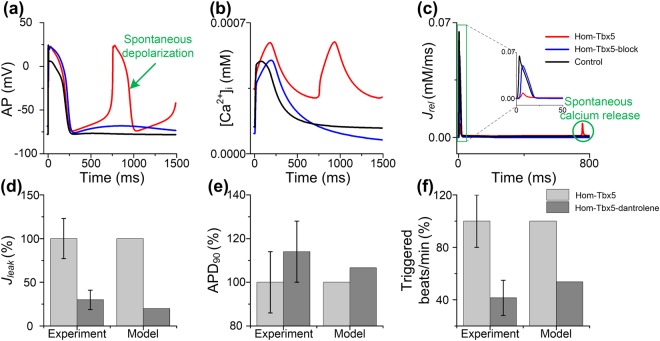
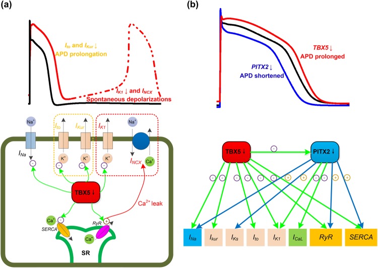
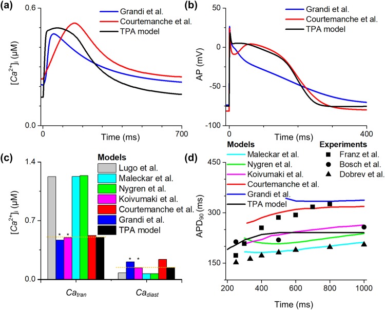
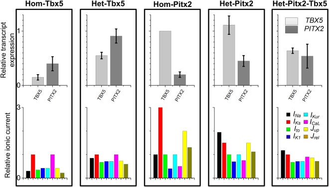
Transcription factors TBX5 and PITX2 involve in the regulation of gene expression of ion channels and are closely associated with atrial fibrillation (AF), the most common cardiac arrhythmia in developed countries. The exact cellular and molecular mechanisms underlying the increased susceptibility to AF in patients with TBX5/PITX2 insufficiency remain unclear. In this study, we have developed and validated a novel human left atrial cellular model (TPA) based on the ten Tusscher-Panfilov ventricular cell model to systematically investigate how electrical remodeling induced by TBX5/PITX2 insufficiency leads to AF. Using our TPA model, we have demonstrated that spontaneous diastolic depolarization observed in atrial myocytes with TBX5-deletion can be explained by altered intracellular calcium handling and suppression of inward-rectifier potassium current (IK1). Additionally, our computer simulation results shed new light on the novel cellular mechanism underlying AF by indicating that the imbalance between suppressed outward current IK1 and increased inward sodium-calcium exchanger current (INCX) resulted from SR calcium leak leads to spontaneous depolarizations. Furthermore, our simulation results suggest that these arrhythmogenic triggers can be potentially suppressed by inhibiting sarcoplasmic reticulum (SR) calcium leak and reversing remodeled IK1. More importantly, this study has clinically significant implications on the drugs used for maintaining SR calcium homeostasis, whereby drugs such as dantrolene may confer significant improvement for the treatment of AF patients with TBX5/PITX2 insufficiency.


Conflict of interest statement

The authors declare no competing interests.

Figures

Figure 1
Figure 1
Simulated action potential (AP) in human atrial cells and experimental AP in mouse atrial cells. (a) Comparison between APs from our human atrial model (Model) and those from the experimental adult mouse heart by Nadadur et al. under control (black) and homozygous TBX5-knockout (Hom-Tbx5; red) conditions. Representative abnormal depolarization events (i.e., spontaneous depolarization is indicated by the arrow) were observed in mouse atrial myocytes and reproduced using our human atrial cell model. Compared to control atrial cells, the resting membrane potential (RMP) and the number of triggered events were increased under the Hom-Tbx5 condition. (b) The simulated APs using our human atrial cell model were compared to representative APs of mouse atrial myocytes under control (Control-Clamp) and Hom-Tbx5 (Hom-Tbx5-Clamp) conditions. Triggered activity in Hom-Tbx5 atrial myocytes was suppressed under the Hom-Tbx5-Clamp condition. RMP and action potential duration (APD90) were increased, compared to Control-Clamp. This figure demonstrates that our human atrial model is capable of reproducing experimentally obtained AP characteristics of the Hom-Tbx5 atrial myocytes.
Figure 2
Figure 2
Effects of individual ionic remodeling targets on the cytosolic calcium concentration ([Ca2+]i) and action potential (AP). (a) Characteristics of control atrial cells with each TBX5-remodeled cellular component i.e., fast sodium current, INa; transient outward potassium current, Ito; ultrarapid delayed rectifier potassium current, IKur; calcium flow through the sarcoplasmic reticulum calcium ATPase (SERCA), Jup, calcium flow through the ryanodine receptor (RyR), Jrel and inward rectifier potassium current, IK1, respectively. Spontaneous depolarizations are induced in human atrial cells with the remodeled IK1. AP prolongation, diastolic calcium elevation and increase in resting membrane potential (RMP) occur in human atrial cells with remodeled IKur, Jup and IK1, respectively. (bd) Diastolic calcium concentration (Cadiast), RMP and action potential duration (APD90) for all cell variants are compared to control atrial myocytes (red bar). Main components which contribute to AP abnormalities in Hom-Tbx5 atrial cells are marked with red rectangles. Biomarkers of spontaneous depolarizations are marked with stars.
Figure 3
Figure 3
Effects of reversing remodeling of individual ionic channels on the cytosolic calcium concentration ([Ca2+]i) and action potential (AP). (a) Characteristics of homozygous TBX5-knockout (Hom-Tbx5) atrial cells without each remodeled cellular component, i.e., INa, Ito, IKur, Jup, Jrel and IK1, respectively. Spontaneous depolarizations were suppressed in human atrial cells without the remodeled IK1. Action potential duration (APD90) was well restored when the remodeling of INa, IKur and Jup were individually reversed. Diastolic calcium was decreased in Hom-Tbx5 atrial cells without effects of the remodeled Jrel and Jup. (bd) The diastolic calcium concentration (Cadiast), resting membrane potential (RMP) and APD90 for all cell variants were compared to those of Hom-Tbx5 atrial myocytes (red bar). Main components which contribute to AP abnormalities in Hom-Tbx5 atrial cells are marked with green rectangles. Biomarkers of the normal action potential are marked with stars.
Figure 4
Figure 4
Role of the TBX5-PITX2 regulatory loop in spontaneous depolarization generation. (a) Cytosolic calcium concentration ([Ca2+]i) and action potential (AP) characteristics in homozygous TBX5-knockout (Hom-Tbx5), heterozygous TBX5-knockout (Het-Tbx5), homozygous PITX2-knockout (Hom-Pitx2), heterozygous PITX2-knockout (Het-Pitx2) and heterozygous knockout of both PITX2 and TBX5 (Het-Pitx2-Tbx5) atrial cells. TBX5 insufficiency (e.g., Hom-Tbx5) led to AP prolongation and diastolic calcium elevation, whereas PITX2 insufficiency (e.g., Hom-Pitx2) caused AP abbreviation and a decrease in diastolic calcium. AP abnormalities induced by TBX5 haploinsufficiency were rescued by PITX2 haploinsufficiency. (bd) The diastolic calcium concentration (Cadiast), resting membrane potential (RMP) and action potential duration (APD90) for all cell variants were compared to control atrial myocytes (black bar).
Figure 5
Figure 5
Role of sarcoplasmic reticulum (SR) calcium leak (Jleak) in spontaneous depolarization generation. Simulated action potential (AP) (a), cytosolic calcium concentration [Ca2+]i (b) and calcium flow Jrel (c) through the ryanodine receptor (RyR) are displayed under control, homozygous TBX5-knockout (Hom-Tbx5) and Hom-Tbx5 with inhibition of Jleak (Hom-Tbx5-block). According to the effect of dantrolene on Jleak (d), when Jleak was inhibited by 80% (Hom-Tbx5-dantrolene), changes in APD90 (e) and triggered beats/min (f) compared to Hom-Tbx5 are shown. Simulated results were compared to the experimental data by Hartmann et al..
Figure 6
Figure 6
The role of electrical remodeling of TBX5 insufficiency in atrial fibrillation. (a) The schematic illustration of the impact of TBX5 loss-of-function mutation on ionic currents/action potential duration (APD). TBX5-deletion leads to reductions in INa, Ito, IKur, IK1, SERCA and RyR. Reduced repolarizing potassium currents (e.g., Ito and IKur) lead to prolonged APD. Suppression of IK1 and increased INCX due to cytosolic calcium overload lead to phase 4 depolarization and predispose to spontaneous depolarizations. (b) TBX5 regulates PITX2 expression, and TBX5 and PITX2 antagonistically regulate downstream targets. Reduced PITX2 leads to upregulation of INa, IKs, SERCA and RyR, and downregulation of ICaL and IK1. Loss of TBX5 leads to prolonged APD, whereas loss of PITX2 leads to shortened APD. Therefore, reduced PITX2 in TBX5-mutant atria contributes to a protective mechanism.
Figure 7
Figure 7
Comparison of the TPA model with different human atrial models. The myocyte was stimulated at a pacing frequency of 1 Hz. The intracellular calcium ([Ca2+]i, (a)) and action potentials (AP, (b)) of Grandi et al. model (blue lines), Courtemanche et al.model (red lines) and the current model (black lines) are shown. (c) The amplitude of calcium transient (Catran) and diastolic calcium concentration (Cadiast) (black bar) are compared to simulation results of Lugo et al. model (gray bar), Maleckar et al. model (cyan bar), Nygren et al. model (green bar), Koivumaki et al. model (magenta bar), Courtemanche et al. model (red bar) and Grandi et al. model (blue bar). (d) The action potential duration at 90% repolarization (APD90) restitution curve (black lines) is compared to simulated APD90 restitution of Maleckar et al. model (cyan lines), Nygren et al. model (green lines), Koivumaki et al. model (magenta lines), Courtemanche et al. model (red lines) and Grandi et al. model (blue lines), and experimental data of Franz et al. (▪), Bosch et al. (•) and Dobrev et al. (▲). All the data are adapted from Wilhelms et al..
Figure 8
Figure 8
Ionic current changes in homozygous TBX5-knockout (Hom-Tbx5), heterozygous TBX5-knockout (Het-Tbx5), homozygous PITX2-knockout (Hom-Pitx2), heterozygous PITX2-knockout (Het-Pitx2), and heterozygous knockout of both PITX2 and TBX5 (Het-Pitx2-Tbx5) atrial cells. Relative transcript expression of TBX5 and PITX2, and relative ionic current of INa, IKs, Ito, IK1, IKur, ICaL, Jup and Jrel in each condition.

Similar articles

Cited by

References

    1. Lip GYH, et al. Atrial fibrillation. Nat Rev Dis Primers. 2016;2:16016. doi: 10.1038/nrdp.2016.16. - DOI - PubMed
    1. Lau DH, Nattel S, Kalman JM, Sanders P. Modifiable risk factors and atrial fibrillation. Circulation. 2017;136:583–596. doi: 10.1161/CIRCULATIONAHA.116.023163. - DOI - PubMed
    1. Xiong Z, et al. A Machine Learning Aided Systematic Review and Meta-Analysis of the Relative Risk of Atrial Fibrillation in Patients With Diabetes Mellitus. Frontiers in physiology. 2018;9:835. doi: 10.3389/fphys.2018.00835. - DOI - PMC - PubMed
    1. Olesen MS, Nielsen MW, Haunsø S, Svendsen JH. Atrial fibrillation: the role of common and rare genetic variants. Eur J Hum Genet. 2013;22:297–306. doi: 10.1038/ejhg.2013.139. - DOI - PMC - PubMed
    1. Zhou M, Liao Y, Tu X. The role of transcription factors in atrial fibrillation. J Thorac Dis. 2015;7:152–158. doi: 10.3978/j.issn.2072-1439.2015.01.21. - DOI - PMC - PubMed

Publication types