Review
. 2019 Feb;49(2):254-262.
doi: 10.1007/s00247-018-4270-1. Epub 2018 Oct 23.

Introduction to contrast-enhanced ultrasound of the brain in neonates and infants: current understanding and future potential

Affiliations
Review

Introduction to contrast-enhanced ultrasound of the brain in neonates and infants: current understanding and future potential

Misun Hwang. Pediatr Radiol. 2019 Feb.

Abstract

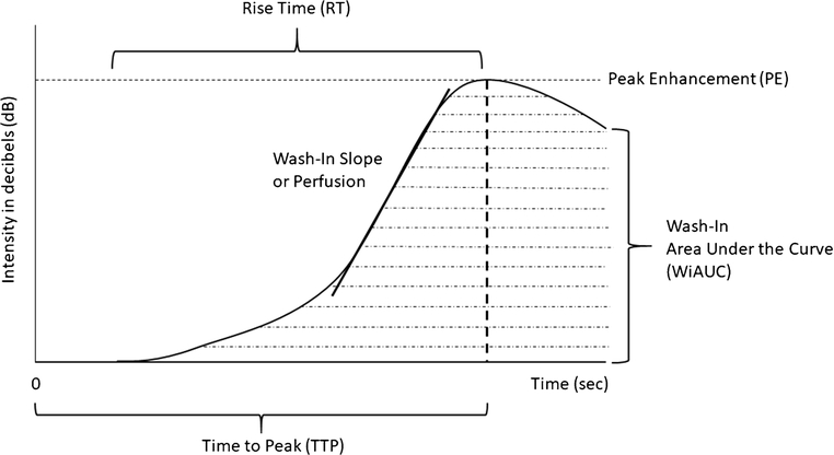
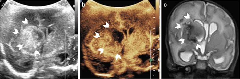
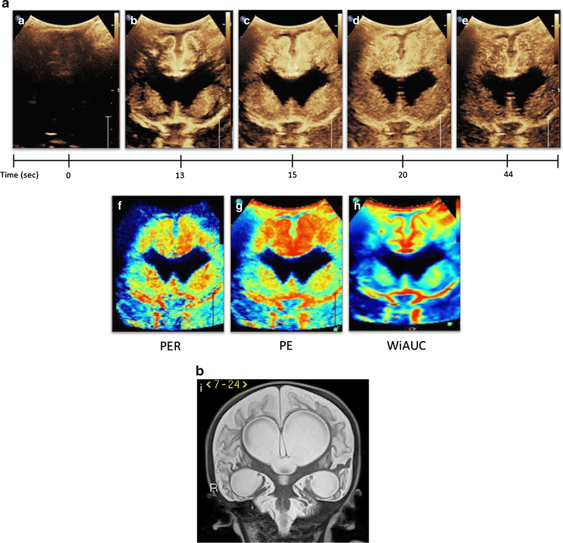
Contrast-enhanced ultrasound (CEUS) is a valuable bedside imaging technique that enables both qualitative and quantitative assessment of cerebral perfusion. In neonates and infants whose fontanelles remain open, the technique is particularly useful as it delineates cerebral pathology with high soft-tissue contrast. The technique has the potential to be a valuable alternative to computed tomography (CT) or magnetic resonance imaging (MRI) in critically ill neonates and infants in need of bedside imaging. While further studies are needed to validate the technique, preliminary data in this regard appear promising. This review introduces the current understanding and future potential of brain CEUS.

Keywords: Brain; Clinical protocol; Contrast-enhanced ultrasound; Neonate; Perfusion; Ultrasound.


Figures

Figure 1.
Figure 1.. Microbubble Wash-In Time Intensity Curve (TIC)
The graph represents a time intensity curve generated based on the dynamic microbubble wash-in data. Time 0 represents the time of microbubble injection. Peak enhancement (PE) refers to maximum microbubble intensity (in decibels or dB) during the wash-in phase. The time (sec) it takes from injection to reach PE is called time to peak (TTP). The time from the start of curve rise to peak enhancement is called rise time (RT). The area under the TIC is called wash-in area under the curve (WiAUC) and represents microbubble volume. The slope of the wash-in curve is called wash-in slope or perfusion.
Figure 2.
Figure 2.. Dynamic Microbubble Wash-In on Mid-Coronal Brain Scan in a Normal Subject
Figure 2A denotes a mid-coronal grayscale ultrasound scan through the brain of a normal subject showing bilateral frontal lobes (orange arrows), frontal horns (yellow arrowheads), basal ganglia (red arrows), and temporal lobes (white arrows). Figure 2B-G demonstrates dynamic microbubble wash-in through the mid-coronal slice through the brain on a contrast specific mode from the time of injection (time 0) to 1 min. Note that the microbubbles flow into the partially visualized Circle of Willis by 13 seconds. By 15 seconds, relatively more avid enhancement to the basal ganglia with respect to the remainder of the brain is seen. Further enhancement of the cortex but with relative hyperenhancement of the basal ganglia is noted at 20 seconds. Wash-out of contrast from both the basal ganglia and cortex begins at 25 seconds and further wash-out noted at 60 seconds. Note that Figure 2 images were obtained with EPIQ scanner (Phillips Healthcare, Bothell, WA) and C5–1 transducer with settings of 12 Hz and MI of 0.06.
Figure 3.
Figure 3.. Multifocal and Symmetric, Diffuse Perfusion Abnormalities on CEUS-B
Figure 3A depicts a coronal scan through the posterior parietooccipital lobes in a neonate with hypoxic ischemic injury. The image was obtained at 27 seconds after microbubble injection at peak intensity. Generalized hypoperfusion with multifocal perfusion abnormalities noted as evidenced by paucity of microbubbles in scattered areas (white arrows). Figure 3B and 3C, both obtained at 18 seconds after microbubble injection, are neonatal cases of symmetric, diffuse hypoxic ischemic injury resulting in generalized hyperperfusion to the brain in the immediate post-injury period. Figure 3D is an infant post prolonged cardiac arrest demonstrating diffuse hypoperfusion to the brain. Note that Figure 3 images were obtained with EPIQ scanner (Phillips Healthcare, Bothell, WA). Figure 3A was obtained with C5–1 transducer and settings of 12 Hz, MI 0.06, Figure 3B with C9–2 transducer and settings of 13 Hz, MI 0.06, Figure 3C with C9–2 transducer and settings of 7 Hz, MI 0.06, and Figure 3D with C9–2 transducer and settings of 12 Hz, MI 0.06. This figure is adapted from Hwang et al.[2] and Shin et al.[27] with permission.
Figure 4.
Figure 4.. Central Gray Nuclei to Cortex Ratio (GNC)
Central gray nuclei to cortex ratio in a 1-month-old male (a, b) and a 14-day-old male (c). The central gray nuclei (basal ganglia and thalami) (red) and cortex (areas excluding central gray nuclei) (yellow) are outlined on a mid-coronal CEUS. The central gray nuclei to cortex (GNC) ratio equals perfusion in central gray nuclei (red) over perfusion in cortex (yellow). Figure (a) and(b) demonstrates a representative case with the normally seen hyperperfusion to the central gray nuclei, with (a) obtained at 10 seconds after microbubble administration and (b) obtained at 15 seconds after administration at peak enhancement. (c) demonstrates a hypoxic ischemic injury case in which the normally seen GNC ratio greater than 1 is absent due to diffuse hyperperfusion to the brain in the immediate post injury setting. In this case, the GNC ratio is noted to be approximately equal to 1. The image was obtained at 18 seconds after microbubble administration at peak enhancement. Note that all the images were obtained with EPIQ scanner (Philips Healthcare, Bothell, WA) and C5–1 transducer with settings of 12 Hz and mechanical index of 0.06.
Figure 5.
Figure 5.. Intracranial Hemorrhage on CEUS-B
Figure 5 is a CEUS-B of a neonate with heterogeneous right germinal matrix hemorrhage with associated periventricular edema/infarct (arrowheads) on grayscale ultrasound (A), CEUS-B (B), T2-weighted MR imaging (C). Heterogeneous hypoperfusion is noted in the right periventricular region in the area of known hemorrhage (B). There is mild leftward midline shift and ventricular dilatation with asymmetric effacement of the right frontal horn due to the hemorrhage. Note that the cerebral perfusion in the remainder of the right hemisphere as well as the left hemisphere is preserved (arrows). Note that Figure 5(B) was obtained with EPIQ scanner (Phillips Healthcare, Bothell, WA) and C5–1 transducer with settings of 12 Hz and MI of 0.06.
Figure 6.
Figure 6.. Generation of Ultrasound Perfusion Maps from CEUS-B
Figure 6a (A-E) depicts CEUS-B of an infant status post multiple cardiac arrests and extracorporeal membrane oxygenation. Dynamic CEUS-B images from time 0 to 44 seconds demonstrate abnormally avid perfusion to the cortical and subcortical regions of the cerebral cortex as well as the central gray nuclei, resulting in abnormal GNC ratio Higher peak intensity and area under the curve, as well as delayed washout was seen in both regions as compared to normal subjects. Figure 6a (F-H) represents ultrasound perfusion maps reconstructed from the original CEUS-B. Black denotes no perfusion, blue low perfusion, yellow/green moderate perfusion, and red high perfusion relative to the remainder of the brain. Figure 6a-F is a map of wash-in or perfusion and shows fast rate of wash-in (red) in the cortical/subcortical regions as well as the central gray nuclei. Figure 6a-G is a map of peak enhancement and shows accentuation of increased flow in the cortical/subcortical regions greater than the central gray nuclei. Figure 6a-H is a map of wash-in area under the curve or WiAUC which similarly demonstrates increased flow in the cortical/subcortical regions and central gray nuclei. Figure 6I is a coronal T2-weighted MR sequence of the same patient 4 months following the shown CEUS-B scan and demonstrates interval development of marked encephalomalacia and ventriculomegaly due to the severe hypoxic ischemic injury. This figure is adapted from Shin et al.[27] with permission. Note that Figure 6(A-E) was obtained with EPIQ scanner (Phillips Healthcare, Bothell, WA) and C9–2 transducer with settings of 7 Hz and MI of 0.06.

References

    1. Hwang M, de Jong RM, Herman S et al. (2016) Novel Contrast Ultrasound Evaluation in Neonatal Hypoxic Ischemic Injury: Case Series and Future Directions. J Ultrasound Med 36:2379–2386 - PubMed
    1. Hwang M, Riggs BJ, Katz J et al. (2018) Advanced Pediatric Neurosonography Techniques: Contrast-Enhanced Ultrasonography, Elastography, and Beyond. J Neuroimaging 28:150–157. - PubMed
    1. Hwang M, Sridharan A, Darge K et al. (2018) Novel Quantitative Contrast-Enhanced Ultrasound Detection of Hypoxic Ischemic Injury in Neonates and Infants: Pilot Study I. - PubMed
    1. Ilves P, Lintrop M, Talvik I et al. (2009) Low cerebral blood flow velocity and head circumference in infants with severe hypoxic ischemic encephalopathy and poor outcome. Acta Paediatr 98:459–465 - PubMed
    1. Okereafor A, Allsop J, Counsell SJ et al. (2008) Patterns of brain injury in neonates exposed to perinatal sentinel events. Pediatrics 121:906–914 - PubMed