. 2019 Feb;34(1):153-164.
doi: 10.1007/s11011-018-0331-2. Epub 2018 Oct 23.

Increased CD74 binding and EAE treatment efficacy of a modified DRα1 molecular construct

Affiliations

Increased CD74 binding and EAE treatment efficacy of a modified DRα1 molecular construct

Roberto Meza-Romero et al. Metab Brain Dis. 2019 Feb.

Abstract

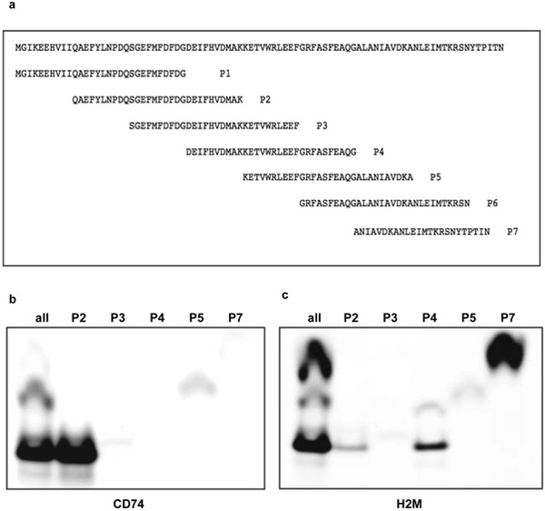
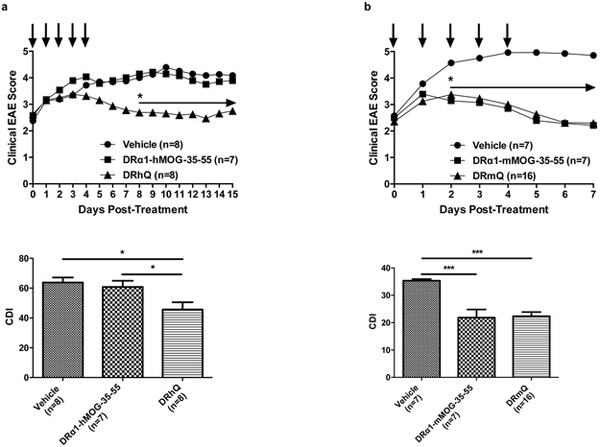
Multiple sclerosis (MS) is a demyelinating and degenerative disease of the central nervous system (CNS) with a strong inflammatory component that affects more than 2 million people worldwide (and at least 400,000 in the United States). In MS, macrophage migration inhibitory factor (MIF) and D-dopachrome tautomerase (D-DT) enhance the inflammatory event as a result of their interaction with their cognate receptor CD74. Therefore, the search for new agents aimed at blocking this interaction is critical for therapeutic purposes and will be of paramount importance for the treatment of MS. DRα1-MOG-35-55 constructs have been demonstrated to be effective in the treatment of experimental autoimmune encephalomyelitis (EAE) a mouse model for MS. This effect is directly correlated with the binding to its cell surface receptor, CD74, apparently preventing or blocking the binding of two inflammatory factors, MIF and D-DT. Here we report that a single amino acid substitution (L50Q) in the DRα1 domain of the human and mouse DRα1-MOG-35-55 constructs (notated as DRhQ and DRmQ, respectively) possessed increased affinity for CD74, a greater capacity to block MIF binding, the ability to inhibit pERK1/2 signaling and increased therapeutic activity in mice with EAE. These data suggest that binding affinity for CD74 could serve as an in vitro indicator of biological potency of DRhQ and thus support its possible clinical utility as an effective therapy for MS and perhaps other diseases in which there is an inflammatory reaction driven by MIF and D-DT.

Keywords: CD74; D-dopachrome tautomerase; EAE; MIF; Multiple sclerosis; pERK1/2.


Conflict of interest statement

Conflict of Interest: Drs. Vandenbark, Offner, Benedek, Meza-Romero and OHSU have a significant financial interest in Artielle ImmunoTherapeutics, Inc., a company that may have a commercial interest in the results of this research and technology. This potential conflict of interest has been reviewed and managed by the OHSU and VA Portland Health Care System Conflict of Interest in Research Committees. The other authors (Grant Gerstner, Gail Kent, Ha Nguyen) declare no conflicts of interest.

Figures

Fig. 1
Fig. 1
Class II α1 domain alignment showing a Q residue at position 18 (indicated by an arrow). The amino acid residue Q18 (nomenclature taken from the sequences reported in protein data bank) is highly conserved among the class II α chains, except with DRαl. Here position 18 was changed from L to Q by site-directed mutagenesis
Fig. 2
Fig. 2
Binding of DRα1-hMOG-35-55, human MHC Class II DR-derived RTL302, human Class II DR-derived RTL312, human Class II-derived DP2- and DP4-derived constructs. In addition, we tested the binding of a mouse MHC Class II-derived RTL551 to coated rhCD74(C27S). Binding experiment was carried out using equimolar concentration (250nm) of each protein ligand (Fig. 2a). Alignment showing the final amino acid sequence of the Q mutants compared to the WT versions created in this work. In addition to the DRα1-hMOG-35-55 and the DRhQ we synthesized the DRα-1mMOG-35-55 and DRmQ. The arrow indicates the positions of the mutagenesis (Fig. 2b)
Fig. 3
Fig. 3
DRα1-derived constructs DRα1-hMOG-35-55, DRα1-mMOG-35-55 and DRhQ were probed with the Fab G4 (Fig. 3a) in order to evaluate immunological differences among them (the characterization of the DRmQ is not shown). Circular dichroism was used to evaluate differences in secondary structure content as described before (Fig. 3b)
Fig. 4
Fig. 4
Strategy was designed to locate the DRα1 binding site for CD74. Overlapping peptides used in this experiment to identify the binding region to CD74 (Fig. 4a). Mouse CD74 or mouse H2M were immunoprecipitated with specific monoclonal antibodies adsorbed to Protein A/G beads and then a pool of the overlapping peptides or individual peptides were added. Immune complexes were washed extensively and bound peptides were eluted with electrophoresis sample buffer containing 1% SDS. Eluted material was analyzed by electrophoresis by SDS-peptide gels in Tris-Tricine. Gels were then scanned for the fluorophore in a BioRad Molecular Imager FX (Fig. 4b and 4c)
Fig. 5
Fig. 5
Two of the overlapping peptides were tested individually for their ability to bind immunoprecipitated mouse CD74 or Fab G4 adsorbed to Protein A/G beads and Protein L beads, respectively. Only P2 was able to bind rhCD74 and Fab G4. P7 was unable to bind to any of the targets (Fig. 5a). Graphic representation of the P2 location in the DRα1 domain structure. A schematic view of the DRα1 domain (colored in cyan) with the P2 peptide location (colored in green) (Fig. 5b). Competition experiment showed that DRα1-hMOG-35-55 was able to outcompete the binding of the FITC- labeled P2 peptide to immunoprecipitated CD74 with a relative affinity (KD) of 750nM (Fig. 5c)
Fig. 6
Fig. 6
Constructs DRα1-hMOG-35-55 and the DRhQ (Fig. 6a) were assessed for direct binding to rhCD74 onto ELISA plates by direct binding assay with or without G4 Fab during the binding. The KD calculated polypeptides were 0.65uM for the DRα1-hMOG-35-55 and 0.089 for the DRhQ. In a competitive experiment DRhQ showed higher activity against rhMIF to bind CD74 compared to the DRα1-hMOG 35-55 (Fig. 6b) and Table 1
Fig. 7
Fig. 7
C57BL/6 WT male mice between 8 and 12wks of age were purchased from the Jackson Laboratory and immunized as described in Material and Methods. DRα1-hMOG-35-55 and DRhQ (Fig. 7a) or DRα1-mMOG-35-55 and DRmQ (Fig. 7b) proteins (100 μg in 0.1ml) was injected s.c. daily for 5 days beginning at an EAE score of ≥2.0 and the mice were scored for clinical signs of EAE. Mean EAE scores and SDs for mouse groups were calculated for each day from day 8 through day 27 post-immunization and summed for each mouse by numerically integrating the EAE score curve over the entire experiment (CDI, represents total disease load)
Fig. 8
Fig. 8
ERK1/2 phosphorylation assay. 2 million cells from EAE mice were treated with vehicle, DRhQ or DRα1-hMOG-35-55 for 30 min and then cells were lysed in the presence of protease and phosphatase inhibitor. Supernatants were analyzed by electrophoresis and WB to evaluate P-ERK1/2 and total ERK1/2. DRα1-hMOG-35-55 and DRhQ were able to downregulate ERK1/2 phosphorylation in vitro
Fig. 9
Fig. 9
Ribbon-rendered structure model. Shows the position of relevant amino acid residues in the DRhQ construct described in this work and that were shown by the docking model to contact CD74. The Q is highlighted with a stick-drawn side chain at position 14. The antigenic MOG peptide is shown in white and the side chain for W3 in the MOG peptide is displayed (Fig. 9a). Several α1 domains from molecules described in Fig. 1 were recovered from the Protein Data Bank and their β-strand 1-loop- β-strand 2 regions aligned using PyMOL Molecular Graphics System, Version 2.1 INTEL-10.25.19 Schrödinger, LLC. Position 14 has been highlighted as sticks. The PDB entries are shown in the inset (Fig. 9b). Note: the amino acid residues numbering in the Fig. 1 differs from the numbering in the crystal structures: Q18 in Figure 1 corresponds to amino acid residue 14 in most of the crystal structures and is located at the end of the β-strand 1 of the domain

Similar articles

Cited by

References

    1. Benedek G et al. (2013) Partial MHC class II constructs inhibit MIF/CD74 binding and downstream effects Eur J Immunol 43:1309–1321 doi:10.1002/eji.201243162 - DOI - PMC - PubMed
    1. Benedek G, Meza-Romero R, Jordan K, Keenlyside L, Offner H, Vandenbark AA (2015) HLA-DRalpha1-mMOG-35-55 treatment of experimental autoimmune encephalomyelitis reduces CNS inflammation, enhances M2 macrophage frequency, and promotes neuroprotection J Neuroinflammation 12:123 doi:10.1186/s12974-015-0342-4 - DOI - PMC - PubMed
    1. Benedek G et al. (2017) MIF and D-DT are potential disease severity modifiers in male MS subjects Proc Natl Acad Sci U S A 114:E8421–E8429 doi:10.1073/pnas.1712288114 - DOI - PMC - PubMed
    1. Chitnis T (2007) The role of CD4 T cells in the pathogenesis of multiple sclerosis Int Rev Neurobiol 79:43–72 doi:10.1016/S0074-7742(07)79003-7 - DOI - PMC - PubMed
    1. Frohman EM, Racke MK, Raine CS (2006) Multiple sclerosis--the plaque and its pathogenesis The New England journal of medicine 354:942–955 doi:10.1056/NEJMra052130 - DOI - PubMed

Publication types

MeSH terms

Substances

LinkOut - more resources