

Relative skeletal distribution of proliferating marrow in the adult dog determined using 3'-deoxy-3'-[18 F]fluorothymidine
- PMID: 30353574
- PMCID: PMC6587773
- DOI: 10.1111/ahe.12410
Relative skeletal distribution of proliferating marrow in the adult dog determined using 3'-deoxy-3'-[18 F]fluorothymidine
Abstract
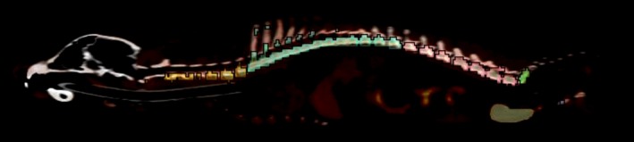
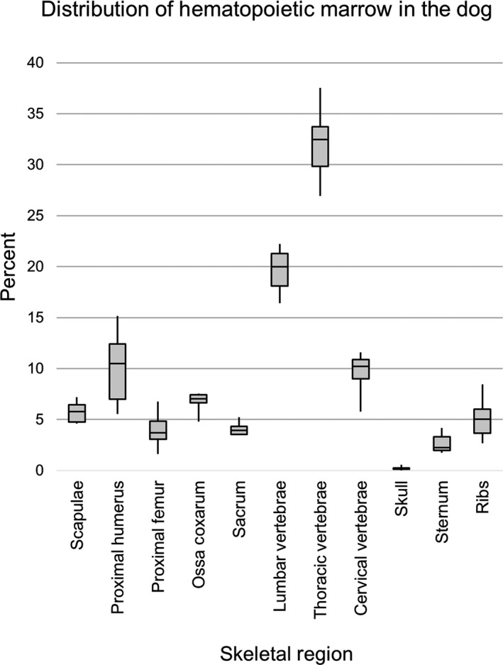
3'-deoxy-3'-[18 F]fluorothymidine (18 FLT) is a radiopharmaceutical tracer used with positron emission tomography (PET), often in combination with computed tomography (CT), to image DNA synthesis, and thus, cellular proliferation. Characteristic accumulation of the tracer within haematopoietic bone marrow provides a noninvasive means to assess marrow activity and distribution throughout the living animal. The present study utilizes three-dimensional analysis of 18 FLT-PET/CT scans to quantify the relative skeletal distribution of active marrow by anatomic site in the dog. Scans were performed on six healthy, adult (3-6 years of age), mixed-breed dogs using a commercially available PET/CT scanner consisting of a 64-slice helical CT scanner combined with an integrated four ring, high-resolution LSO PET scanner. Regions of interest encompassing 11 separate skeletal regions (skull, cervical vertebral column, thoracic vertebral column, lumbar vertebral column, sacrum, ribs, sternum, scapulae, proximal humeri, ossa coxarum, and proximal femora) were manually drawn based on CT images and thresholded by standardized uptake value to delineate bone marrow activity. Activity within each skeletal region was then divided by the total skeletal activity to derive the per cent of overall marrow activity within an individual site. The majority of proliferative marrow was located within the vertebral column. Of the sites traditionally accessed clinically for marrow sampling, the proximal humerus contained the largest percentage, followed by the ossa coxarum, proximal femur, and sternum, respectively. This information may be used to guide selection of traditional marrow sampling sites as well as inform efforts to spare important sites of haematopoiesis in radiation therapy planning.
Keywords: bone marrow; computed tomography; dogs; haematopoiesis; positron emission tomography; radiopharmaceuticals.
© 2018 The Authors. Anatomia, Histologia, Embryologia Published by Blackwell Verlag GmbH.
Conflict of interest statement
The University of Tennessee maintains research collaborations with Siemens Healthineers.
Figures

Similar articles
-
Whole-body biodistribution of 3'-deoxy-3'-[(18) f]fluorothymidine ((18) FLT) in healthy adult cats.Vet Radiol Ultrasound. 2013 May-Jun;54(3):299-306. doi: 10.1111/vru.12024. Epub 2013 Mar 7. Vet Radiol Ultrasound. 2013. PMID: 23464567
-
More advantages in detecting bone and soft tissue metastases from prostate cancer using 18F-PSMA PET/CT.Hell J Nucl Med. 2019 Jan-Apr;22(1):6-9. doi: 10.1967/s002449910952. Epub 2019 Mar 7. Hell J Nucl Med. 2019. PMID: 30843003
-
Distribution Atlas of Proliferating Bone Marrow in Non-Small Cell Lung Cancer Patients Measured by FLT-PET/CT Imaging, With Potential Applicability in Radiation Therapy Planning.Int J Radiat Oncol Biol Phys. 2015 Aug 1;92(5):1035-1043. doi: 10.1016/j.ijrobp.2015.04.027. Epub 2015 Apr 22. Int J Radiat Oncol Biol Phys. 2015. PMID: 26194679
-
PET-Computed Tomography in Veterinary Medicine.Vet Clin North Am Small Anim Pract. 2016 May;46(3):515-33, vi. doi: 10.1016/j.cvsm.2015.12.008. Vet Clin North Am Small Anim Pract. 2016. PMID: 27068445 Review.
-
FLT: measuring tumor cell proliferation in vivo with positron emission tomography and 3'-deoxy-3'-[18F]fluorothymidine.Semin Nucl Med. 2007 Nov;37(6):429-39. doi: 10.1053/j.semnuclmed.2007.08.001. Semin Nucl Med. 2007. PMID: 17920350 Review.
Cited by
-
Advanced Cancer Imaging Applied in the Comparative Setting.Front Oncol. 2020 Feb 7;10:84. doi: 10.3389/fonc.2020.00084. eCollection 2020. Front Oncol. 2020. PMID: 32117739 Free PMC article.
References
-
- Agool, A. , Schot, B. W. , Jager, P. L. , & Vellenga, E. (2006). 18F‐FLT PET in hematologic disorders: A novel technique to analyze the bone marrow compartment. Journal of Nuclear Medicine, 47(10), 1592–1598. 47/10/1592 [pii] - PubMed
-
- Agool, A. , Slart, R. H. , Kluin, P. M. , de Wolf, J. T. , Dierckx, R. A. , & Vellenga, E. (2011). F‐18 FLT PET: A noninvasive diagnostic tool for visualization of the bone marrow compartment in patients with aplastic anemia: A pilot study. Clinical Nuclear Medicine, 36(4), 286–289. 10.1097/RLU.0b013e31820aa1a1 - DOI - PubMed
-
- Akula, M. , Collier, T. , Kabalka, G. , Wall, J. , Kennel, S. , Stuckey, A. , & LeBlanc, A. (2010). Microfluidic synthesis of [18F]FLT. Journal of Nuclear Medicine, 51(Suppl. 2), 1473.
MeSH terms
Substances
LinkOut - more resources
Full Text Sources
