. 2019 Jan;110(1):334-344.
doi: 10.1111/cas.13847. Epub 2018 Dec 13.

Conophylline suppresses pancreatic cancer desmoplasia and cancer-promoting cytokines produced by cancer-associated fibroblasts

Affiliations

Conophylline suppresses pancreatic cancer desmoplasia and cancer-promoting cytokines produced by cancer-associated fibroblasts

Norihiro Ishii et al. Cancer Sci. 2019 Jan.

Abstract

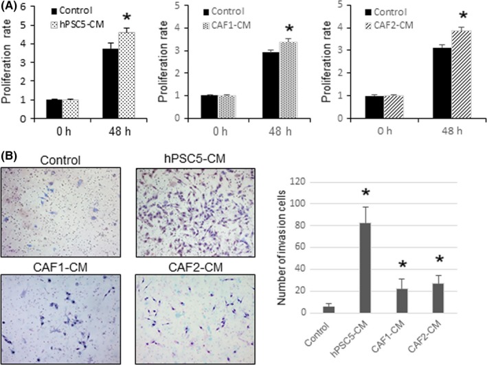
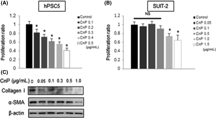
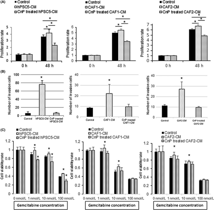
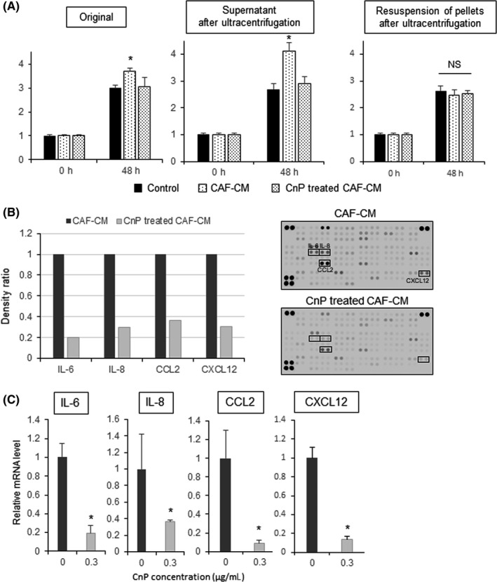
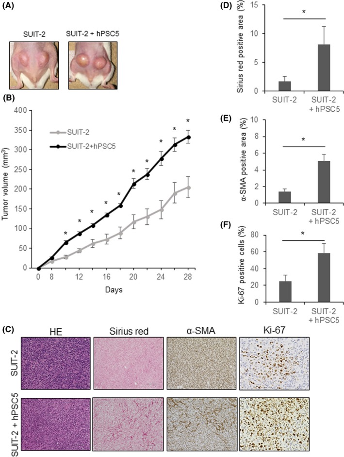
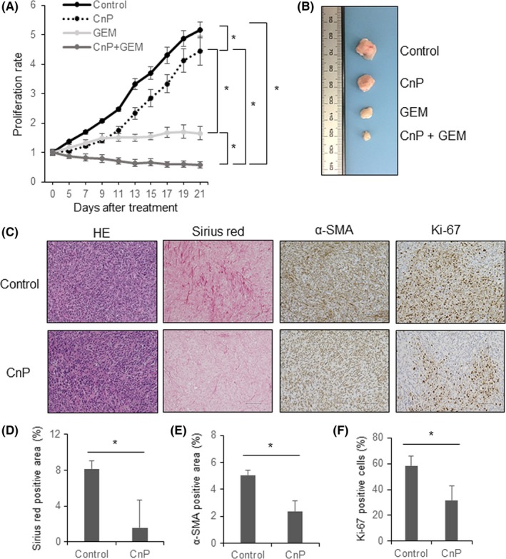
Despite recent advances in cancer treatment, pancreatic cancer is a highly malignant tumor type with a dismal prognosis and it is characterized by dense desmoplasia in the cancer tissue. Cancer-associated fibroblasts (CAF) are responsible for this fibrotic stroma and promote cancer progression. We previously reported that a novel natural compound conophylline (CnP) extracted from the leaves of a tropical plant reduced liver and pancreatic fibrosis by suppression of stellate cells. However, there have been no studies to investigate the effects of CnP on CAF, which is the aim of this work. Here, we showed that CAF stimulated indicators of pancreatic cancer malignancy, such as proliferation, invasiveness, and chemoresistance. We also showed that CnP suppressed CAF activity and proliferation, and inhibited the stimulating effects of CAF on pancreatic cancer cells. Moreover, CnP strongly decreased the various cytokines involved in cancer progression, such as interleukin (IL)-6, IL-8, C-C motif chemokine ligand 2 (CCL2), and C-X-C motif chemokine ligand 12 (CXCL12), secreted by CAF. In vivo, CAF promoted tumor proliferation and desmoplastic formation in a mouse xenograft model, CnP reduced desmoplasia of tumors composed of pancreatic cancer cells + CAF, and combination therapy of CnP with gemcitabine remarkably inhibited tumor proliferation. Our findings suggest that CnP is a promising therapeutic strategy of combination therapy with anticancer drugs to overcome refractory pancreatic cancers.

Keywords: conophylline; cytokine; fibroblast; microenvironment; stellate cell.


Figures

Figure 1
Figure 1
Effects of cancer‐associated fibroblasts conditioned medium (CAF‐CM) on pancreatic cancer cells (SUIT‐2 cells). A, Conditioned media from hPSC5, CAF1, and CAF2 enhance the proliferation of SUIT‐2 cells. B, Conditioned media from hPSC5, CAF1, and CAF2 enhance the invasive ability of SUIT‐2 cells. Representative photomicrographs are shown to the left and quantitative plots are shown to the right. *P < .05 (vs control). hPSC5, human pancreatic stellate cell line
Figure 2
Figure 2
Effects of conophylline (CnP) on cancer‐associated fibroblasts (CAF) and SUIT‐2 cells. A, CnP inhibits the proliferation of hPSC5 concentration‐dependently. B, CnP inhibits the proliferation of SUIT‐2 cells only at concentrations >1.0 μg/mL. C, Collagen I and α‐smooth muscle actin (SMA) expression in hPSC5 cells treated by CnP is evaluated by western blotting. β‐Actin was used as the internal control. Representative blots are shown. *P < .05 (vs control). hPSC5, human pancreatic stellate cell line
Figure 3
Figure 3
Conophylline (CnP) inhibits the promoting effects of cancer‐associated fibroblasts (CAF) on pancreatic cancer cells. A, Proliferation‐enhancing effects of CAF‐conditioned medium (CM) on SUIT‐2 are eliminated by CnP‐treated CAF‐CM. B, Invasiveness‐enhancing effects of CAF‐CM on SUIT‐2 are decreased by CnP‐treated CAF‐CM. C, Enhanced gemcitabine resistance of SUIT‐2 cells by CAF‐CM is improved by using CnP‐treated CAF‐CM. Left, hPSC5; middle, CAF1; right, CAF2, *P < .05 (vs control). hPSC5, human pancreatic stellate cell line
Figure 4
Figure 4
Conophylline (CnP) decreases the cytokines produced by cancer‐associated fibroblasts (CAF). A, After ultracentrifugation, supernatants showed the same phenotype as the original solutions. The resuspended pellets did not show this effect. B, Cytokine array comparing CAF‐conditioned medium (CM) and CnP‐treated CAF‐CM. Interleukin (IL)‐6, IL‐8, C‐C motif chemokine ligand 2 (CCL2), and C‐X‐C motif chemokine ligand 12 (CXCL12) were remarkably reduced by CnP treatment. Array images are shown to the right of the plotted data. C, mRNA levels of IL‐6, IL‐8, CCL2, and CXCL12. mRNA expression levels were quantified by RT‐qPCR and were normalized to 18S rRNA levels. *P < .05 (vs control)
Figure 5
Figure 5
Cancer‐associated fibroblasts (CAF) promote tumor proliferation and desmoplastic formation in a mouse xenograft model. A, Representative photographs of tumors consisting of SUIT‐2 cells (LEFT), and SUIT‐2 + hPSC5 cells combined (right). B, Tumor growth curve of SUIT‐2 cells and SUIT‐2 + hPSC5 cells. Tumor growth of SUIT‐2 + hPSC5 cells was much greater compared with that of SUIT‐2 cells alone. C, Representative photomicrographs of histological evaluations of tumor tissue; original magnification (HE, Sirius red and α‐smooth muscle actin [SMA], ×200; scale bar, 100 μm; original magnification [Ki‐67], ×400). D, Sirius red‐positive stained area of tumors consisting of SUIT‐2 + hPSC5 cells was significantly higher than that of SUIT‐2 cells alone. E, α‐SMA‐positive stained area of tumors consisting of SUIT‐2 + hPSC5 cells was significantly higher than that of SUIT‐2 cells alone. F, Proportion of Ki‐67‐positive cells was significantly higher in tumors consisting of SUIT‐2 + hPSC5 cells compared with that of SUIT‐2 cells alone. *P < .05. hPSC5, human pancreatic stellate cell line
Figure 6
Figure 6
Conophylline (CnP) reduces desmoplasia of tumors consisting of SUIT‐2 + hPSC5 cells, and combination therapy with gemcitabine markedly inhibits tumor proliferation. A, Tumor growth curve showing that combination therapy with CnP plus gemcitabine suppresses the growth of tumors consisting of SUIT‐2 + hPSC5 cells to the greatest extent. B, Representative photographs of tumors from the four treatment groups (control, CnP, gemcitabine [GEM], CnP + GEM). C, Representative photomicrographs of histological examinations comparing control and CnP treatment (original magnification, ×200; scale bar, 100 μm); D‐F, CnP treatment significantly reduces Sirius red‐positive staining (D), α‐smooth muscle actin (SMA)‐positive staining (E), and percentage of Ki‐67‐positive cells (F) in tumors consisting of SUIT‐2 + hPSC5 cells. *P < .05. hPSC5, human pancreatic stellate cell line

Similar articles

Cited by

References

    1. Ilic M, Ilic I. Epidemiology of pancreatic cancer. World J Gastroenterol. 2016;22:9694‐9705. - PMC - PubMed
    1. Hidalgo M. Pancreatic cancer. N Engl J Med. 2010;362:1605‐1617. - PubMed
    1. Apte MV, Wilson JS, Lugea A, Pandol SJ. A starring role for stellate cells in the pancreatic cancer microenvironment. Gastroenterology. 2013;144:1210‐1219. - PMC - PubMed
    1. Pan B, Liao Q, Niu Z, Zhou L, Zhao Y. Cancer‐associated fibroblasts in pancreatic adenocarcinoma. Future Oncol. 2015;11:2603‐2610. - PubMed
    1. Hwang RF, Moore T, Arumugam T, et al. Cancer‐associated stromal fibroblasts promote pancreatic tumor progression. Cancer Res. 2008;68:918‐926. - PMC - PubMed

MeSH terms