Review
. 2018 Nov;17(11):1016-1024.
doi: 10.1016/S1474-4422(18)30318-1.

The glymphatic pathway in neurological disorders

Affiliations
Review

The glymphatic pathway in neurological disorders

Martin Kaag Rasmussen et al. Lancet Neurol. 2018 Nov.

Abstract

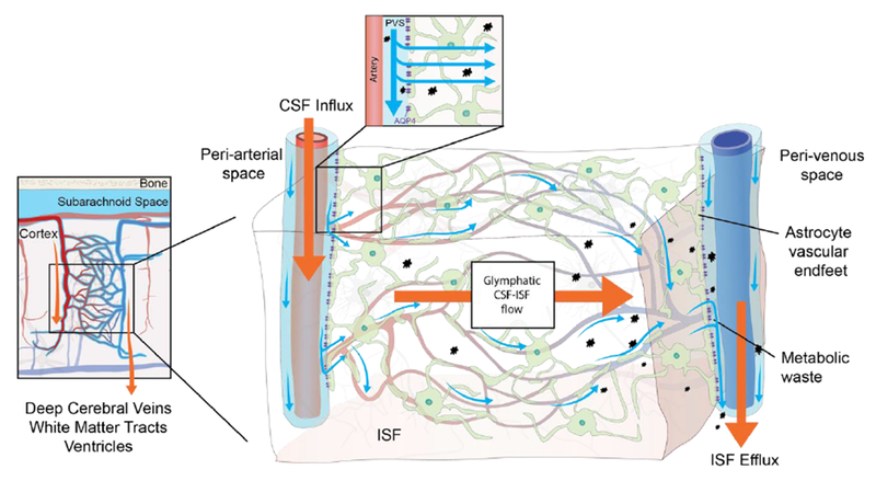
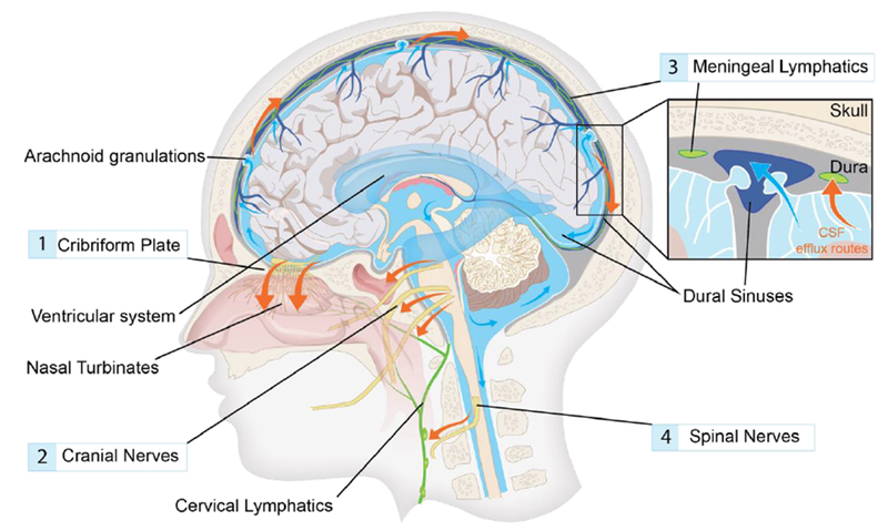
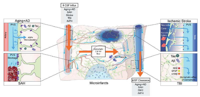
Background: The glymphatic (glial-lymphatic) pathway is a fluid-clearance pathway identified in the rodent brain in 2012. This pathway subserves the flow of CSF into the brain along arterial perivascular spaces and subsequently into the brain interstitium, facilitated by aquaporin 4 (AQP4) water channels. The pathway then directs flow towards the venous perivascular and perineuronal spaces, ultimately clearing solutes from the neuropil into meningeal and cervical lymphatic drainage vessels. In rodents, the glymphatic pathway is predominantly active during sleep, when the clearance of harmful metabolites such as amyloid β (Aβ) increases two-fold relative to the waking state. Glymphatic dysfunction, probably related to perturbed AQP4 expression, has been shown in animal models of traumatic brain injury, Alzheimer's disease, and stroke. The recent characterisations of the glymphatic and meningeal lymphatic systems in rodents and in humans call for revaluation of the anatomical routes for CSF-interstitial fluid flow and the physiological role that these pathways play in CNS health.

Recent developments: Several features of the glymphatic and meningeal lymphatic systems have been shown to be present in humans. MRI scans with intrathecally administered contrast agent show that CSF flows along pathways that closely resemble the glymphatic system outlined in rodents. Furthermore, PET studies have revealed that Aβ accumulates in the healthy brain after a single night of sleep deprivation, suggesting that the human glymphatic pathway might also be primarily active during sleep. Other PET studies have shown that CSF clearance of Aβ and tau tracers is reduced in patients with Alzheimer's disease compared with healthy controls. The observed reduction in CSF clearance was associated with increasing grey-matter concentrations of Aβ in the human brain, consistent with findings in mice showing that decreased glymphatic function leads to Aβ accumulation. Altered AQP4 expression is also evident in brain tissue from patients with Alzheimer's disease or normal pressure hydrocephalus; glymphatic MRI scans of patients with normal pressure hydrocephalus show reduced CSF tracer entry and clearance. WHERE NEXT?: Research is needed to confirm whether specific factors driving glymphatic flow in rodents also apply to humans. Longitudinal imaging studies evaluating human CSF dynamics will determine whether a causal link exists between reduced brain solute clearance and the development of neurodegenerative diseases. Assessment of glymphatic function after stroke or traumatic brain injury could identify whether this function correlates with neurological recovery. New insights into how behaviour and genetics modify glymphatic function, and how this function decompensates in disease, should lead to the development of new preventive and diagnostic tools and novel therapeutic targets.


Conflict of interest statement

Declaration of interests

The authors declare no conflict of interest.

Figures

Figure 1.
Figure 1.. The glymphatic pathway.
Rodent studies have shown that CSF from the subarachnoid space is driven into the perivascular space of major cerebral arteries on the brain surface from where it flows along the artery as it branches into penetrating arteries.,,,A similar pattern of CSF flow has been shown in patients undergoing MRI in combination with intrathecal contrast agent., In these patients it was observed that the CSF contrast agent flows along the large leptomeningeal cerebral arteries in an anterograde fashion, and that presence of contrast agent in the subarachnoid space precedes parenchymal uptake in adjacent brain regions. The microscopic details of CSF flow within the brain thus far all stem from animal research. These studies have shown that the perivascular space runs along the entire penetrating artery, known as the Virchow-Robin space, and continues to follow the vessel as it branches into arterioles and capillaries.,, In the murine brain, CSF influx into the extracellular space happens at every level of the perivascular space after entry to the brain parenchyma and is facilitated by a polarized expression of the AQP4 water channel towards the astrocytic end-feet that line the perivascular space. Whether a similar parenchymal CSF flow occurs in human brain has not yet been proven, but humans also harbor intracerebral perivascular spaces and polarized AQP4 expression towards astrocytic end-feet., The basis of fluid movement within the interstitium is still a matter of debate. Bulk flow clearance of ISF is a long-standing observation, which could be driven by multiple factors such as CSF inflow, arterial pulsatility, hydrostatic pressure gradients between the arterial and venous perivascular spaces, and osmotic gradients. Rodent studies show that ISF and its solutes move towards the venous perivascular space, where the fluid is taken up and drained by convection out of the brain parenchyma. This directional flow removes solutes from the brain parenchyma accumulated during neural activity. CSF, cerebrospinal fluid; ISF interstitial fluid, PVS: perivascular space; AQP4, aquaporin-4.
Figure 2.
Figure 2.. Cerebrospinal fluid efflux in humans.
Cerebrospinal fluid (CSF) produced in the choroid plexus flows from the ventricles to the subarachnoid space of the brain and spinal cord. CSF contained in the subarachnoid space keeps the central nervous system buoyant and serves as a fluid source for glymphatic influx. Egress sites of cranial cerebrospinal fluid (red arrows) fall into three functionally distinct categories, namely the perineural sheaths surrounding cranial and spinal nerves,, dural lymphatic vessels,,, and arachnoid granulations. The contribution and significance of each egress pathway is still a matter of debate. A main perineural egress site in both rodents and human is along the olfactory nerve through the cribriform plate (1) towards lymphatic vessels of the nasal mucosa., From here the CSF is drained to the cervical lymph nodes. Other significant perineural efflux pathways in rodents are the trigeminal, the glossopharyngeal, vagal, and spinal accessory nerves (2). Dural lymphatic vessels have also been shown to carry CSF towards the cervical lymphatic system (3). In rodents, these vessels exit the skull along the pterygopalatine artery, the veins that drain the sigmoid sinus and retroglenoid vein, and the foramina of the cranial nerves., In humans, meningeal lymphatic vessels have been visualized with MRI and were located around the dural sinuses, middle meningeal artery and cribiform plate. Arachnoid granulations are protrusions of the arachnoid membrane where CSF flows into the sagittal sinus, and constitute the only known egress site that drains directly to the blood stream. Traditionally, this site was thought to be the main cerebrospinal fluid egress site, but evidence suggests that under physiological intracranial pressure virtually no CSF leaves to the blood stream. The main egress site of CSF in the spinal cord is along the spinal nerves (4).
Figure 3.
Figure 3.. Glymphatic imaging modalities.
Analysis of the glymphatic pathway can be accomplished through multiple approaches. Ex vivo imaging is performed with micrometer-thick brain sections prepared from animals after injection of tracer into the CSF compartment., This method provides information on parenchymal distribution of CSF in the glymphatic pathway both on a brain-wide and regional scale, as well as at the cellular level, depending on the selected microscopic field of view. Combined with immunohistochemistry, the CSF tracer distribution can be compared to expression patterns of specific molecules e.g. AQP4 polarization towards the astrocytic end-feet, In vivo imaging of the glymphatic pathway has been performed with 2-photon laser scanning microscopy (2PLSM) to map out flow mechanisms in the perivascular space in rodents. Optical access is acquired by surgically implanting an imaging window, i.e. a craniotomy covered with a glass coverslip. Affording high spatial resolution and the utilization of fluorescent tracers along with transgenic labeling of anatomical structures, 2PLSM is ideally suited for detailed study of perivascular space flow patterns and the factors impacting flow.,, For more global approaches, transcranial macroscopic imaging is used in combination with fluorescent CSF tracers. This process is less invasive since imaging is done through the intact skull bone of a mouse; it provides information on CSF flow patterns across the entire dorsal cortex. MRI and PET/CT is used in both basic as well as clinical neuroimaging studies of glymphatic flow. These are the least invasive methods, and the only approaches by which a 4-dimensional view of the CSF flow can be acquired in the entire brain of living animals and humans. ,,, Ultrafast MR encephalography provides 3D imaging of the entire human brain volume in ~100 ms intervals, which provides sufficient temporal resolution to study pulsations propagating in brain parenchyma. The left panel and the two middle panels show representative images from mouse brain obtained by imaging methods only applicable in animal research. The right panel displays neuroimaging of glymphatic function in human brain, thus presenting techniques that are applicable in both clinical and basic science. [Left panel ex vivo section is reprinted with permission from Kress et al., right panel MRI image is reprinted with permission from Ringstad et al.]
Figure 4.
Figure 4.. Pathological changes to the glymphatic pathway.
Aging and several diseases have been associated with a decrease in CSF influx to the glymphatic pathway and/or reduced clearance efficacy both in animals and in humans. In aging mice, the flow changes are likely caused by reduced vascular compliance, increased AQP4 expression and AQP4 mislocalization away from the astrocytic end-feet, which all cause reduced parenchymal influx of CSF. In a human postmortem study, AQP4 expression increased with age, albeit without AQP4 mislocalization. In murine models of AD, soluble and insoluble Aβ plaques provoke AQP4 mislocalization and impaired CSF influx., In AD patients, CSF clearance rate is reduced and exhibits an inverse relationship with Aβ levels. Post-mortem studies of AD patients identified AQP4 mislocalization and an increase in total AQP4 expression in AD patients compared to non-AD subjects. In hemorrhagic stroke in mice and gyrenchephalic non-human primates, blood components leaking into the PVS, especially fibrin/fibrinogen deposits, occlude the PVS, which leads to reduced CSF influx. In rodent models of ischemic stroke, necrotic cores are formed within the brain parenchyma, around which reactive astrocytes create a barrier (glial scar) to contain the injury and the toxic agents that form upon necrosis. Contents of the necrotic core leak through the permeable glial scar into the PVS. In mice, cerebral microinfarcts lead to a transient global reduction in glymphatic influx, and prolonged trapping of solutes within the infarct cores, probably due to reduced interstitial fluid turnover. TBI in mice leads to reduced glymphatic clearance, and biomarkers of the injured parenchyma are transported through the glymphatic pathway towards the cervical lymphatic system. In iNPH patients, glymphatic function is broadly impaired and characterized by both a delayed influx and a reduced clearance rate following intrathecal contrast injection. In rat models of diabetes mellitus type 2 (DM2), CSF tracers remain trapped within the brain parenchyma for prolonged periods, suggesting that perivenous efflux is decreased. This finding has not yet been replicated in humans, but we speculate that reduced brain clearance could contribute to the cognitive decline that is often seen in DM2 patients. CSF, cerebrospinal fluid; ISF interstitial fluid, PVS: perivascular space; AQP4, aquaporin-4, AD, Alzheimer’s Disease; SAH, subarachnoid hemorrhage; TBI, traumatic brain injury; iNPH, idiopathic normalpressure hydrocephalus; DM2, diabetes mellitus type 2; GFAP, glial fibrillary acidic protein; S100B, S100 calcium binding protein; NSE, neuron-specific enolase.

Comment in

  • Making Maiken Nedergaard.
    Burton A. Burton A. Lancet Neurol. 2018 Nov;17(11):935. doi: 10.1016/S1474-4422(18)30365-X. Lancet Neurol. 2018. PMID: 30353865 No abstract available.

References

    1. Pollay M. The function and structure of the cerebrospinal fluid outflow system. Cerebrospinal Fluid Res 2010; 7: 9. - PMC - PubMed
    1. Brown RH, Al-Chalabi A. Amyotrophic Lateral Sclerosis. N Engl J Med 2017; 377(2): 162–72. - PubMed
    1. Sevigny J, Chiao P, Bussiere T, et al. The antibody aducanumab reduces Abeta plaques in Alzheimer’s disease. Nature 2016; 537(7618): 50–6. - PubMed
    1. Peng C, Gathagan RJ, Covell DJ, et al. Cellular milieu imparts distinct pathological alpha-synuclein strains in alpha-synucleinopathies. Nature 2018; 557(7706): 558–63. - PMC - PubMed
    1. Labbadia J, Morimoto RI. The biology of proteostasis in aging and disease. Annu Rev Biochem 2015; 84: 435–64. - PMC - PubMed

Publication types

MeSH terms