

A momentum-based diffeomorphic demons framework for deformable MR-CT image registration
- PMID: 30353886
- PMCID: PMC9136583
- DOI: 10.1088/1361-6560/aae66c
A momentum-based diffeomorphic demons framework for deformable MR-CT image registration
Abstract
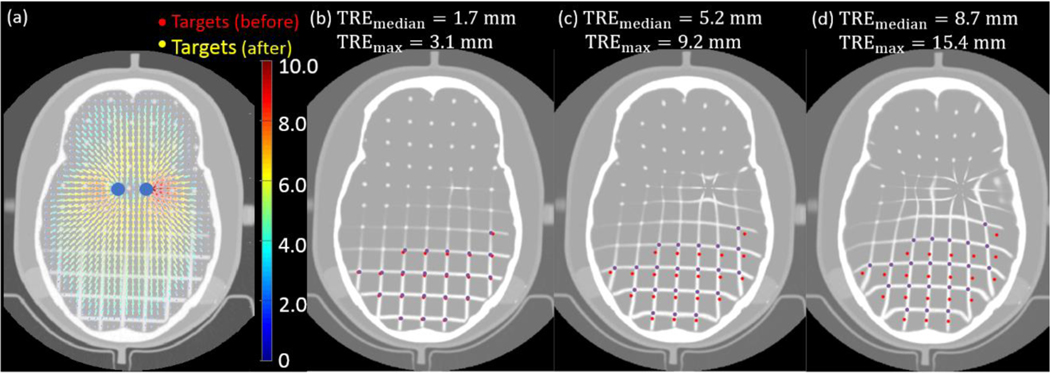
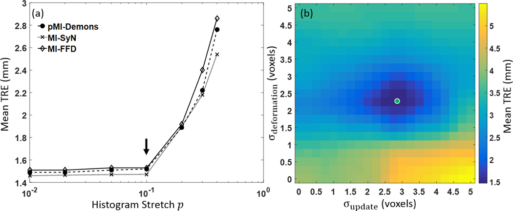
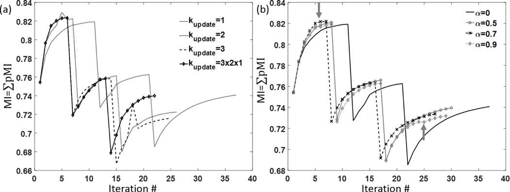
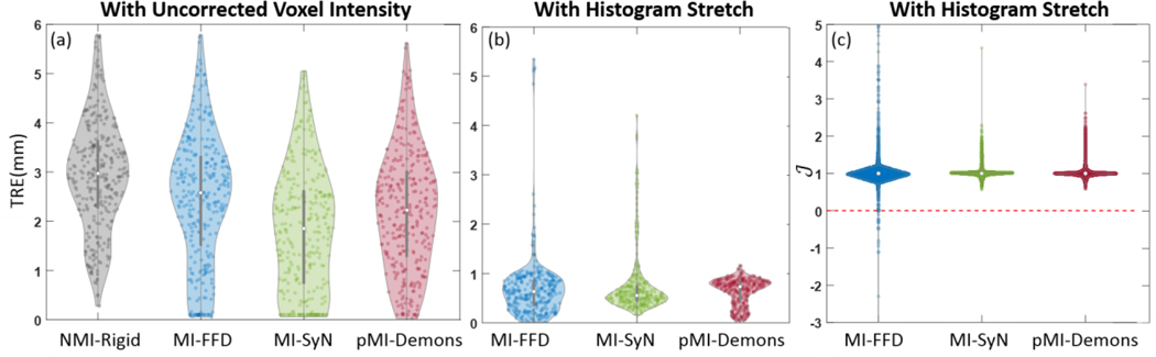
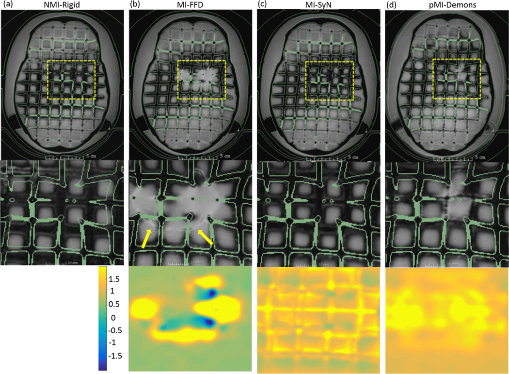
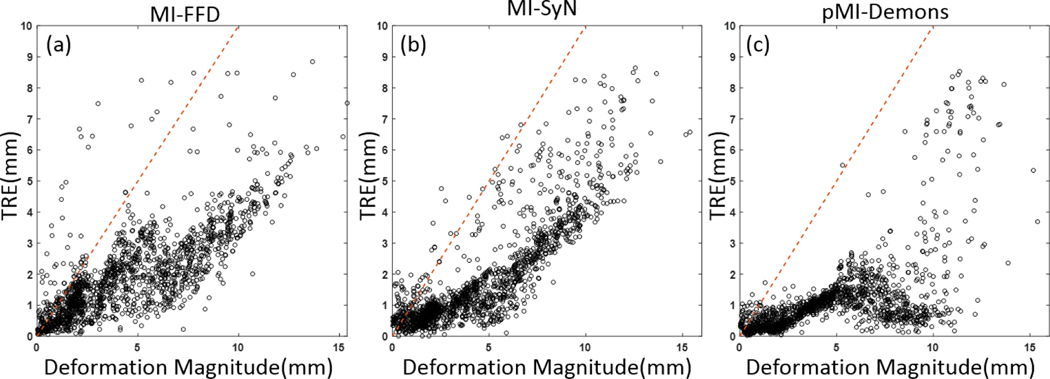
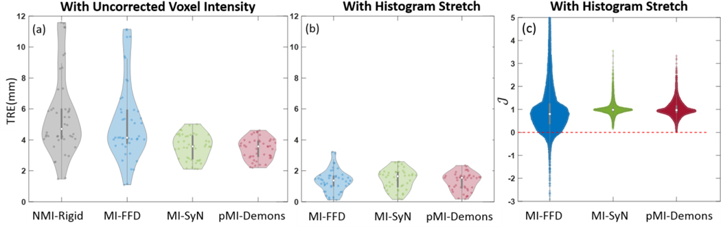
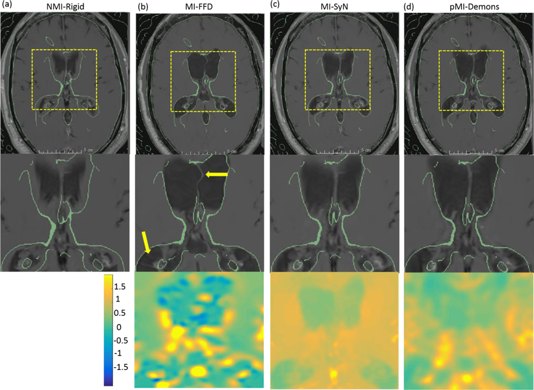
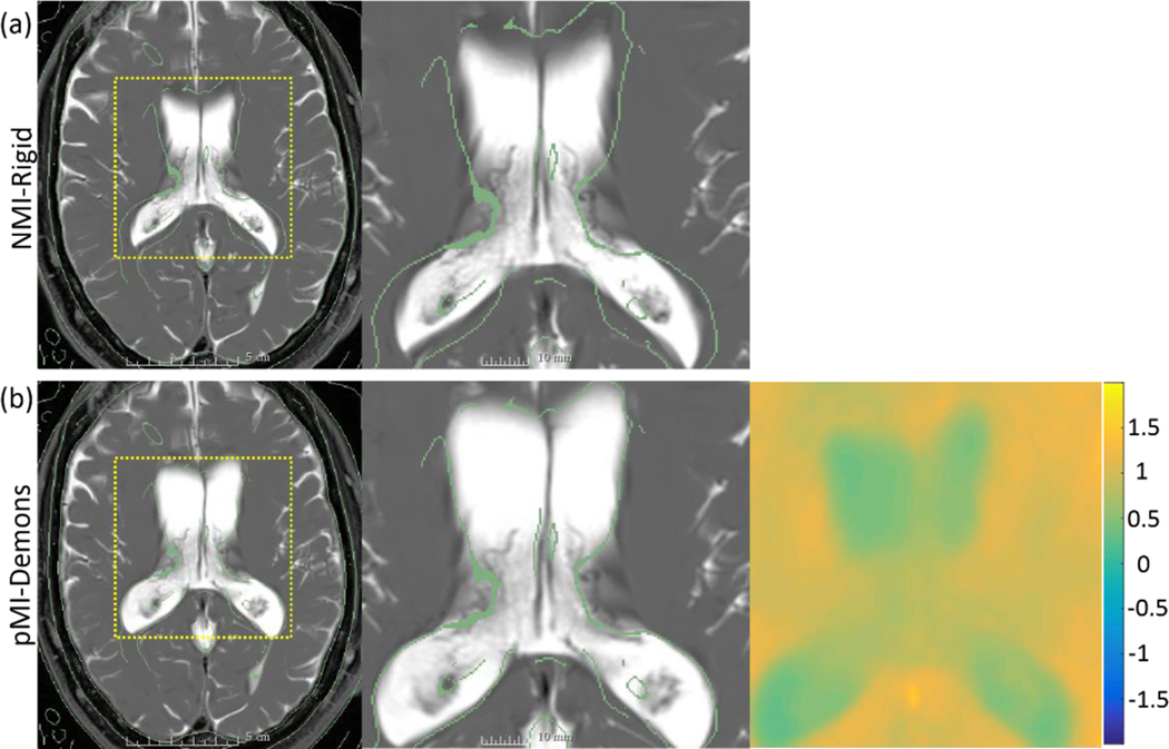
Neuro-navigated procedures require a high degree of geometric accuracy but are subject to geometric error from complex deformation in the deep brain-e.g. regions about the ventricles due to egress of cerebrospinal fluid (CSF) upon neuroendoscopic approach or placement of a ventricular shunt. We report a multi-modality, diffeomorphic, deformable registration method using momentum-based acceleration of the Demons algorithm to solve the transformation relating preoperative MRI and intraoperative CT as a basis for high-precision guidance. The registration method (pMI-Demons) extends the mono-modality, diffeomorphic form of the Demons algorithm to multi-modality registration using pointwise mutual information (pMI) as a similarity metric. The method incorporates a preprocessing step to nonlinearly stretch CT image values and incorporates a momentum-based approach to accelerate convergence. Registration performance was evaluated in phantom and patient images: first, the sensitivity of performance to algorithm parameter selection (including update and displacement field smoothing, histogram stretch, and the momentum term) was analyzed in a phantom study over a range of simulated deformations; and second, the algorithm was applied to registration of MR and CT images for four patients undergoing minimally invasive neurosurgery. Performance was compared to two previously reported methods (free-form deformation using mutual information (MI-FFD) and symmetric normalization using mutual information (MI-SyN)) in terms of target registration error (TRE), Jacobian determinant (J), and runtime. The phantom study identified optimal or nominal settings of algorithm parameters for translation to clinical studies. In the phantom study, the pMI-Demons method achieved comparable registration accuracy to the reference methods and strongly reduced outliers in TRE (p [Formula: see text] 0.001 in Kolmogorov-Smirnov test). Similarly, in the clinical study: median TRE = 1.54 mm (0.83-1.66 mm interquartile range, IQR) for pMI-Demons compared to 1.40 mm (1.02-1.67 mm IQR) for MI-FFD and 1.64 mm (0.90-1.92 mm IQR) for MI-SyN. The pMI-Demons and MI-SyN methods yielded diffeomorphic transformations (J > 0) that preserved topology, whereas MI-FFD yielded unrealistic (J < 0) deformations subject to tissue folding and tearing. Momentum-based acceleration gave a ~35% speedup of the pMI-Demons method, providing registration runtime of 10.5 min (reduced to 2.2 min on GPU), compared to 15.5 min for MI-FFD and 34.7 min for MI-SyN. The pMI-Demons method achieved registration accuracy comparable to MI-FFD and MI-SyN, maintained diffeomorphic transformation similar to MI-SyN, and accelerated runtime in a manner that facilitates translation to image-guided neurosurgery.
Figures

Similar articles
-
MIND Demons: Symmetric Diffeomorphic Deformable Registration of MR and CT for Image-Guided Spine Surgery.IEEE Trans Med Imaging. 2016 Nov;35(11):2413-2424. doi: 10.1109/TMI.2016.2576360. Epub 2016 Jun 2. IEEE Trans Med Imaging. 2016. PMID: 27295656 Free PMC article.
-
MIND Demons for MR-to-CT Deformable Image Registration In Image-Guided Spine Surgery.Proc SPIE Int Soc Opt Eng. 2016 Feb 27;9786:97860H. doi: 10.1117/12.2208621. Epub 2016 Mar 18. Proc SPIE Int Soc Opt Eng. 2016. PMID: 27330239 Free PMC article.
-
Performance evaluation of MIND demons deformable registration of MR and CT images in spinal interventions.Phys Med Biol. 2016 Dec 7;61(23):8276-8297. doi: 10.1088/0031-9155/61/23/8276. Epub 2016 Nov 3. Phys Med Biol. 2016. PMID: 27811396 Free PMC article.
-
Normal vibration distribution search-based differential evolution algorithm for multimodal biomedical image registration.Neural Comput Appl. 2023 May 30:1-23. doi: 10.1007/s00521-023-08649-z. Online ahead of print. Neural Comput Appl. 2023. PMID: 37362574 Free PMC article. Review.
-
Comparison of physics-based deformable registration methods for image-guided neurosurgery.Front Digit Health. 2023 Dec 8;5:1283726. doi: 10.3389/fdgth.2023.1283726. eCollection 2023. Front Digit Health. 2023. PMID: 38144260 Free PMC article. Review.
Cited by
-
Deformable MR-CT image registration using an unsupervised, dual-channel network for neurosurgical guidance.Med Image Anal. 2022 Jan;75:102292. doi: 10.1016/j.media.2021.102292. Epub 2021 Oct 29. Med Image Anal. 2022. PMID: 34784539 Free PMC article.
-
Joint synthesis and registration network for deformable MR-CBCT image registration for neurosurgical guidance.Phys Med Biol. 2022 Jun 10;67(12):10.1088/1361-6560/ac72ef. doi: 10.1088/1361-6560/ac72ef. Phys Med Biol. 2022. PMID: 35609586 Free PMC article.
-
A comprehensive comparative study of generative adversarial network architectures for synthetic computed tomography generation in the abdomen.Med Phys. 2025 Aug;52(8):e18038. doi: 10.1002/mp.18038. Med Phys. 2025. PMID: 40804793 Free PMC article.
References
-
- Brun CC, Lepore N, Pennec X, Chou Y-Y, Lee AD, de Zubicaray G, McMahon KL, Wright MJ, Gee JC and Thompson PM 2011. A nonconservative Lagrangian framework for statistical fluid registration-SAFIRA IEEE Trans Med Imaging 30 184–202 - PubMed
-
- Cao K, Du K, Ding K, Reinhardt JM and Christensen GE Regularized Nonrigid Registration of Lung CT Images by Preserving Tissue Volume and Vesselness Measure 12
-
- Choi Y and Lee S 2000. Injectivity Conditions of 2D and 3D Uniform Cubic B-Spline Functions Graphical Models 62 411–27
Publication types
MeSH terms
Grants and funding
LinkOut - more resources
Full Text Sources
Medical
Miscellaneous