

Using C-Arm X-ray images from marker insertion to confirm the gold fiducial marker identification in an MRI-only prostate radiotherapy workflow
- PMID: 30354010
- PMCID: PMC6236813
- DOI: 10.1002/acm2.12478
Using C-Arm X-ray images from marker insertion to confirm the gold fiducial marker identification in an MRI-only prostate radiotherapy workflow
Abstract
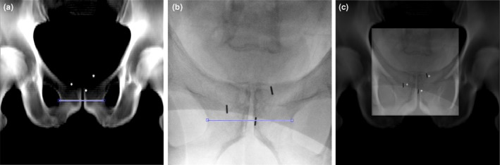
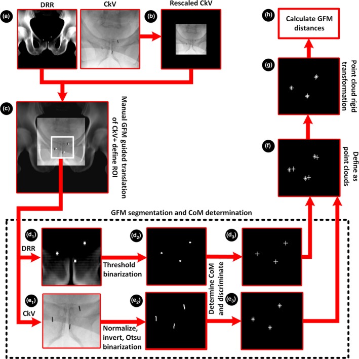
Prostate cancer radiotherapy workflows, solely based on magnetic resonance imaging (MRI), are now in clinical use. In these workflows, intraprostatic gold fiducial markers (GFM) show similar signal behavior as calcifications and bleeding in T2-weighted MRI-images. Accurate GFM identification in MRI-only radiotherapy workflows is therefore a major challenge. C-arm X-ray images (CkV-images), acquired at GFM implantation, could provide GFM position information and be used to confirm correct identification in T2-weighted MRI-images. This would require negligible GFM migration between implantation and MRI-imaging. Marker migration was therefore investigated. The aim of this study was to show the feasibility of using CkV-images to confirm GFM identification in an MRI-only prostate radiotherapy workflow. An anterior-posterior digitally reconstructed radiograph (DRR)-image and a mirrored posterior-anterior CkV-image were acquired two weeks apart for 16 patients in an MRI-only radiotherapy workflow. The DRR-image originated from synthetic CT-images (created from MRI-images). A common image geometry was defined between the DRR- and CkV-image for each patient. A rigid registration between the GFM center of mass (CoM) coordinates was performed and the distance between each of the GFM in the DRR- and registered CkV-image was calculated. The same methodology was used to assess GFM migration for 31 patients in a CT-based radiotherapy workflow. The distance calculated was considered a measure of GFM migration. A statistical test was performed to assess any difference between the cohorts. The mean absolute distance difference for the GFM CoM between the DRR- and CkV-image in the MRI-only cohort was 1.7 ± 1.4 mm. The mean GFM migration was 1.2 ± 0.7 mm. No significant difference between the measured total distances of the two cohorts could be detected (P = 0.37). This demonstrated that, a C-Arm X-ray image acquired from the GFM implantation procedure could be used to confirm GFM identification from MRI-images. GFM migration was present but did not constitute a problem.
Keywords: MRI-only prostate; MRI-only radiotherapy; gold fiducial markers; synthetic CT.
© 2018 The Authors. Journal of Applied Clinical Medical Physics published by Wiley Periodicals, Inc. on behalf of American Association of Physicists in Medicine.
Figures

References
-
- Tenhunen M, Korhonen J, Kapanen M, et al. MRI‐only based radiation therapy of prostate cancer: workflow and early clinical experience. Acta Oncol. 2018;57:902–907. - PubMed
-
- Roberson PL, McLaughlin PW, Narayana V, Troyer S, Hixson GV, Kessler ML. Use and uncertainties of mutual information for computed tomography/magnetic resonance (CT/MR) registration post permanent implant of the prostate. Med Phys. 2005;32:473–482. - PubMed
-
- Köhler M, Vaara T, Van Grootel M, Hoogeveen R, Kemppainen R, Renisch S. MR‐Only Simulation for Radiotherapy Planning Treatment Planning. White paper: Philips MRCAT for prostate dose calculations using only MRI data; 2015:1–16. http://www.philips.se.
MeSH terms
Substances
Grants and funding
LinkOut - more resources
Full Text Sources
Medical
