. 2018 Oct;72(4):909-917.
doi: 10.1161/HYPERTENSIONAHA.118.11666.

Extracellular Ubiquitin(1-76) and Ubiquitin(1-74) Regulate Cardiac Fibroblast Proliferation

Affiliations

Extracellular Ubiquitin(1-76) and Ubiquitin(1-74) Regulate Cardiac Fibroblast Proliferation

Edwin K Jackson et al. Hypertension. 2018 Oct.

Abstract

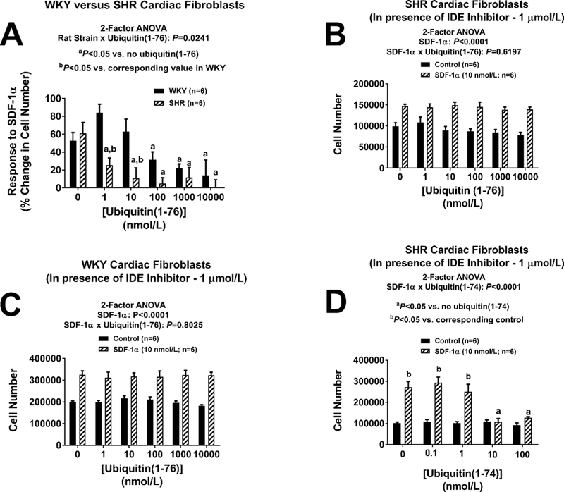
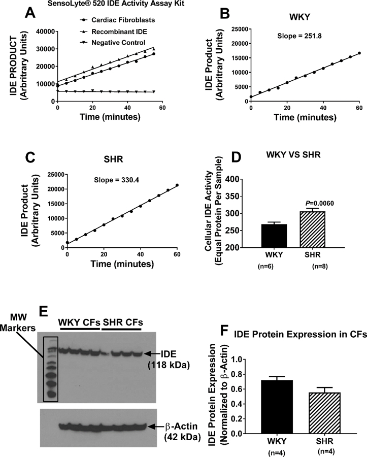
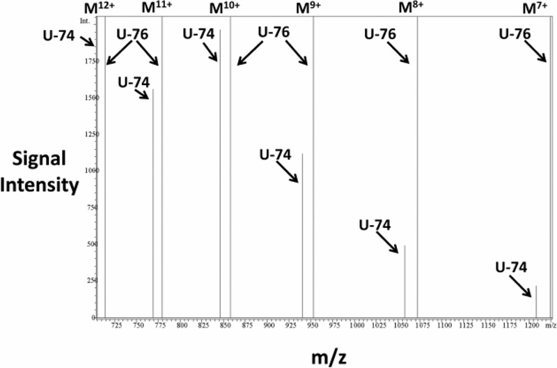
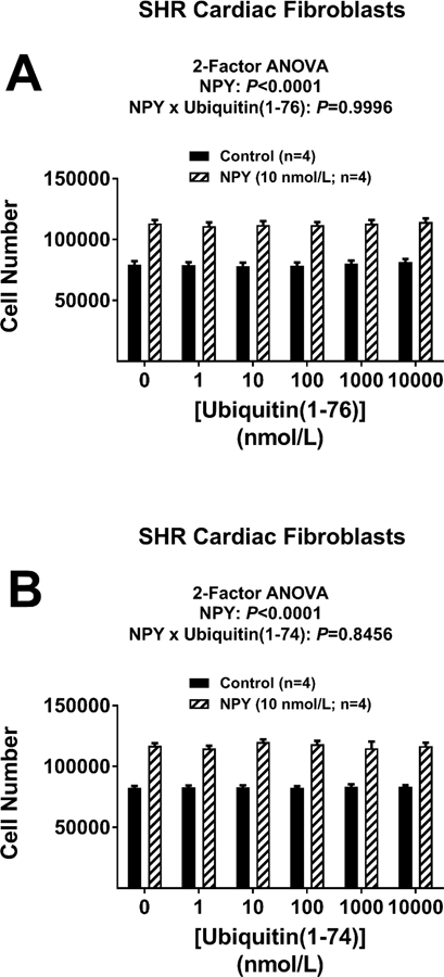
SDF-1α (stromal cell-derived factor-1α) is a CXCR4-receptor agonist and DPP4 (dipeptidyl peptidase 4) substrate. SDF-1α, particularly when combined with sitagliptin to block the metabolism of SDF-1α by DPP4, stimulates proliferation of cardiac fibroblasts via the CXCR4 receptor; this effect is greater in cells from spontaneously hypertensive rats versus Wistar-Kyoto normotensive rats. Emerging evidence indicates that ubiquitin(1-76) exists in plasma and is a potent CXCR4-receptor agonist. Therefore, we hypothesized that ubiquitin(1-76), similar to SDF-1α, should increase proliferation of cardiac fibroblasts. Contrary to our working hypothesis, ubiquitin(1-76) did not stimulate cardiac fibroblast proliferation, yet unexpectedly antagonized the proproliferative effects of SDF-1α combined with sitagliptin. In this regard, ubiquitin(1-76) was more potent in spontaneously hypertensive versus Wistar-Kyoto cells. In the presence of 6bk (selective inhibitor of insulin-degrading enzyme [IDE]; an enzyme known to convert ubiquitin(1-76) to ubiquitin(1-74)), ubiquitin(1-76) no longer antagonized the proproliferative effects of SDF-1α/sitagliptin. Ubiquitin(1-74) also antagonized the proproliferative effects of SDF-1α/sitagliptin, and this effect of ubiquitin(1-74) was not blocked by 6bk and was >10-fold more potent compared with ubiquitin(1-76). Neither ubiquitin(1-76) nor ubiquitin(1-74) inhibited the proproliferative effects of the non-CXCR4 receptor agonist neuropeptide Y (activates Y1 receptors). Cardiac fibroblasts expressed IDE mRNA, protein, and activity and converted ubiquitin(1-76) to ubiquitin(1-74). Spontaneously hypertensive fibroblasts expressed greater IDE activity. Extracellular ubiquitin(1-76) blocks the proproliferative effects of SDF-1α/sitagliptin via its conversion by IDE to ubiquitin(1-74), a potent CXCR4 antagonist. Thus, IDE inhibitors, particularly when combined with DPP4 inhibitors or hypertension, could increase the risk of cardiac fibrosis.

Keywords: CXCR4 receptor; SDF-1α; fibroblasts; hypertension; neuropeptide Y; ubiquitin.


Figures

Figure 1.
Figure 1.. Ubiquitin(1–76) and ubiquitin(1–74) antagonize the pro-proliferative effects of the CXCR4-receptor agonist SDF-1α in SHR and WKY cardiac fibroblasts.
Figure illustrates the concentration-dependent effects of ubiquitin(1–76) (panels A and B) and ubiquitin (1–74) (panels C and D) on proliferation of SHR (panels A and C) and WKY (panels B and D) cardiac fibroblasts stimulated with SDF-1α (10 nmol/L). In the absence of ubiquitins, a four-day treatment with SDF-1α significantly increased proliferation (cell number) of both SHR and WKY cardiac fibroblasts. In the absence of SDF-1α, a four-day treatment with either of the two ubiquitins did not affect cardiac fibroblast proliferation in either SHR or WKY cardiac fibroblasts. However, both ubiquitin(1–76) and ubiquitin(1–74) significantly antagonized the pro-proliferative effect of SDF-1α. Values are means ± SEM.
Figure 2.
Figure 2.. Panel A: The inhibitory potency of ubiquitin(1–76) on the pro-proliferative effects of the CXCR4-receptor agonist SDF-1α is greater in SHR compared with WKY cardiac fibroblasts.
Panel A illustrates the proliferative response (expressed as the percentage change in cell number) to SDF-1α (10 nmol/L) at different concentrations of ubiquitin(1–76) (1 to 10,000 nmol/L). In SHR cardiac fibroblasts, ubiquitin(1–76) significantly inhibited the growth effects of SDF-1α at concentrations as low as 1 nmol/L. In contrast, in WKY cardiac fibroblasts a ubiquitin(1–76) concentration of 100 nmol/L was required to significantly inhibit the growth effects of SDF-1α. The calculated IC50 for ubiquitin(1–76) in SHR was 0.4 nmol/L; in contrast the IC50 for ubiquitin(1–76) in WKY was 55 nmol/L. Panels B and C: Ubiquitin(1–76) does not antagonize the pro-proliferative effects of the CXCR4-receptor agonist SDF-1α in SHR and WKY cardiac fibroblasts treated with an inhibitor of insulin-degrading enzyme. Cardiac fibroblast from SHR (panel B) and WKY (panel C) rats were treated with the insulin-degrading enzyme (IDE) inhibitor 6bk (1 μmol/L) for four days with or without SDF-1α (10 nmol/L) and with or without ubiquitin(1–76) (1 to 10,000 nmol/L). A four-day treatment with SDF-1α significantly increased proliferation (cell number) of both SHR and WKY cardiac fibroblasts, and this response was not affected by ubiquitin(1–76). Panels D: Ubiquitin(1–74) antagonizes the pro-proliferative effects of the CXCR4-receptor agonist SDF-1α in cardiac fibroblasts even in the presence of the insulin-degrading enzyme inhibitor 6bk. Cardiac fibroblasts from SHR rats were treated with the insulin-degrading enzyme (IDE) inhibitor 6bk (1 μmol/L) for four days with or without SDF-1α (10 nmol/L) and with or without ubiquitin(1–74) (1 to 100 nmol/L). A four-day treatment with SDF-1α significantly increased proliferation (cell number) of both SHR and WKY cardiac fibroblasts, and this response was inhibited by ubiquitin(1–74). In all panels, values are means ± SEM.
Figure 3.
Figure 3.. Panel A: Validation of the Anaspec SensoLyte® 520 insulin-degrading enzyme (IDE) assay:
As active IDE cleaves a FRET substrate, 5-carboxyfluorescein is released, and its release is monitored over time by measuring fluorescence at excitation/emission of 490 nm/520 nm. IDE activity is proportional to the slope of the time versus product relationship. In the presence of recombinant IDE or a homogenate of cardiac fibroblasts, IDE product accumulated. In the absence of a source of IDE (negative control), there was no signal. Panels B, C, and D: IDE activity is greater in SHR compared with WKY cells. SHR and WKY cardiac fibroblasts were washed with phosphate-buffered saline, lysed in assay buffer, homogenized, and centrifuged. Cellular IDE activity was determined in 50 μl of supernatant. Panels B and C show typical results in WKY and SHR cardiac fibroblasts, respectively, and Panel D shows that the cellular IDE activity is significantly enhanced in SHR cardiac fibroblasts. Panels E and F. Both WKY and SHR cardiac fibroblasts express IDE protein. Panel E shows image of western blot for IDE in four batches of WKY and SHR cardiac fibroblast. Panel F shows the densitometry results from the image in panel E (normalized to β-actin). For all panels, values are means ± SEM.
Figure 4.
Figure 4.. Cardiac fibroblasts convert ubiquitin(1–76) to ubiquitin(1–74).
SHR cardiac fibroblasts were treated with ubiquitin(1–76) (U-76; 1000 nmol/L) for 24 hours, and the medium was collected and processed for analysis by mass spectrometry. Samples were loaded onto a polar reverse phase column connected to a Shimadzu HPLC/LCMS-2010 system. Separation was conducted with a linear gradient from 20% acetonitrile to 60% acetonitrile in the presence of 0.1% formic acid at flow rate 0.4 ml/min. The mass spectrometer was operated in the positive mode with selected ion monitoring. The chromatographic peak (total ion current) corresponding to the retention time of ubiquitin(1–76) and ubiquitin(1–74) (7.46 to 9.00 minutes) was scanned for multiple charged ions (M12+ to M7+): for ubiquitin(1–76), 714.80, 779.80, 857.80, 952.80, 1071.80, and 1224.80 m/z, respectively; for ubiquitin(1–74), 705.30, 769.50, 846.20, 940.20, 1057.50, 1208.50 m/z, respectively). The m/z versus signal intensity plot of the selected ions unequivocally identifies ubiquitin(1–74) as a product of ubiquitin(1–76).
Figure 5.
Figure 5.. Neither ubiquitin(1–76) nor ubiquitin(1–74) affects the pro-proliferative effects of neuropeptide Y in cardiac fibroblasts.
Figure illustrates the lack of effects of ubiquitin(1–76) (panel A) and ubiquitin(1–74) (panel B) (1 to 10,000 nmol/L) on cardiac fibroblasts stimulated with neuropeptide Y (NPY; 10 nmol/L). A four-day treatment with NPY significantly increased proliferation (cell number) of cardiac fibroblasts, and this effect was not attenuated by either ubiquitin(1–76) or ubiquitin(1–74). Values are means ± SEM.
Figure 6.
Figure 6.. Implications of the current findings.
The current findings uncover a potentially harmful interaction between DPP4 inhibitors and IDE inhibitors that may be exacerbated in hypertension.

References

    1. Chu P- Y, Zatta A, Kiriazis H, Chin-Dusting J, Du X- J, Marshall T, Kaye DM. CXCR4 antagonism attenuates the cardiorenal consequences of mineralocorticoid excess. Circ Heart Fail 2011;4:651–658. - PubMed
    1. Shao S, Cai W, Sheng J, Yin L. Role of SDF-1 and Wnt signaling pathway in the myocardial fibrosis of hypertensive rats. Am J Transl Res 2015;7:1345–1356. - PMC - PubMed
    1. Chu PY, Walder K, Horlock D, Williams D, Nelson E, Byrne M, Jandeleit-Dahm K, Zimmet P, Kaye DM. CXCR4 antagonism attenuates the development of diabetic cardiac fibrosis. PLoS ONE 2015;10:e0133616. - PMC - PubMed
    1. Kazakov A, Hall R, Jagoda P, Bachelier K, Muller-Best P, Semenov A, Lammert F, Bohm M, Laufs U. Inhibition of endothelial nitric oxide synthase induces and enhances myocardial fibrosis. Cardiovasc Res 2013;100:211–221. - PubMed
    1. Schober A, Knarren S, Lietz M, Lin EA, Weber C. Crucial role of stromal cell-derived factor-1α in neointima formation after vascular injury in apolipoprotein E-deficient mice. Circulation 2003;108:2491–2497. - PubMed

Publication types

MeSH terms

LinkOut - more resources