. 2018 Nov;72(5):e43-e52.
doi: 10.1161/HYPERTENSIONAHA.118.11485.

Sex Differences in Renal Inflammation and Injury in High-Fat Diet-Fed Dahl Salt-Sensitive Rats

Affiliations

Sex Differences in Renal Inflammation and Injury in High-Fat Diet-Fed Dahl Salt-Sensitive Rats

Roxanne Fernandes et al. Hypertension. 2018 Nov.

Abstract

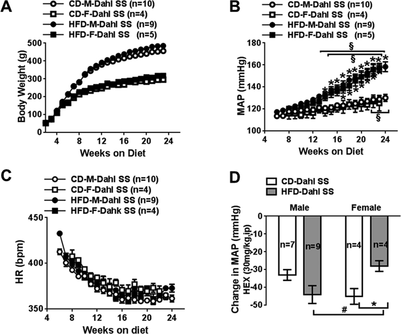
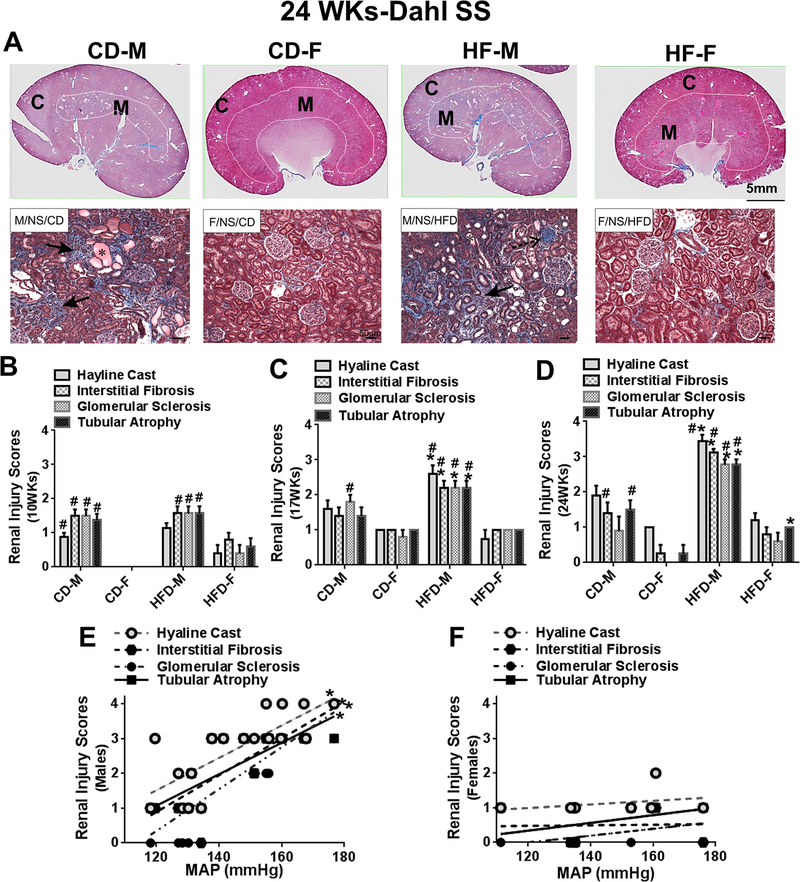
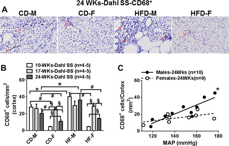
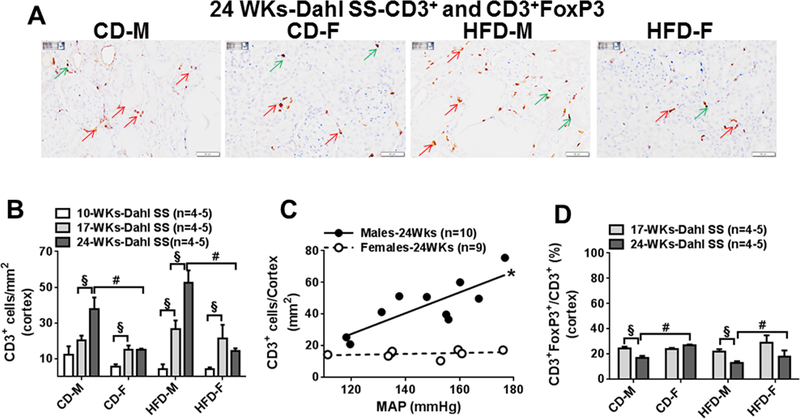
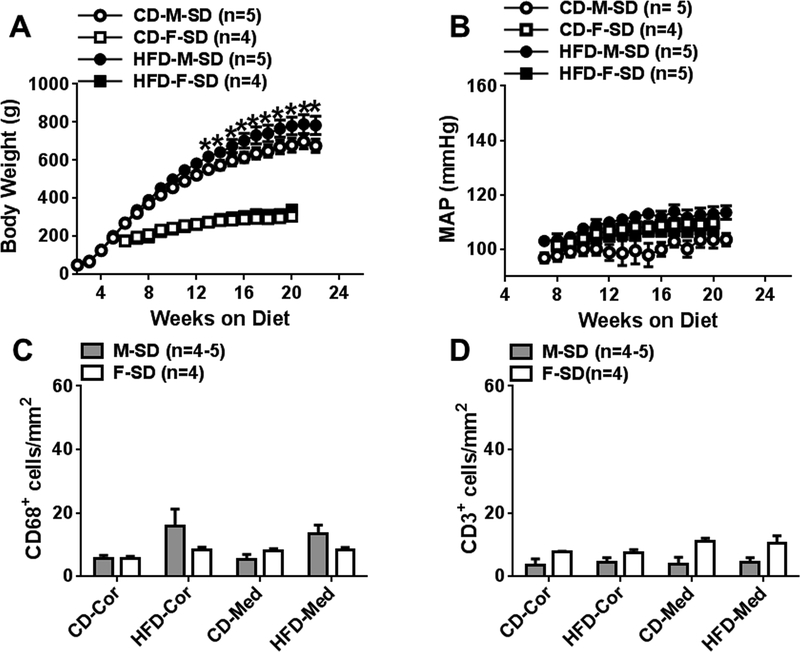
We examined the impact of sex on high-fat diet (HFD)-induced renal alterations in Dahl salt-sensitive and Sprague Dawley rats. In Dahl rats, HFD (60% kcal from fat for 24-26 weeks starting at weaning) significantly and equally increased blood pressure in males and females when compared with rats fed a control diet (10% kcal from fat). Male Dahl rats on HFD exhibited progressive renal histological injury and moderately increased renal macrophage infiltration at 10 and 24 weeks of feeding when compared with males on control diet. Female Dahl rats had lower grade renal injury and less macrophage infiltration (except at 17 weeks) than males regardless of diet. Male Dahl rats on both diets showed progressively increasing numbers of renal T-cells, a pattern not observed in females. HFD per se did not significantly affect renal T-cell number. Male Dahl rats had lower renal regulatory T-cells cell ratio than females at 24 weeks. Renal macrophage and T-cell infiltrations were highly correlated to final mean arterial pressure levels in males but not in females. Sprague Dawley rats fed HFD were normotensive without significant renal injury/inflammation after 24 weeks of feeding. In summary, HFD feeding fails to increase arterial blood pressure in Sprague Dawley rats but strongly promotes hypertension in both male and female Dahl salt-sensitive rats. Only Dahl males, however, exhibited blood pressure-associated renal inflammation and injury. Maintenance of regulatory T-cells ratio may protect against hypertension-associated renal injury/inflammation but not HFD-induced hypertension.

Keywords: diet; hypertension; immunity; kidney; sex.


Conflict of interest statement

Conflict(s) of Interest/Disclosure(s) Statement

None

Figures

Fig 1:
Fig 1:
Comparison of body weight (A) and weekly 24-h mean arterial blood pressure (B), and heart rate (C) measured following 24 weeks of control and HFD with normal salt in Dahl salt sensitive (SS) male and female rats. D, maximal changes of MAP after injection of hexamethonium (HEX) in Dahl SS rats at 24 weeks. CD, control fed; HFD, high fat fed; MAP, mean arterial blood pressure; HR, heart rate, M, male; F, female. Data are presented as mean ± SE. *P<0.05, HFD vs CD; #, P<0.05 M vs F; § P<0.05, vs MAP at their 10 weeks.
Fig 2:
Fig 2:
A, Representative light photomicrographs taken from Masson’s Trichrome-stained whole renal sections and higher magnified cortical areas from male and female Dahl SS rats at 24 weeks (24WKs). Comparisons of semi-quantified renal injury scores in CD and HFD Dahl SS male and female rats at 10 (B), 17 (C), and 24 (D) weeks. Indicating the occurrence of histological changes in kidney sections: *hyaline cast; interstitial fibrosis (peritubular); stippled arrow, glomerular sclerosis. C, cortex; M, medulla. Data are mean ± SE. *P<0.05, HFD vs CD; #P<0.05 M vs F.
Fig 3:
Fig 3:
A, Representative images (400x) of renal CD68+ positive cells in renal cortex taken from CD and HFD Dahl SS males and females at 24 weeks. B, Comparisons of cortical macrophages in CD and HFD males and females at 10 (10WKs), 17 (17WKs) and 24 weeks (24WKs). C Correlation between blood pressure and cortical CD68+ infiltrations in male and female Dahl SS rats at 24 weeks. Red arrow indicates CD68+ positive cells. Data are mean ± SE. *P<0.05, HFD vs CD; #P<0.05 M vs F, §; P<0.05, 10WKs vs 17WKs or 24WKs.
Fig 4:
Fig 4:
A, Representative images (400x) of renal CD3+ and CD3+FoxP3+ positive cells in cortex from CD and HFD males and females at 24 weeks. B, Comparisons of cortical CD3+ cells in CD and HFD males and females at 10 (10WKs), 17 (17WKs) and 24 weeks (24WKs). C, Correlation between blood pressure and cortical CD3+ infiltrations in male and female Dahl SS rats at 24 weeks. D, Comparisons of the ratio of T-reg cells (CD3+FoxP3+/CD3+ cells) in cortex area in CD and HFD Dahl SS male and female rats at 17 (17WKs) and 24 weeks (24WKs). Red arrow indicates CD3+ positive cells; green arrow indicates CD3+FoxP3 positive cells. Data are mean ± SE. #P<0.05 M vs F, §; P<0.05, 10WKs vs 17WKs or 24WKs
Fig 5:
Fig 5:
A and B, Comparisons of cortical mRNA expression of cytokine and MCP-1 in CD and HF males and females at 17 (17WKs) and 24 weeks (24WKs). Data are mean ± SE. #P<0.05 M vs F, §; P<0.05, 17WKs vs 24WKs
Fig 6:
Fig 6:
Comparison of body weight (A) and weekly 24-h mean arterial blood pressure (B) measured following 24 weeks of CD and HF feeding with normal salt in Sprague-Dawley (SD) male and female rats. C and D, comparisons of cortical and medullar CD68+ and CD3+ cells in CD and HFD males and females at 24 weeks. Data are mean ± SE.

Comment in

  • Cutting the Fat.
    Taylor LE, Sullivan JC. Taylor LE, et al. Hypertension. 2018 Nov;72(5):1081-1083. doi: 10.1161/HYPERTENSIONAHA.118.11680. Hypertension. 2018. PMID: 30354831 Free PMC article. No abstract available.

References

    1. Brands MW, Hall JE. Insulin resistance, hyperinsulinemia, and obesity-associated hypertension. Journal of the American Society of Nephrology : JASN. 1992;3:1064–1077. - PubMed
    1. Odermatt A The western-style diet: A major risk factor for impaired kidney function and chronic kidney disease. American journal of physiology. Renal physiology 2011;301:F919–931. doi: 10.1152/ajprenal.00068.2011. - DOI - PubMed
    1. Lambert EA, Phillips S, Belski R, Tursunalieva A, Eikelis N, Sari CI, Dixon JB, Straznicky N, Grima M, Head GA, Schlaich M, Lambert GW. Endothelial function in healthy young individuals is associated with dietary consumption of saturated fat. Frontiers in physiology. 2017;8:876. doi: 10.3389/fphys.2017.00876. - DOI - PMC - PubMed
    1. Aroor AR, Jia G, Sowers JR. Cellular mechanisms underlying obesity-induced arterial stiffness. American journal of physiology. Regulatory, integrative and comparative physiology. 2018;314:R387–R398. doi:10.1152/ajpregu.00235.2016. - DOI - PMC - PubMed
    1. Lim K, Jackson KL, Sata Y, Head GA. Factors responsible for obesity-related hypertension. Current hypertension reports. 2017;19:53. doi: 10.1007/s11906-017-0750-1. - DOI - PubMed

Publication types

Substances