Review
. 2019 Jan;98(1):36-45.
doi: 10.1177/0022034518805978. Epub 2018 Oct 24.

Genome Editing: A New Horizon for Oral and Craniofacial Research

Affiliations
Review

Genome Editing: A New Horizon for Oral and Craniofacial Research

N Yu et al. J Dent Res. 2019 Jan.

Abstract

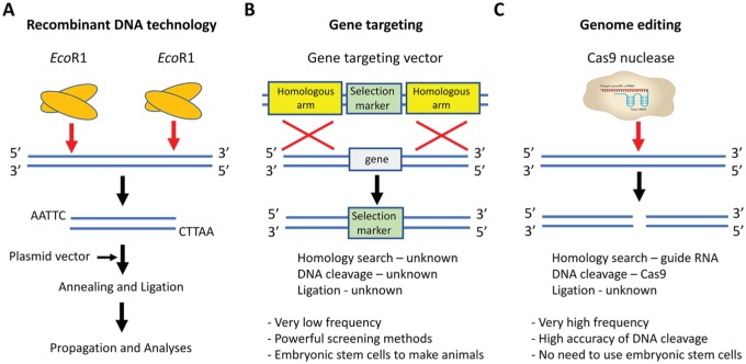
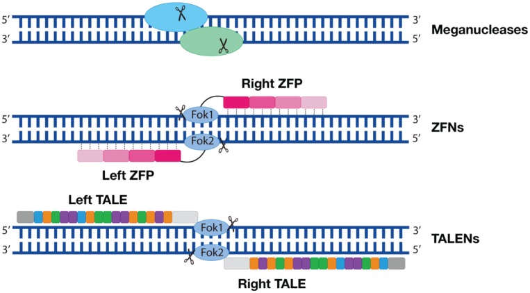
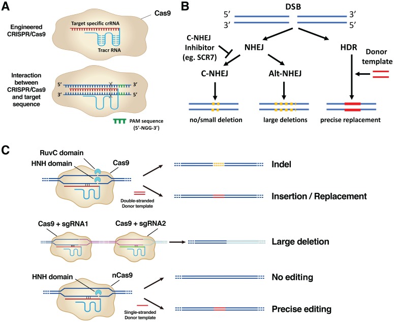
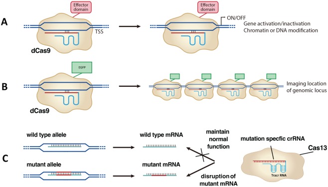
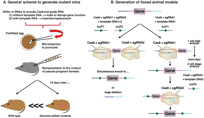
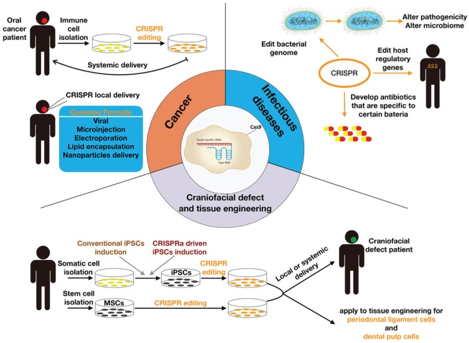
Precise and efficient genetic manipulations have enabled researchers to understand gene functions in disease and development, providing a platform to search for molecular cures. Over the past decade, the unprecedented advancement of genome editing techniques has revolutionized the biological research fields. Early genome editing strategies involved many naturally occurring nucleases, including meganucleases, zinc finger nucleases, and transcription activator-like effector-based nucleases. More recently, the clustered regularly interspaced short palindromic repeats (CRISPR) / CRISPR-associated nucleases (CRISPR/Cas) system has greatly enriched genetic manipulation methods in conducting research. Those nucleases generate double-strand breaks in the target gene sequences and then utilize DNA repair mechanisms to permit precise yet versatile genetic manipulations. The oral and craniofacial field harbors a plethora of diseases and developmental defects that require genetic models that can exploit these genome editing techniques. This review provides an overview of the genome editing techniques, particularly the CRISPR/Cas9 technique, for the oral and craniofacial research community. We also discuss the details about the emerging applications of genome editing in oral and craniofacial biology.

Keywords: CRISPR; DNA cleavage; DNA repair; dentistry; gene editing; genetic therapy.


Conflict of interest statement

The authors received no financial support and declare no potential conflicts of interest with respect to the authorship and/or publication of this article.

Figures

Figure 1.
Figure 1.
A historical view of genetic modification. (A) Recombinant DNA technology. The restriction enzyme EcoRI cuts the donor DNA and plasmid vector. Modified plasmid vector and donor DNA are annealed and ligated to facilitate the propagation and analyses of the newly constructed DNA fragment. (B) Gene targeting. A gene-targeting vector contains a selection marker flanked by 2 homologous arms; the target gene is flanked by the same arms. Through a homologous recombination, the selection marker replaces the target locus. The exact mechanisms of homology search, DNA cleavage, and ligation are poorly understood in gene targeting and are therefore labeled as “unknown.” Gene targeting also presents with low frequency and requires powerful screening methods for embryonic stem cells. (C) Genome editing. Cas9 enzyme and sgRNA containing crRNA and tracrRNA can cleave DNA at the specific sites. In this method, the homology search relies on the guide RNA, and DNA cleavage relies on Cas9 enzyme; the ligation mechanism is still unknown. This genetic manipulation method presents with high frequency and the accuracy of DNA cleavage. It also surpasses the need to use the embryonic stem cells. crRNA, CRISPR RNA; sgRNA, single-guide RNA; tracrRNA, transactivating crRNA.
Figure 2.
Figure 2.
Engineered early genome editing nucleases. Top row: Meganucleases. The oval shapes symbolize a pair of nucleases; the scissors symbolize DNA cleavage. Middle row: ZFNs are composed of a FokI nuclease domain (oval shapes) and a ZFP domain (a tandem of rectangular shapes). Each ZFP recognizes 3–base pair DNA. Four ZFPs are illustrated on 1 DNA strand. Bottom row: TALENs are composed of a FokI nuclease domain and a tandem of DNA-binding domain (rectangular shapes). Each rectangular shape symbolizes 1 protein repeat that contains 3 amino acids. Each protein repeat binds to 1 nucleotide. TALEN, transcription activator–like effector-based nuclease; ZFN, zinc finger nuclease; ZFP, zinc finger protein.
Figure 3.
Figure 3.
Mechanisms of the CRISPR/Cas9 system. (A) Top row: Engineered CRISPR/Cas9 is composed of a sgRNA that contains ~20-bp crRNA (red sequence) fused with a tracrRNA (blue sequence) and a Cas9 enzyme component (beige irregular circle). Bottom row: sgRNA binds to the specific target DNA sequences, and Cas9 makes a double-strand break (DSB) at 3 bp upstream of a conserved protospacer-adjacent motif (PAM) region (highlighted in green). Cas9 requires a PAM sequence of 5′-NGG-3′ sequence (N represents any nucleotide base). (B) Repair mechanisms after DSB. Two DNA repair pathways—nonhomologous end-joining (NHEJ) or homology-directed repair (HDR)— kick in after DSB. The NHEJ mechanism is further divided into a canonical (C-NHEJ) or alternative (alt-NHEJ) pathway. C-NHEJ results in no deletion or a small deletion; alt-NHEJ results in the large deletions. Ligase IV inhibitor SCR7 inhibits the C-NHEJ pathway. The HDR pathway is promoted with the presence of a donor template DNA leading to precise replacement of genomic sequences. (C) The application of CRISPR/Cas9 in genome editing. Top row: NHEJ after DSB results in insertion/deletion (indel) mutations; HDR after DSB prompted by a double-stranded donor template DNA results in precise insertion/replacement. Middle row: NHEJ results in a large deletion (pink sequence) when 2 distant DSBs are created by CRISPR/Cas9 systems containing sgRNA1 and sgRNA2. Bottom row: CRISPR/Cas9 with a mutation in the RuvC domain generates nick instead of DSB. DNA repair from a nick with a single-stranded donor template is highly accurate. bp, base pair; crRNA, CRISPR RNA; sgRNA, single-guide RNA; tracrRNA, transactivating crRNA.
Figure 4.
Figure 4.
Application of CRISPR beyond genome editing. (A) dCas9 fused with effector domains reveals gene activation/inactivation or chromatin or DNA modification of these effectors. (B) dCas9 fused with enhanced green fluorescent protein (EGFP) illuminates genomic loci. (C) CRISPR/Cas13 can edit RNA precisely. Mutation-specific crRNA can bind to the mutant mRNA and disrupt its functions.
Figure 5.
Figure 5.
Applications of CRISPR/Cas9 to generate mutant animals. (A) General scheme to generate mutant mouse models. DNAs or RNAs encoding Cas9 and single-guide RNA (sgRNA) with or without template DNA are microinjected into the pronuclei of the fertilized egg. These modified zygotes are reimplanted to the oviduct of pseudo-pregnant surrogate mice. After 19 gestational days, mutant mice offspring are born. (B) Generation of floxed animal models. The left arm represents the simultaneous method. This method involves coinjecting nucleotides encoding Cas9, 2 sgRNAs, and 2 individual loxP template DNAs to zygote. It simultaneously knocks in 2 loxP alleles to flank the target exon gene. However, large deletions can occur as a result. The right arm represents the sequential method. This method involves coinjecting Cas9, first sgRNA, and first loxP template DNA at 1–cell stage embryo to knock in the first loxP allele. On the next day, the second group of Cas9, second sgRNA, and second loxP template DNA are injected into 2–cell stage embryos, leading to knock-in of the second loxP allele.
Figure 6.
Figure 6.
Applications of the CRISPR system in oral and craniofacial biology. Cancer: Immune cells are isolated from patients with oral cancer and receive CRISPR genome editing in vitro. Genome-edited immune cells are then systemically delivered back to the patients for cancer therapy. CRISPR components can be locally delivered to the hot spot of the cancer lesion, following various delivery formats. Craniofacial defect and tissue regeneration: Somatic cells are isolated from human subjects. Through either conventional induced pluripotent stem cell (iPSC) induction or CRISPR activation (CRISPRa) iPSC induction, somatic cells become iPSCs. Specific stem cell lineages, such as mesenchymal stem cells (MSCs), can be isolated and receive CRISPR editing. Both stem cells can be delivered either systemically or locally to the patient with craniofacial defects. Genome-edited stem cells also contribute to tissue engineering of periodontal ligament cells or dental pulp cells. Infectious diseases: CRISPR may edit the bacterial genome to alter either pathogenicity or the microbiome; CRISPR may edit host regulatory genes to fight against infections; CRISPR may also contribute to developing pathogen-specific antibiotics.

References

    1. Abudayyeh OO, Gootenberg JS, Essletzbichler P, Han S, Joung J, Belanto JJ, Verdine V, Cox DBT, Kellner MJ, Regev A, et al. 2017. RNA targeting with CRISPR-Cas13. Nature. 550(7675):280–284. - PMC - PubMed
    1. American Cancer Society. 2017. Cancer facts and figures. Atlanta (GA): American Cancer Society.
    1. Barrangou R, Marraffini LA. 2014. CRISPR-Cas systems: prokaryotes upgrade to adaptive immunity. Mol Cell. 54(2):234–244. - PMC - PubMed
    1. Barros SP, Offenbacher S. 2014. Modifiable risk factors in periodontal disease: epigenetic regulation of gene expression in the inflammatory response. Periodontology 2000. 64(1):95–110. - PubMed
    1. Betermier M, Bertrand P, Lopez BS. 2014. Is non-homologous end-joining really an inherently error-prone process? PLoS Genet. 10(1):e1004086. - PMC - PubMed