. 2020 Jan;40(1):163-176.
doi: 10.1177/0271678X18806893. Epub 2018 Oct 24.

Involvement of caveolin-1 in neurovascular unit remodeling after stroke: Effects on neovascularization and astrogliosis

Affiliations

Involvement of caveolin-1 in neurovascular unit remodeling after stroke: Effects on neovascularization and astrogliosis

Camille Blochet et al. J Cereb Blood Flow Metab. 2020 Jan.

Abstract

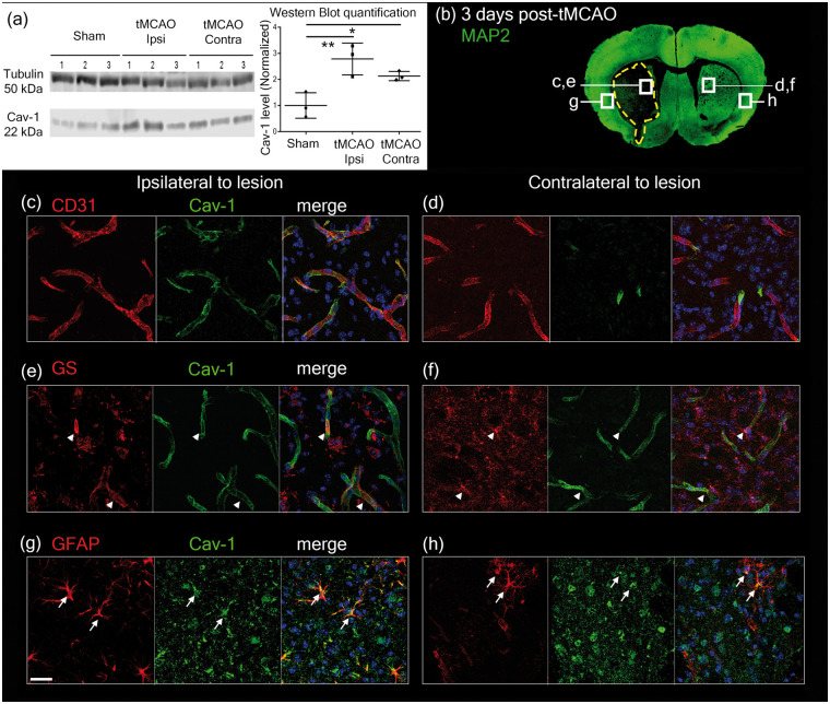
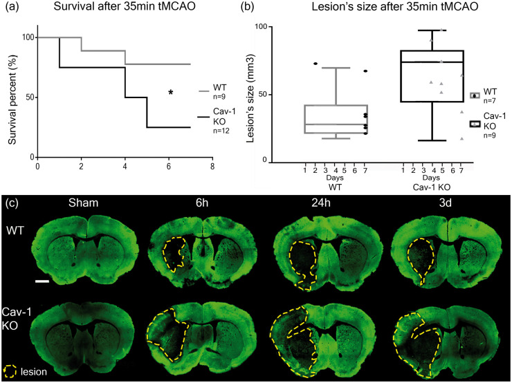
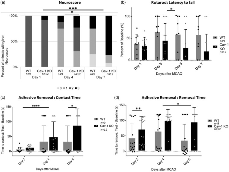
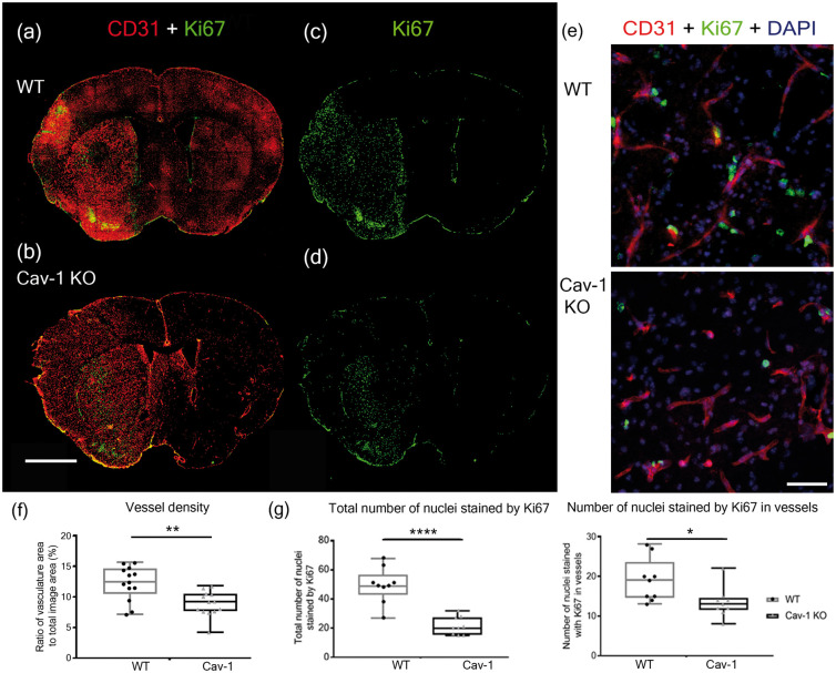
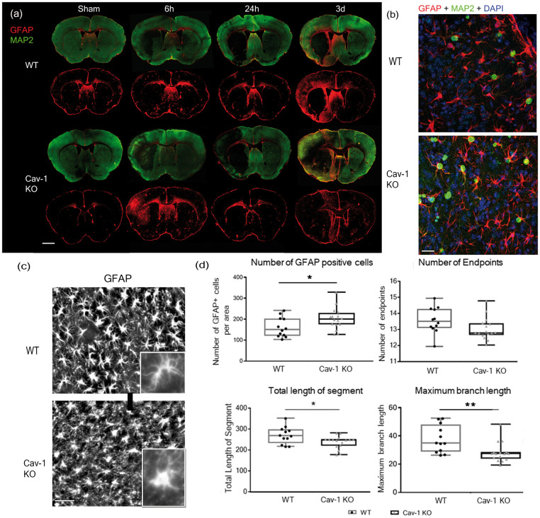
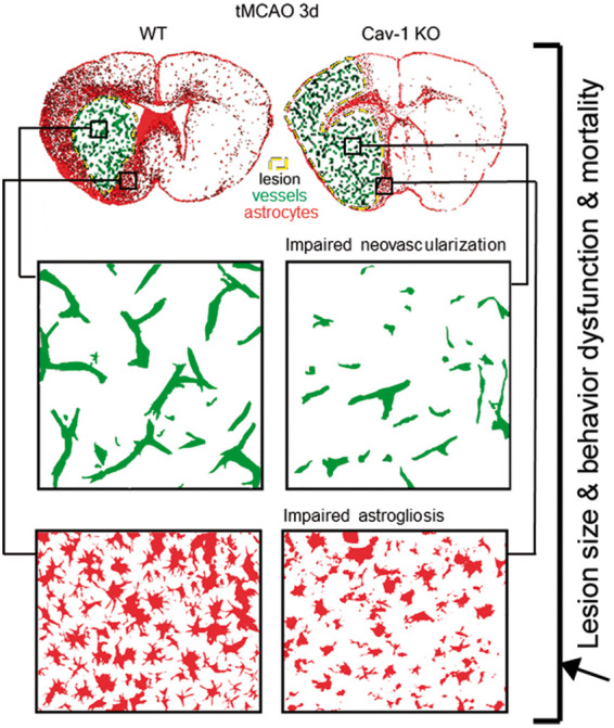
Complex cellular and molecular events occur in the neurovascular unit after stroke, such as blood-brain barrier (BBB) dysfunction and inflammation that contribute to neuronal death, neurological deterioration and mortality. Caveolin-1 (Cav-1) has distinct physiological functions such as caveolae formation associated with endocytosis and transcytosis as well as in signaling pathways. Cav-1 has been proposed to be involved in BBB dysfunction after brain injury; however, its precise role is poorly understood. The goal of this study was to characterize the expression and effect of Cav-1 deletion on outcome in the first week in a transient Middle Cerebral Artery Occlusion stroke model. We found increased Cav-1 expression in new blood vessels in the lesion and in reactive astrocytes in the peri-lesion areas. In Cav-1 KO mice, the lesion volume was larger and the behavioral outcome worse than in WT mice. Cav-1 KO mice exhibited reduced neovascularization and modified astrogliosis, without formation of a proper glial scar around the lesion at three days post injury, coinciding with aggravated outcomes. Altogether, these results point towards a potential protective role of endogenous Cav-1 in the first days after ischemia by promoting neovascularization, astrogliosis and scar formation.

Keywords: Caveolin-1; astrogliosis; ischemic stroke; neovascularization; neuroprotection.


Figures

Figure 1.
Figure 1.
(a) Western blot showing caveolin-1 protein levels (Cav-1, 22 kDa band) in WT sham and tMCAO ipsilateral (tMCAO Ipsi) and contralateral (tMCAO Contra) side to the lesion. α-Tubulin (50 kDa band) was used as loading control. Quantification was done using mean grey values and normalized against the mean value for Sham. n = 3 animals per condition. (b) Image showing the locations where pictures were taken for Cav-1 and cell-marker expression in relation to the lesion (dotted line showing loss of neuronal MAP-2 staining) on a coronal section in immunofluorescence analysis after tMCAO. (c–d) Cav-1 (green) co-localizes with CD31-labeled (red) endothelial cells in the striatum ipsilateral (c) and contralateral (d) to the ischemic lesion at 3 dpi. (e–f) In the ipsilateral (e) and in the contralateral hemisphere (f), Cav-1 (green) was also observed (arrowheads) in GS-positive astrocytes (red). (g–h) In the cortex, Cav-1 (green) was found to co-localize (arrows) in reactive astrocytes stained with GFAP (red). Respective control staining was done using the same markers on Cav-1 KO tissue and is available in Supplementary Figure 1. Scale bar = 20 µm.
Figure 2.
Figure 2.
Survival and lesion size assessment after 35 min tMCAO in WT and Cav-1 KO mice. (a) Kaplan–Meier survival plot and (b) lesion size at time of sacrifice measured on cresyl violet-stained sections (not shown). Box plots show the mean lesion volumes ± min/max for WT and Cav-1 KO mice after 35 min tMCAO and the dots correspond to individual animals with the time of sacrifice specified on the x-axis. (c) Immunofluorescence staining using MAP-2 (neuronal marker) on coronal slices of WT and Cav-1 KO mice after tMCAO or sham surgery collected 6 h, 24 h and three days after stroke, with lesion delimited by the yellow dotted line. Scale bar = 1 mm.
Figure 3.
Figure 3.
Behavioral assessment (a) Neuroscore assessed on a scale from 0 (no deficit) to 3 (sacrifice/death) at 1, 4 and 7 dpi. (b) Latency to fall from the Rotarod apparatus assessed at one, three, five and seven days post-injury (dpi) and expressed as percent of the best baseline performance value (mean ± SD). (c) Time before contact and (d) before removal of the adhesive placed under each paw of the mouse assessed at 2, 4 and 6 dpi and expressed as the difference between best baseline performance and best test time for each mouse (mean ± SD).
Figure 4.
Figure 4.
Neovascularization in the lesion. (a–d) Immunofluorescence staining with CD31 (red) and Ki67 (green) in WT (a, c) and Cav-1 KO (b, d) mice at three days post-MCAO. Scale bar = 1 mm. (e) Immunofluorescence staining with CD31 (red), Ki67 (green) and DAPI counterstaining (blue) in the striatum ipsilateral to the lesion in the WT (top image) and in the Cav-1 KO (bottom image). Scale bar = 20 µm. (f) Quantification of the vessel density showing the ratio of vasculature area to the total image area (individual points, mean and SD) in WT and Cav-1 KO mice (g) Quantification (individual points, mean and SD) of the total number of nuclei stained with Ki67 per area (left plot) and the number of nuclei labeled by Ki67 in vessels per area (right plot) in WT and Cav-1 KOs. n = 3 areas per animal with three animals per group, 20× sections.
Figure 5.
Figure 5.
Astrocyte reactivity and morphology. (a) Astrocyte reactivity time-course on coronal brain sections from WT and Cav-1 KO mice stained with GFAP and MAP-2. Sections were collected from sham animals and 6 h, one and three days after tMCAO. Scale bar = 1 mm (b) Immunofluorescence staining with proliferating cells-marker Ki67 (green), reactive astrocytes marker GFAP (red) and DAPI nuclear counterstaining (blue) in the striatum ipsilateral to the lesion in the WT (top image) and in the Cav-1 KO (bottom image). Scale bar = 20 µm (c) High magnification (40×) panels from sections obtained at 3 dpi illustrating GFAP-positive reactive astrocytes (astrocytes white on black background) in the striatal peri-lesion in WT (top image) and Cav-1 KO (bottom image). Scale bar = 20 µm. Inserts with zoom on a single astrocyte from each group. (d) Analysis of astrocyte number and astrocytic morphology by skeletonization: plot of the number of GFAP-positive cells, number of endpoints, total length of the segment and the maximum branch length in WT and Cav-1 KO astrocytes in the peri-lesion. n = 4 areas per animal with three animals per group.
Figure 6.
Figure 6.
Summary figure of the differences observed between WT and Cav-1 KO mice. Increased lesion sizes associated with behavioral dysfunction such as sensorimotor deficits were observed in parallel to impaired neovascularization in the core of the lesion and altered astrogliosis, more specifically changes in astrocytic morphology preventing proper scar formation in the peri-lesion.

References

    1. Mozaffarian D, Benjamin EJ, Go AS, et al.Heart disease and stroke statistics – 2016 update: a report from the American Heart Association. Circulation 2016; 133: e38–360. - PubMed
    1. Meadows KL. Experimental models of focal and multifocal cerebral ischemia: a review. Rev Neurosci 2018; 29: 661–674. - PubMed
    1. Goyal M, Menon BK, van Zwam WH, et al.Endovascular thrombectomy after large-vessel ischaemic stroke: a meta-analysis of individual patient data from five randomised trials. Lancet 2016; 387: 1723–1731. - PubMed
    1. Neuhaus AA, Couch Y, Hadley G, et al.Neuroprotection in stroke: the importance of collaboration and reproducibility. Brain 2017; 140: 2079–2092. - PubMed
    1. Zhang JH, Badaut J, Tang J, et al.The vascular neural network – a new paradigm in stroke pathophysiology. Nat Rev Neurol 2012; 8: 711–716. - PMC - PubMed

Publication types