Review
. 2019 Jan 1;99(1):235-309.
doi: 10.1152/physrev.00055.2017.

Thick Ascending Limb Sodium Transport in the Pathogenesis of Hypertension

Affiliations
Review

Thick Ascending Limb Sodium Transport in the Pathogenesis of Hypertension

Agustin Gonzalez-Vicente et al. Physiol Rev. .

Abstract

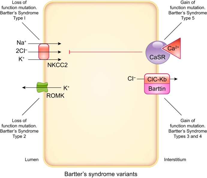
The thick ascending limb plays a key role in maintaining water and electrolyte balance. The importance of this segment in regulating blood pressure is evidenced by the effect of loop diuretics or local genetic defects on this parameter. Hormones and factors produced by thick ascending limbs have both autocrine and paracrine effects, which can extend prohypertensive signaling to other structures of the nephron. In this review, we discuss the role of the thick ascending limb in the development of hypertension, not as a sole participant, but one that works within the rich biological context of the renal medulla. We first provide an overview of the basic physiology of the segment and the anatomical considerations necessary to understand its relationship with other renal structures. We explore the physiopathological changes in thick ascending limbs occurring in both genetic and induced animal models of hypertension. We then discuss the racial differences and genetic defects that affect blood pressure in humans through changes in thick ascending limb transport rates. Throughout the text, we scrutinize methodologies and discuss the limitations of research techniques that, when overlooked, can lead investigators to make erroneous conclusions. Thus, in addition to advancing an understanding of the basic mechanisms of physiology, the ultimate goal of this work is to understand our research tools, to make better use of them, and to contextualize research data. Future advances in renal hypertension research will require not only collection of new experimental data, but also integration of our current knowledge.


Figures

FIGURE 1.
FIGURE 1.
Inverse correlation between certainty of conclusions and applicability to understanding of blood pressure regulation based on experimental settings.
FIGURE 2.
FIGURE 2.
Apical and basolateral transporters expressed by thick ascending limbs thought to be important in blood pressure regulation. Predominant directions of ion movement are depicted by arrows. However, all except the Na+-K+-ATPase can move ions in either direction. The apical transporters are Na+-K+-2Cl cotransporter type 2 (NKCC2), renal outer medullary K+ channel (ROMK), and Na+/H+ exchanger type 3 (NHE). Multiple ROMK isoforms are expressed by thick ascending limbs. Multiple NHE isoforms are expressed by thick ascending limbs, but NHE3 is the most abundant. The basolateral transporters are Na+-K+-ATPase, KCl cotransporter, Cl channels, K+ channels, and aquaporin 1 (AQP). The Na+-K+-ATPase ultimately provides the driving force for all NaCl movement. Three subunits comprise the Na+-K+-ATPase. Although there are several α-, β-, and γ-subunits, α1, β1, γAB splice variants predominate. At least two different Cl channels are expressed by thick ascending limbs. At least 2 different K+ channels are expressed in this segment. AQP is expressed by thick ascending limbs, but other water channels may also be present.
FIGURE 3.
FIGURE 3.
Paracellular movement of Na+ in thick ascending limbs. Early in the medullary thick ascending limb, both luminal and interstitial Na+ concentrations are high. There the lumen positive voltage (V+) drives Na+ out of the lumen into the interstitium. However, as Na+ is reabsorbed, the concentration gradient for Na+ becomes large enough to reverse the paracellular flux of Na+ so that it now enters the lumen from the interstitium. The exact point at which the flip in directions occurs is unclear and depends on many factors.
FIGURE 4.
FIGURE 4.
Arginine vasopressin signaling in thick ascending limbs. Arrows indicate stimulation, and T-lines indicate inhibition. Dashed lines indicate that the complete signaling cascade is unknown. AVP, arginine vasopressin; NHE3, Na+/H+ exchanger type 3; NKCC2, Na+-K+-2Cl cotransporter type 2; PKA, cAMP-dependent protein kinase; ROMK, renal outer medullary K+ channel; V2R, arginine vasopressin type 2 receptor.
FIGURE 5.
FIGURE 5.
Nitric oxide (NO) signaling and known effects on individual transporters involved in transcellular NaCl reabsorption in thick ascending limbs. Arrows indicate stimulation, and T-lines indicate inhibition. Dashed lines indicate that the complete signaling cascade is unknown. AMP, adenosine monophosphate; ClC-K, Cl channel; CLD19, claudin 19; NHE3, Na+/H+ exchanger type 3; NKCC2, Na+-K+-2Cl cotransporter type 2; NOS3, NO synthase type 3; PDE2, cGMP-stimulated phosphodiesterase; PKA, cAMP-dependent protein kinase; PKG, cGMP-dependent protein kinase; ROMK, renal outer medullary K+ channel; sGC, soluble guanylyl cyclase.
FIGURE 6.
FIGURE 6.
Flow-induced nitric oxide (NO) production via the mechanosensitive transient receptor potential vanilloid type 4 (TRPV4) channel requires the activation of both basolateral P2X and luminal P2Y purinergic receptors. These receptors activate the phosphatidylinositol 3-kinase (PI3K), which in turn phosphorylates protein kinase B (Akt), which phosphorylates NO synthase type 3 (NOS3), increasing NO production.
FIGURE 7.
FIGURE 7.
Endothelin signaling in rat thick ascending limbs. Arrows indicate stimulation, and T-lines indicate inhibition. Phosphoinositol-dependent kinase (PDK) is known to be an intermediary in other systems but has not been directly demonstrated in mediating the effects of endothelin in thick ascending limbs and so is in gray. Akt, protein kinase B; ET-1, endothelin 1; ETB, endothelin type B receptor; NKCC2, Na+-K+-2Cl cotransporter type 2; NO, nitric oxide; NOS3, NO synthase type 3; PI3K, phosphatidylinositol 3-kinase; PIP2, phosphatidylinositol (4,5)-bisphosphate; PIP3, phosphatidylinositol (3,4,5)-trisphosphate.
FIGURE 8.
FIGURE 8.
Adrenergic signaling and known effects on transporters involved in NaCl reabsorption in thick ascending limbs. Arrows indicate stimulation, and T-lines indicate inhibition. Dashed lines indicate that the complete signaling cascade is unknown. Relatively lower concentrations of norepinephrine (NE) are needed to stimulate α2-receptors compared with β-receptors. Thus the red triangle representing NE is smaller. It is currently unclear how β-receptors activate mitogen-activated protein kinase (MAPK) in thick ascending limbs. It is also unclear how they activate Na+-K+-2Cl cotransporter type 2 (NKCC2). α2R: α2-adrenergic receptor; βR, β-adrenergic receptor; NKCC2, Na+-K+-2Cl cotransporter type 2; NO, nitric oxide; NOS3, NO synthase type 3; PI3K, phosphatidylinositol 3-kinase; PKA, cAMP-dependent protein kinase.
FIGURE 9.
FIGURE 9.
Superoxide (O2) signaling in thick ascending limbs. Arrows indicate stimulation, and T-lines indicate inhibition. Dashed lines indicate that the complete signaling cascade is unknown. NADPH oxidase (NOX) is likely the primary source of O2 in thick ascending limbs but the isoform activated by a given stimulus may vary. NHE, Na+/H+ exchanger; NKCC2, Na+-K+-2Cl cotransporter type 2; PKCα, protein kinase Cα.
FIGURE 10.
FIGURE 10.
Some of the proposed signaling cascades activated by angiotensin II (ANG II) in thick ascending limbs and reported effects on transporters involved in NaCl reabsorption. ANG II effects and signaling in the thick ascending limb are subjects of considerable controversy, and signaling is quite complex. It is likely that both are affected by environmental factors. AT1R, angiotensin II type 1 receptor; AT2R, angiotensin II type 2 receptor; 20-HETE, 20-hydroxyeicosatetraenoic acid; NHE, Na+/H+ exchanger; NKCC2, Na+-K+-2Cl cotransporter type 2; NO, nitric oxide; NOS3, NO synthase type 3; PI3K, phosphatidylinositol 3-kinase; PKC, protein kinase C; PLA, phospholipase A; PLC, phospholipase C.
FIGURE 11.
FIGURE 11.
A: synthesis and release of tumor necrosis factor-α (TNF-α) by thick ascending limbs. B: auto- and paracrine functions of TNF-α in thick ascending limbs. AT1R, angiotensin II (ANG II) type 1 receptor; CaSR, calcium (Ca2+) sensing receptor; COX2, cyclooxygenase 2; LPS, lipopolysaccharide; NFAT5, nuclear factor of activated T cells 5; NKCC2, Na+-K+-2Cl cotransporter type 2; PGE2, prostaglandin E2; ROMK, renal outer medullary K+ channel; TLR4, Toll-like receptor 4; TNFR, TNF-α receptor. See main text for details.
FIGURE 12.
FIGURE 12.
Diagram representation of the tubulovascular arrangement at the largest transverse level in and around a vascular bundle (VB). The red dashed circle indicates the VB core in which the descending vasa recta (DVR) and ascending vasa recta (AVR) are marked in solid red and blue, respectively. I–III represent the three types of short-looped nephrons (SLN) to show their locations. The yellow dashed curve outlines the long-looped nephron (LLN) thick ascending limbs (TAL) at the margin of the VB. The proportions of the various structures are based on the tracing data. DTL, descending thin limb. [From Ren et al. (574) with permission.]
FIGURE 13.
FIGURE 13.
Paracrine factors released into the lumen or interstitium by thick ascending limbs that could alter vasa recta and collecting duct function. ET-1, endothelin 1; HETE, 5-hydroxyeicosatetraenoic acid; NO, nitric oxide; PGs, prostaglandins.
FIGURE 14.
FIGURE 14.
Paracrine factors released into the lumen of thick ascending limbs that have been shown or could alter macula densa and distal convoluted tubule function. ET-1, endothelin 1; HETE, 5-hydroxyeicosatetraenoic acid; NO, nitric oxide; PGs, prostaglandins.
FIGURE 15.
FIGURE 15.
Paracrine factors released into the interstitium by thick ascending limbs that could alter proximal tubule and thin descending limb function. ET-1, endothelin 1; HETE, 5-hydroxyeicosatetraenoic acid; NO, nitric oxide; PGs, prostaglandins.
FIGURE 16.
FIGURE 16.
Pressure natriuresis relationship in Dahl salt-resistant rats fed normal salt and salt-sensitive rats fed normal and high-salt diets. kw, kidney weight; R/Br, Dahl salt-resistant rats, Brookhaven strain, fed normal salt; S/Br+HS, Dahl salt-sensitive rats, Brookhaven strain, fed high salt; S/Br, Dahl salt-sensitive rats, Brookhaven strain, fed normal salt. Pressure natriuresis was blunted in S/Br by a decrease in slope, while S/Br+HS additionally showed a rightward shift. This indicates that, when hypertension is established after dietary manipulations, renal changes lead to increases in blood pressure that are no longer salt-sensitive. [Adapted from Roman (586).]
FIGURE 17.
FIGURE 17.
Angiotensin (ANG) II type 2 receptor (AT2R)-mediated nitric oxide (NO) production in Dahl rats. A: the signaling cascade whereby AT2R-mediated NO inhibits Na+-K+-2Cl cotransporter type 2 (NKCC2) activity in the R phenotype. B: the changes present in the S phenotype leading to a reduced inhibition of NKCC2 by NO. Arrows indicate stimulation, and T-lines indicate inhibition. O2, superoxide; PDE2, cGMP-stimulated phosphodiesterase; PKG, cGMP-dependent protein kinase.
FIGURE 18.
FIGURE 18.
Thick ascending limb-vasa recta crosstalk after angiotensin II (ANG II) treatment in Dahl rats. Left: the R phenotype, where nitric oxide (NO) production in response to incubation with ANG II blunts ANG II-induced superoxide (O2) production. The increase of NO in the thick ascending limb concomitantly raises NO levels in adjacent vasa recta pericytes and buffers vasoconstriction induced by ANG II-dependent Ca2+ increases. A large part of this buffering effect is mediated by vasa recta purinergic receptors instead of just diffusing NO. Right: the S phenotype, where excess O2 production blunts NO increases in the thick ascending limb and vasa recta. Moreover, increases in O2 are also observed in adjacent vasa recta in response to ANG II. Vasoconstriction buffering is blunted in these animals. AT1R, angiotensin II type I receptor; AT2R, angiotensin II type II receptor; P2, purinergic type 2 receptors.
FIGURE 19.
FIGURE 19.
Proposed mechanisms whereby increased reactive oxygen species (ROS) cause hypertension and renal injury in the Dahl salt-sensitive rat. An imbalance favoring renal medullary ROS in the Dahl salt-sensitive rat would increase Na+-K+-2Cl cotransporter type 2 (NKCC2) activity by both direct stimulation and by inhibition of NO synthase type 3 (NOS3). A more active thick ascending limb would decrease Na+ delivery to the macula, thereby reducing tubuloglomerular feedback (TGF). These changes would increase glomerular perfusion pressure and filtration rate, which over time would damage the glomerulus, thereby reducing the filtration surface. In addition, because ROS production is regulated by luminal flow in thick ascending limbs, an increase in filtration rate would augment fluid delivery to this segment, perpetuating the injury. Given the link between ROS production, luminal flow, and Na+ reabsorption by the thick ascending limb, increased medullary oxidative stress is considered one of the driving forces for the development of salt-sensitive hypertension in the Dahl rat.
FIGURE 20.
FIGURE 20.
Proposed effects of 20-hydroxyeicosatetraenoic acid (20-HETE) signaling on transport in the Dahl rat. A: the 20-HETE physiology in R. B: the ultimate cause for decreased 20-HETE levels in S remains unknown. However, the reported decreased in cytochrome P-450 A2 (CYP450A2) protein expression and defective angiotensin II type 2 receptor (AT2R) signaling are expected to reduce the formation of 20-HETE. Concomitantly, excess superoxide (O2) may further lower 20-HETE bioavailability. Together, these effects could increase thick ascending limb NaCl reabsorption on S, by lack of inhibitory signals on Na+-K+-2Cl cotransporter (NKCC2), Na+/H+ exchanger type 3 (NHE3), and renal outer medullary K+ channel (ROMK).
FIGURE 21.
FIGURE 21.
Combined effects of dietary salt [Salt] and the levels of angiotensin II [ANG II] on the blood pressure increase (ΔBP) during the first week of ANG II infusion. The partial contributions of [Salt] and [ANG II] to ΔBP are represented on the right.
FIGURE 22.
FIGURE 22.
Effect of infusing either pressor or slow-pressor doses of angiotensin II (ANG II) on the cumulative Na+ balance during the first week of treatment.
FIGURE 23.
FIGURE 23.
Changes in nitric oxide (NO) signaling in thick ascending limbs during angiotensin II (ANG II)-induced hypertension. Black arrows and T-lines indicate normal physiological stimulation and inhibition, respectively. Arrows in red indicate the direction (up: or down: ) of pathological changes occurring during ANG II-induced hypertension. These changes result in altered cGMP levels and reduced inhibition of Na+-K+-2Cl cotransporter (NKCC2). NOS3, NO synthase type 3; PDE5, phosphodiesterase 5.
FIGURE 24.
FIGURE 24.
Chart of the increase in blood pressure as a function of the dose of NG-nitro-l-arginine methyl ester (l-NAME) under different dietary Na+ isopleths. NOS, nitric oxide synthase.
FIGURE 25.
FIGURE 25.
Mutations in proteins expressed by thick ascending limbs that cause hypertension. Loss of function mutations in the basolateral Ca2+-sensing receptor (CaSR) would decrease the inhibitory effect of this receptor on Na+-K+-2Cl cotransporter (NKCC2), while gain of function mutations in ClC-Kb channels would also lead to increases in NKCC2 activity. Mutations in the CYP4A11 gene may decrease 20-hydroxyeicosatetraenoic acid (20-HETE) levels and, therefore, its inhibitory effects on NaCl reabsorption in the thick ascending limb.
FIGURE 26.
FIGURE 26.
Mutations in proteins expressed by thick ascending limbs that cause Bartter’s Syndrome. This condition is characterized by salt waste and excessive Ca2+ and Mg2+ excretion and may be due to any loss of function mutation in transporters/channels of the thick ascending limb that decreases Na+-K+-2Cl cotransporter (NKCC2) activity, except for the Ca2+-sensing receptor (CaSR), which leads to NKCC2 inhibition by a gain of function mutation.

Similar articles

Cited by

References

    1. Abassi ZA, Ellahham S, Winaver J, Hoffman A. The intrarenal endothelin system and hypertension. News Physiol Sci 16: 152–156, 2001. - PubMed
    1. Abe M, O’Connor P, Kaldunski M, Liang M, Roman RJ, Cowley AW Jr. Effect of sodium delivery on superoxide and nitric oxide in the medullary thick ascending limb. Am J Physiol Renal Physiol 291: F350–F357, 2006. doi:10.1152/ajprenal.00407.2005. - DOI - PubMed
    1. Abu-Soud HM, Yoho LL, Stuehr DJ. Calmodulin controls neuronal nitric-oxide synthase by a dual mechanism. Activation of intra- and interdomain electron transfer. J Biol Chem 269: 32047–32050, 1994. - PubMed
    1. Ackermann D, Gresko N, Carrel M, Loffing-Cueni D, Habermehl D, Gomez-Sanchez C, Rossier BC, Loffing J. In vivo nuclear translocation of mineralocorticoid and glucocorticoid receptors in rat kidney: differential effect of corticosteroids along the distal tubule. Am J Physiol Renal Physiol 299: F1473–F1485, 2010. doi:10.1152/ajprenal.00437.2010. - DOI - PubMed
    1. Agarwal R, Campbell RC, Warnock DG. Oxidative stress in hypertension and chronic kidney disease: role of angiotensin II. Semin Nephrol 24: 101–114, 2004. doi:10.1016/j.semnephrol.2003.11.008. - DOI - PubMed