Review
. 2018 Sep 14;123(7):825-848.
doi: 10.1161/CIRCRESAHA.118.312563.

Mechanisms of Dysfunction in the Aging Vasculature and Role in Age-Related Disease

Affiliations
Review

Mechanisms of Dysfunction in the Aging Vasculature and Role in Age-Related Disease

Anthony J Donato et al. Circ Res. .

Abstract

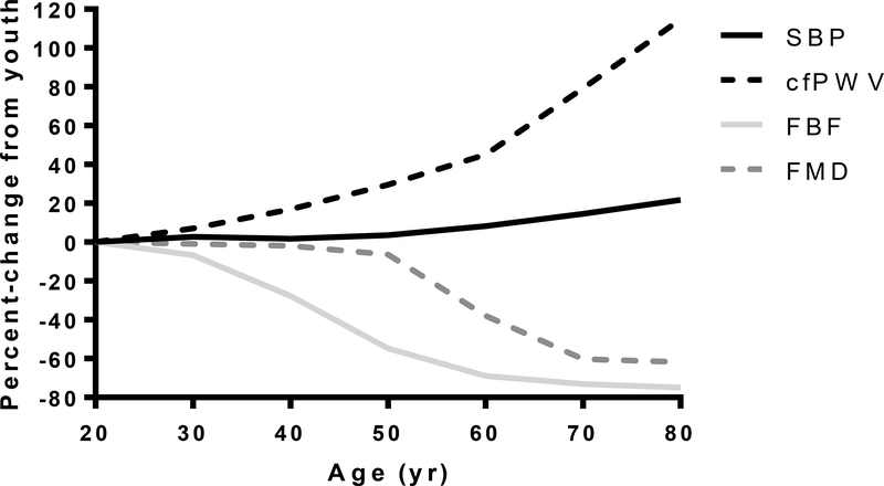
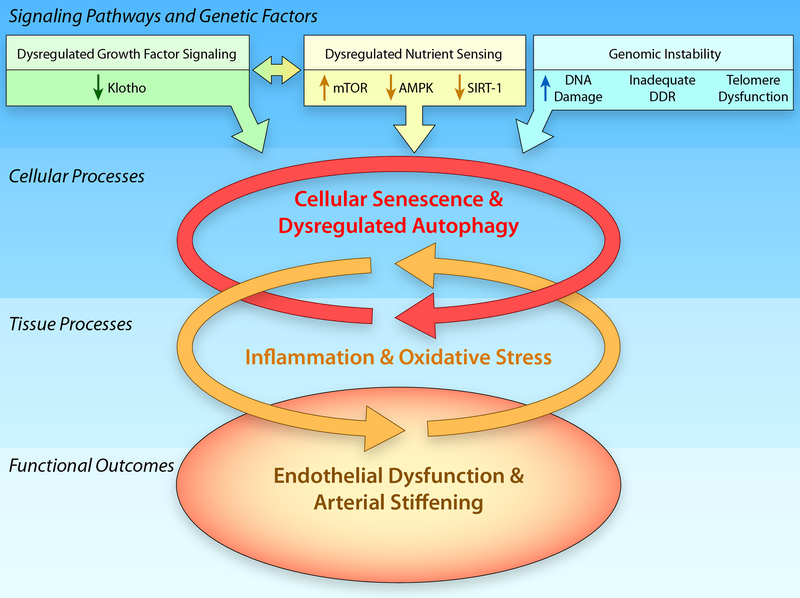
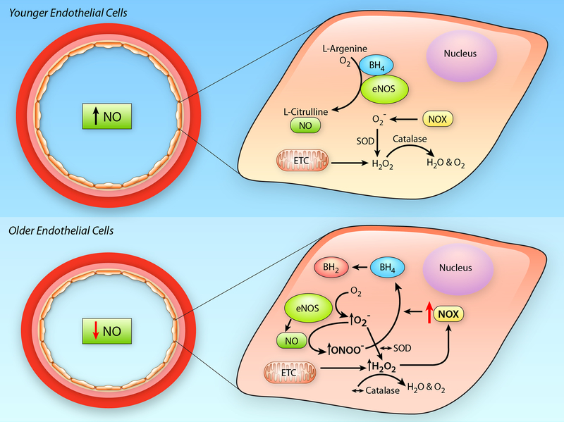
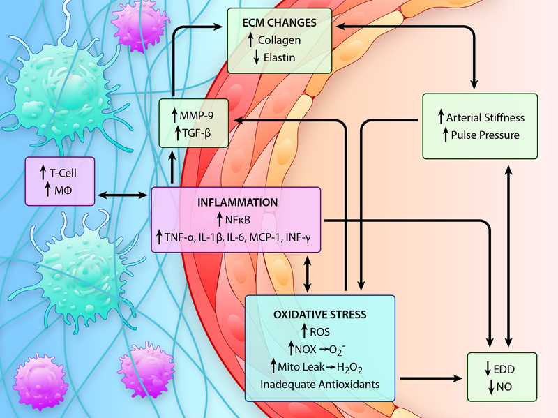
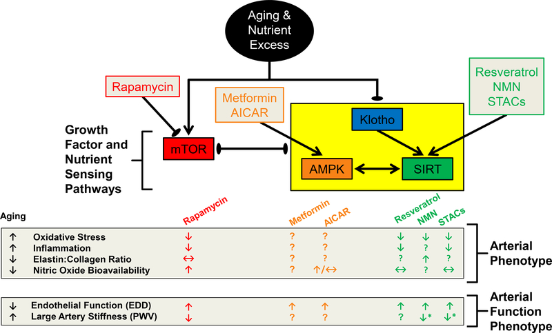
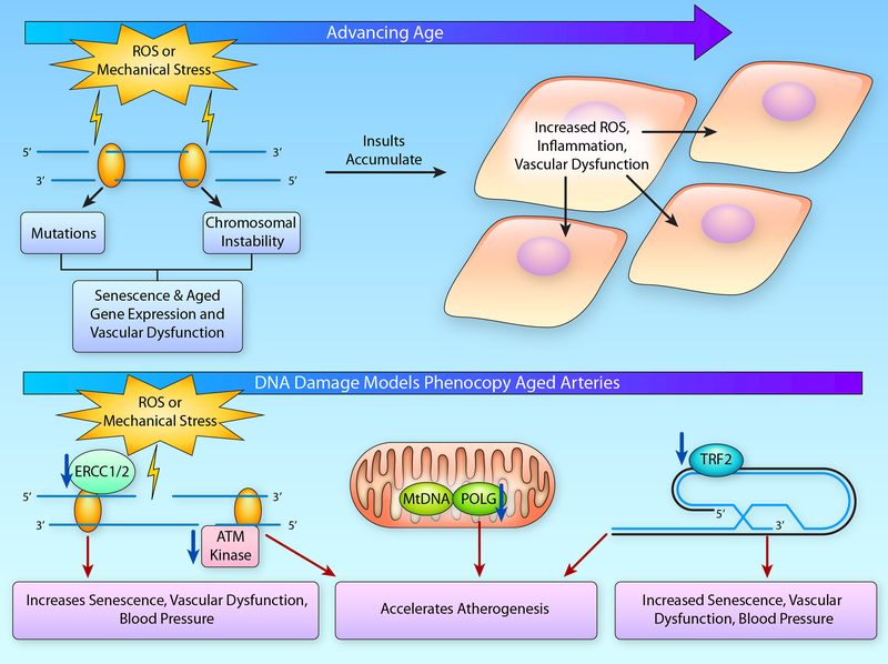
Advancing age promotes cardiovascular disease (CVD), the leading cause of death in the United States and many developed nations. Two major age-related arterial phenotypes, large elastic artery stiffening and endothelial dysfunction, are independent predictors of future CVD diagnosis and likely are responsible for the development of CVD in older adults. Not limited to traditional CVD, these age-related changes in the vasculature also contribute to other age-related diseases that influence mammalian health span and potential life span. This review explores mechanisms that influence age-related large elastic artery stiffening and endothelial dysfunction at the tissue level via inflammation and oxidative stress and at the cellular level via Klotho and energy-sensing pathways (AMPK [AMP-activated protein kinase], SIRT [sirtuins], and mTOR [mammalian target of rapamycin]). We also discuss how long-term calorie restriction-a health span- and life span-extending intervention-can prevent many of these age-related vascular phenotypes through the prevention of deleterious alterations in these mechanisms. Lastly, we discuss emerging novel mechanisms of vascular aging, including senescence and genomic instability within cells of the vasculature. As the population of older adults steadily expands, elucidating the cellular and molecular mechanisms of vascular dysfunction with age is critical to better direct appropriate and measured strategies that use pharmacological and lifestyle interventions to reduce risk of CVD within this population.

Keywords: aging; endothelium; inflammation; oxidative stress; telomere.


Figures

Figure 1.
Figure 1.. Age-Associated Cardiovascular Changes.
Predicted percent-change from youth in markers of vascular function: brachial systolic blood pressure (SBP); carotid-to-femoral pulse wave velocity (cfPWV); maximal forearm blood flow to acetylcholine (FBF); brachial flow-mediated dilation (FMD). All lines are the percent of the predicted fold-change from reference values at 20 yr. Reference values: SBP 110 mmHg; cfPWV 5.9 m/sec; FBF 897 Δ%; FMD 9%.
Figure 2.
Figure 2.. Hallmarks of Vascular Aging.
In addition to increases in genomic instability that are associated with increases in DNA damage, an inadequate DNA damage response (DDR) and telomere dysfunction, dysregulation of signaling pathways, including the growth factor signaling pathway, klotho, and the nutrient sensing pathways, mammalian Target of Rapamycin (mTOR), adenosine monophosphate protein kinase (AMPK), and sirtuin-1 (SIRT-1) will induce increases in cellular senescence and the dysregulation of autophagy that contribute to increased tissue oxidative stress and inflammation that ultimately result in the age-related arterial phenotype characterized by endothelial dysfunction and arterial stiffening. (Illustration Credit: Ben Smith).
Figure 3.
Figure 3.. Impact of Aging on Endothelial Cells.
In younger endothelial cells (Upper Panel), endothelial nitric oxide synthase (eNOS) has adequate cofactor availability, e.g., tetrahydrobiopterin (BH4), and produces nitric oxide (NO) through the conversion of L-arginine to L-citrulline. Reactive oxygen species (ROS), e.g., superoxide (O2) and hydrogen peroxide (H2O2), produced by the mitochondrial electron transport chain (ETC) or cytosolic oxidant enzymes, such as NADPH oxidase (NOX), are quenched by endogenous antioxidant enzymes (superoxide dismutase [SOD] and catalase). In older endothelial cells (Lower Panel), ROS produced in the mitochondria increase NOX mediated O2, this quenches NO bioavailability, through its conversion to peroxynitrite (ONOO), as well as uncouple eNOS by reducing BH4 availability. In the face of unchanged antioxidant defenses, these effects lead to a reduction in NO bioavailability and a pro-oxidant phenotype in the aged endothelium. (Illustration Credit: Ben Smith).
Figure 4.
Figure 4.. Mechanisms of Age-Associated Arterial Dysfunction.
The vascular aging phenotype is characterized by increased large artery stiffness and pulse pressure and reduced endothelium dependent dilation (EDD) and nitric oxide (NO) bioavailability. Oxidative stress, caused by increased production of reactive oxygen species (ROS) in the absence of an adequate antioxidant defense, and inflammation are two interconnected mechanisms that underlie arterial dysfunction in advanced age. Increases in oxidant production with aging are associated with increased NADPH oxidase (NOX)- and mitochondrial-produced ROS, such as superoxide (O2-) and hydrogen peroxide (H2O2). These ROS act to (1) quench bioavailable NO, thus limiting EDD, (2) induce inflammatory signaling through nuclear factor kappa B (NFκB) activation and (3) induce matrix metalloproteinase (MMP)-9 and transforming growth factor beta (TGF-β) that contribute to alterations in the extracellular matrix (ECM) including increases in collagen content and decreases or fragmentation of elastin that contribute to increases in arterial stiffening. In a vicious cycle, increased NFκB activation also exacerbates both oxidative stress and inflammation by transcribing oxidant enzymes such as NOX, pro-inflammatory cytokines and adhesion molecules, such as tumor necrosis factor (TNF)-α, interleukin (IL)-1β, IL-6 and monocyte chemoattractant protein (MCP)-1, and this contributes to downstream increases in MMP-9 and TGF-β expression. Increased adhesion molecule expression contributes to the infiltration of immune cells such as T cells and macrophages (Mφ) to the perivascular tissues and these immune cells further exacerbate inflammation and oxidative stress through the production of cytokines, such as interferon(INF)-γ and TNF-α, as well as O2-. (Illustration Credit: Ben Smith).
Figure 5.
Figure 5.. Summary of the Interdependence of Klotho and Nutrient Sensing Pathways in the Face of Advancing Age and Nutrient Excess, and Strategies to Reverse Age-Related Arterial Phenotypes.
↓ represents a reduction, ↑ represents an increase, ↔ represents weak or conflicting evidence and? Represents a lack of available data for the indicated outcome. Black arrows indicate pathway and drug interactions, with arrow points indicating induction and ovals indicating inhibition. The yellow box indicates that these pathways respond in a similar direction to the interacting pathway indicated by the black lines. AICAR, aminoimidazole carboxamide ribonucleotide; AMPK, AMP-activated protein kinase; mTOR, mammalian target of rapamycin; NMN, nicotamide mononucleotide; SIRT, sirtuin; STACs, sirtuin activating compounds.
Figure 6.
Figure 6.. DNA Damage with Aging.
(A) With advancing age, DNA damage resulting from reactive oxygen species (ROS) or mechanical stress accumulates leading to mutations or chromosomal instability that ultimately contributes to cellular senescence or altered gene expression that drives the age-related pro-inflammatory and pro-oxidant vascular phenotype. This cellular dysfunction acts in an autocrine and paracrine manner affecting the local milieu and exacerbating endothelial dysfunction. (B) Indeed, DNA damage induced by ionizing radiation leads to cellular senescence and vascular dysfunction. Likewise, experimental manipulation genes involved in telomere capping, such as telomeric repeat binding factor (TRF)-2, and those involved in both nuclear DNA repair, including excision repair cross-complementation group (ERCC)-1/2 and ataxia-telangiectasia mutated (ATM) kinase, as well as in mitochondrial (Mt) DNA repair, such as polymerase gamma (POLG), will lead to increases in cellular senescence, vascular dysfunction and atherogenesis. (Illustration Credit: Ben Smith).

References

    1. Heron MP. Deaths: Leading causes for 2011. 2015
    1. Health, united states, 2010: With special feature on death and dying. Hyattsville (MD); 2011. - PubMed
    1. Lakatta EG, Levy D. Arterial and cardiac aging: Major shareholders in cardiovascular disease enterprises: Part i: Aging arteries: A “set up” for vascular disease. Circulation. 2003;107:139–146 - PubMed
    1. Widlansky ME, Gokce N, Keaney JF, Jr., Vita JA. The clinical implications of endothelial dysfunction. J Am Coll Cardiol. 2003;42:1149–1160 - PubMed
    1. Franklin SS, Khan SA, Wong ND, Larson MG, Levy D. Is pulse pressure useful in predicting risk for coronary heart disease? The framingham heart study. Circulation. 1999;100:354–360 - PubMed

Publication types

MeSH terms

Substances